Loading video…


 11 Concepts
 40.2K views

Video Summary: What Is Mechanism of Filopodia Formation
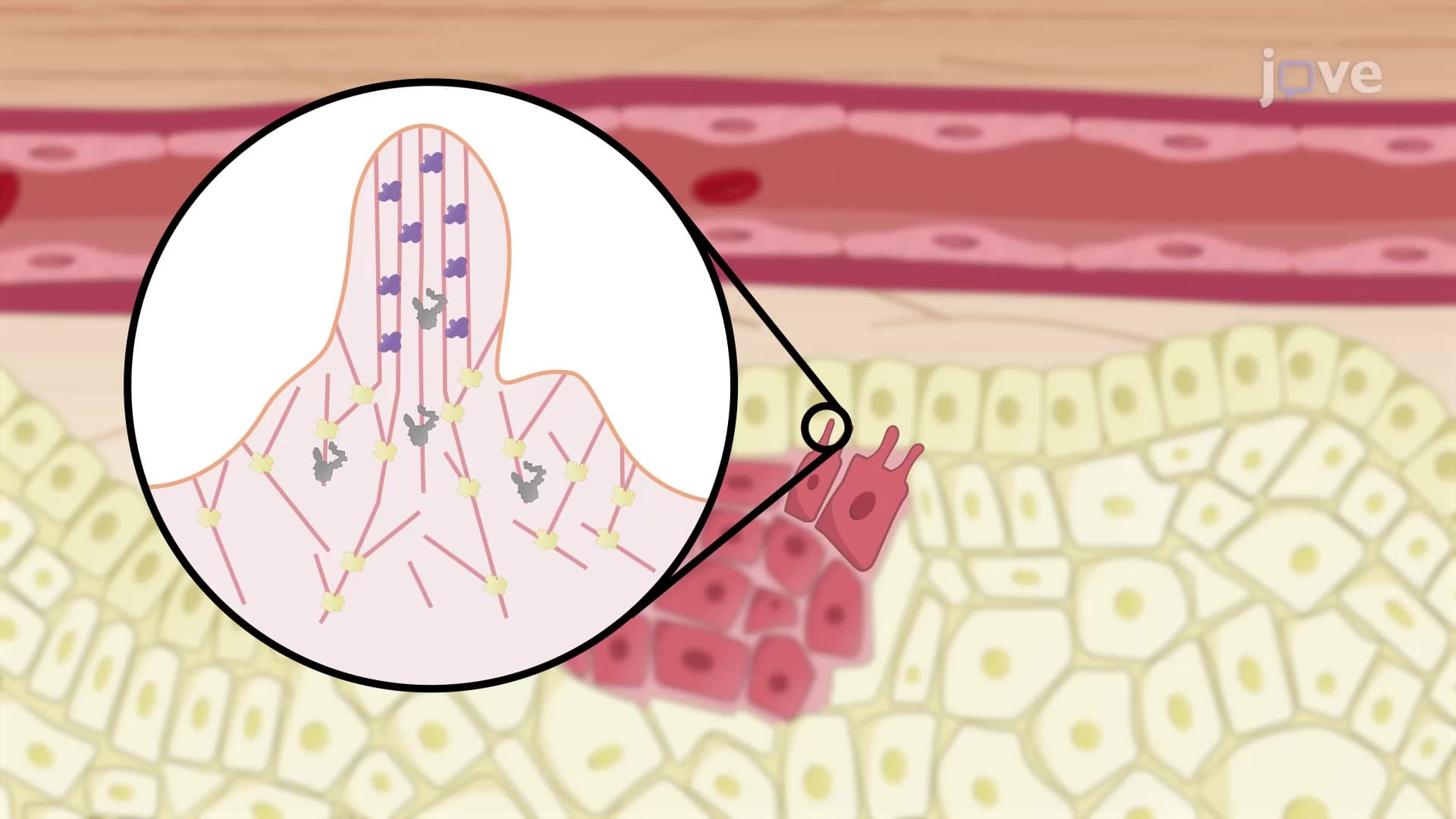
Did you know that cancer cells use tiny finger-like projections to "feel" their way through your body during metastasis? The mechanism of filopodia formation explains how cells create these crucial cellular extensions through a coordinated dance of proteins and membrane reshaping. When researchers at Stanford University study how melanoma cells invade healthy tissue, they're observing this exact process in action. Understanding what is mechanism of filopodia formation reveals how cells navigate, communicate, and move throughout our bodies. Watch the full video on JoVE Coach to master this concept with expert-led visuals and step-by-step explanations.
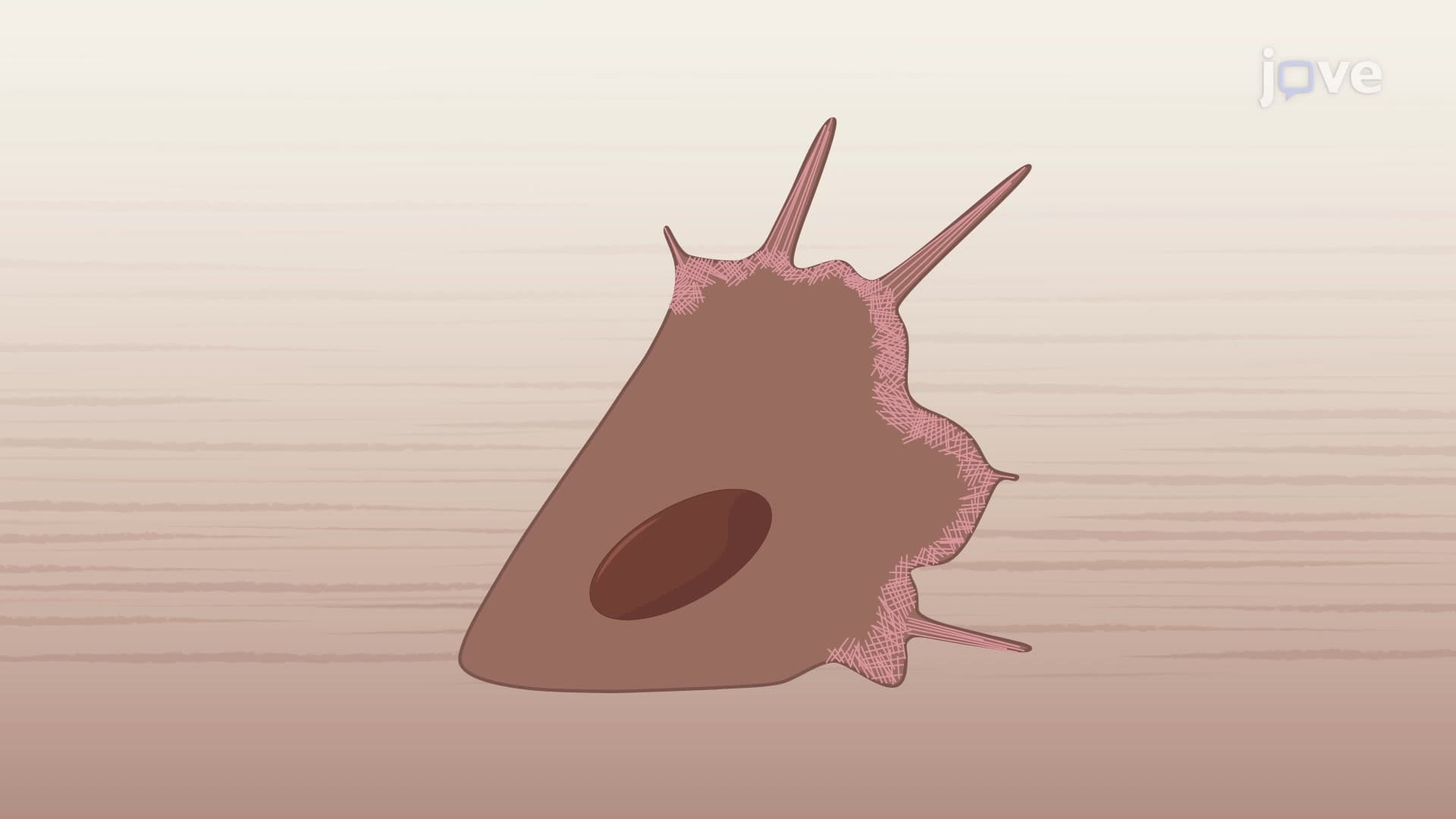
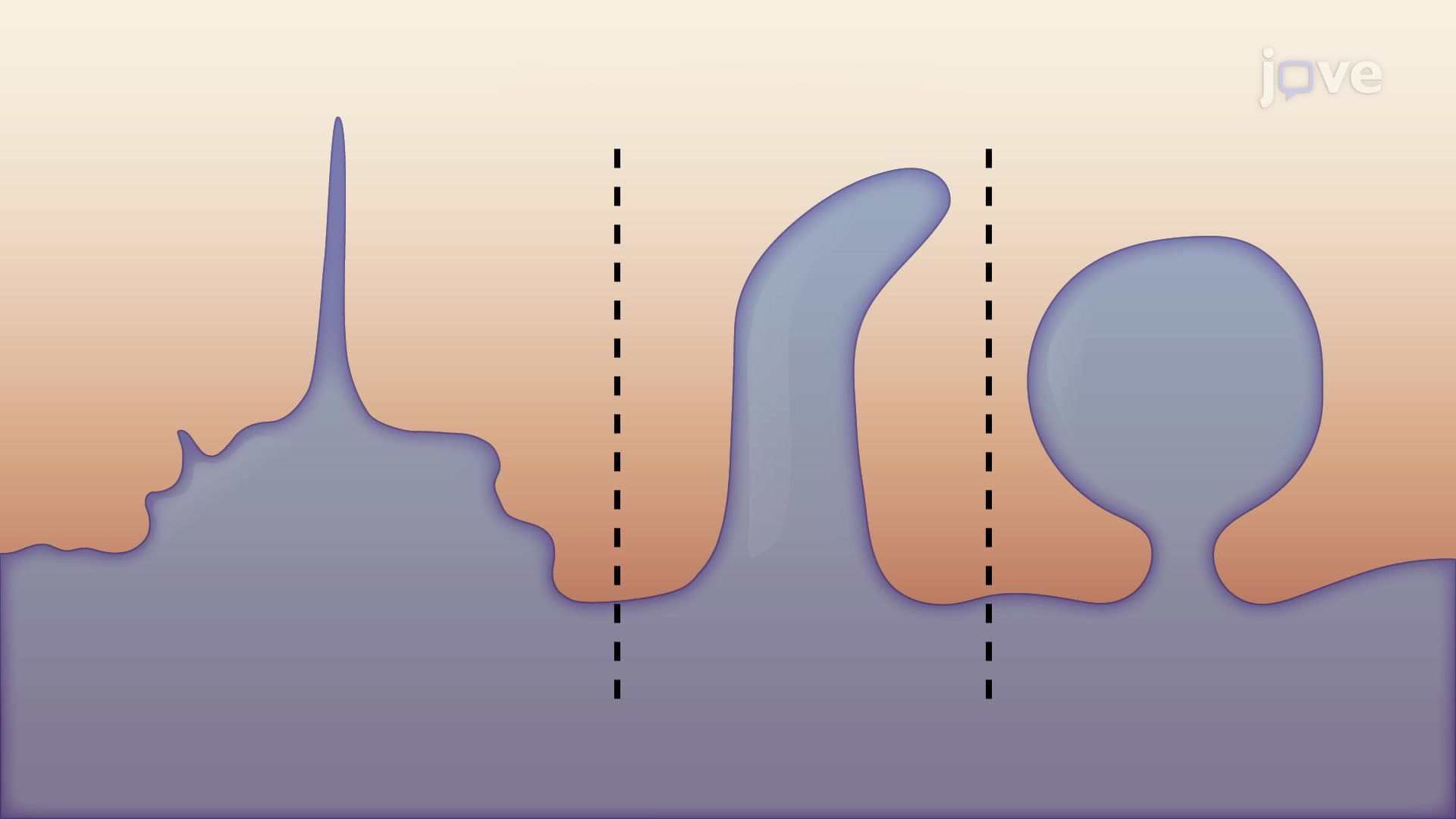
The mechanism of filopodia formation represents one of cell biology's most elegant examples of coordinated molecular machinery. These thin, finger-like cellular projections serve as the cell's sensory antennae, allowing it to probe the environment, detect chemical gradients, and establish directional movement. For students preparing for AP Biology or college-level cell biology courses, mastering this concept provides crucial insight into how cells achieve dynamic behavior through protein interactions.
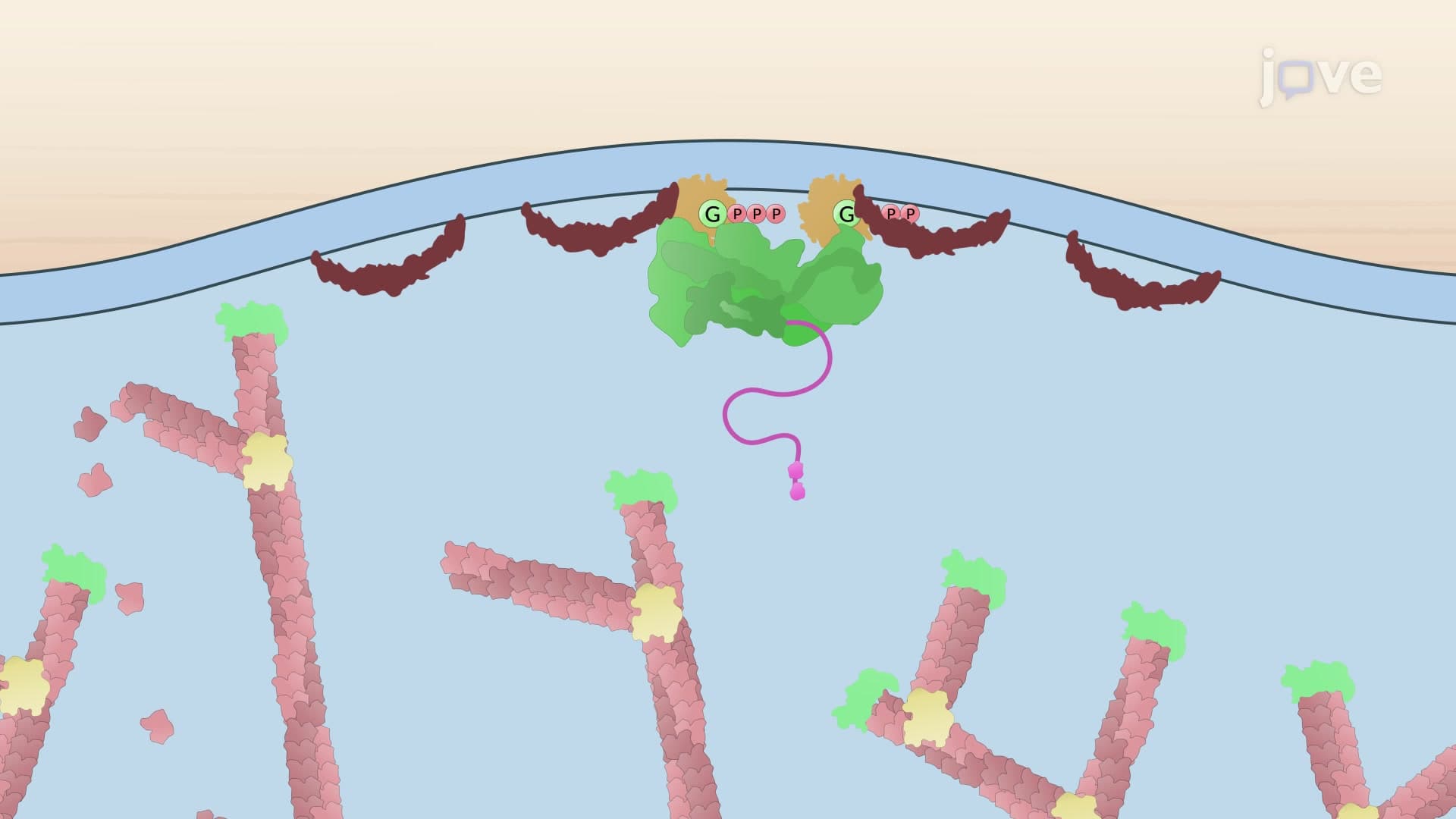
The process begins when cells receive external stimuli-whether from growth factors, mechanical stress, or chemical attractants. This triggers the activation of Cdc42, a small GTPase protein that acts like a molecular switch. When Cdc42 binds GTP (replacing GDP), it undergoes a conformational change that enables it to recruit downstream effector proteins. This activation mechanism appears frequently on MCAT questions, particularly in passages about cell signaling and cancer biology.
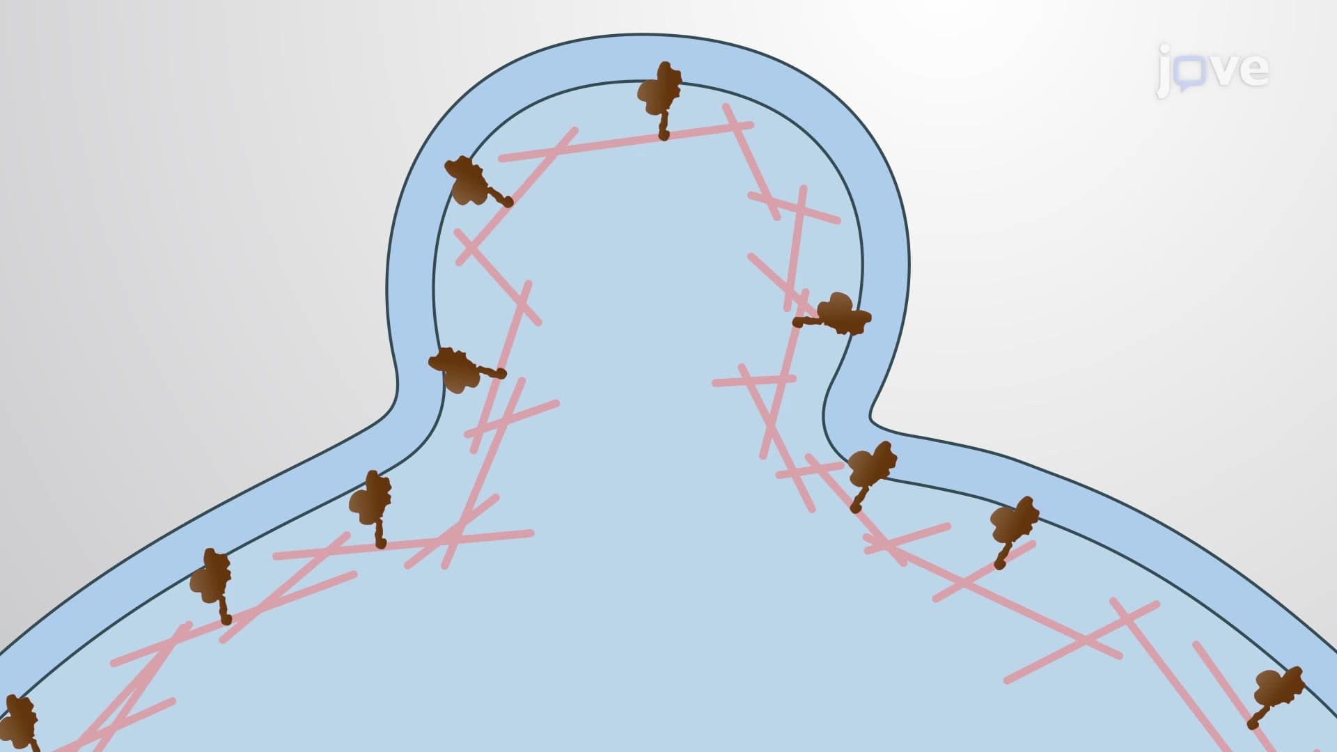
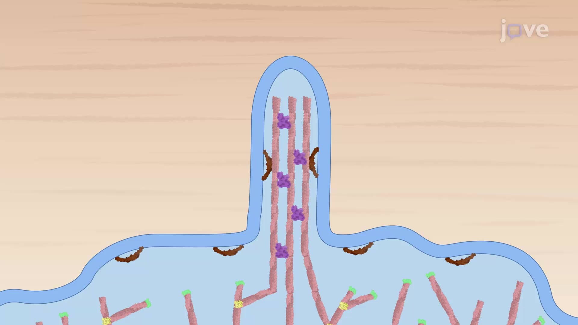
Once activated, GTP-bound Cdc42 recruits IRSp53 dimers to the plasma membrane. IRSp53 serves as a critical scaffolding protein that simultaneously influences both the actin cytoskeleton and membrane structure. The protein's unique ability to bundle actin filaments into parallel arrays while inducing membrane curvature makes it indispensable for filopodium formation. Students studying for the USMLE Step 1 often encounter this protein in pathology contexts, as IRSp53 dysfunction contributes to certain neurological disorders and cancer metastasis.
The recruitment of Ena/VASP proteins to filament ends represents a crucial step in filopodium extension. These proteins displace capping proteins that normally prevent filament growth, allowing rapid actin polymerization. Simultaneously, IRSp53 clusters phosphatidylinositol 4,5-bisphosphate (PIP2) molecules at the membrane's inner surface, creating asymmetric lipid distribution that promotes membrane bending. This dual mechanism-cytoskeletal push and membrane pull-generates the force necessary to protrude the cell membrane outward. The resulting structure gains stability through fascin-mediated cross-linking, which transforms loose actin filaments into rigid bundles capable of withstanding extracellular forces.
Related Micro-courses