Loading video…


 11 Concepts
 40.2K views

Video Summary: What Is Mechanism of Lamellipodia Formation
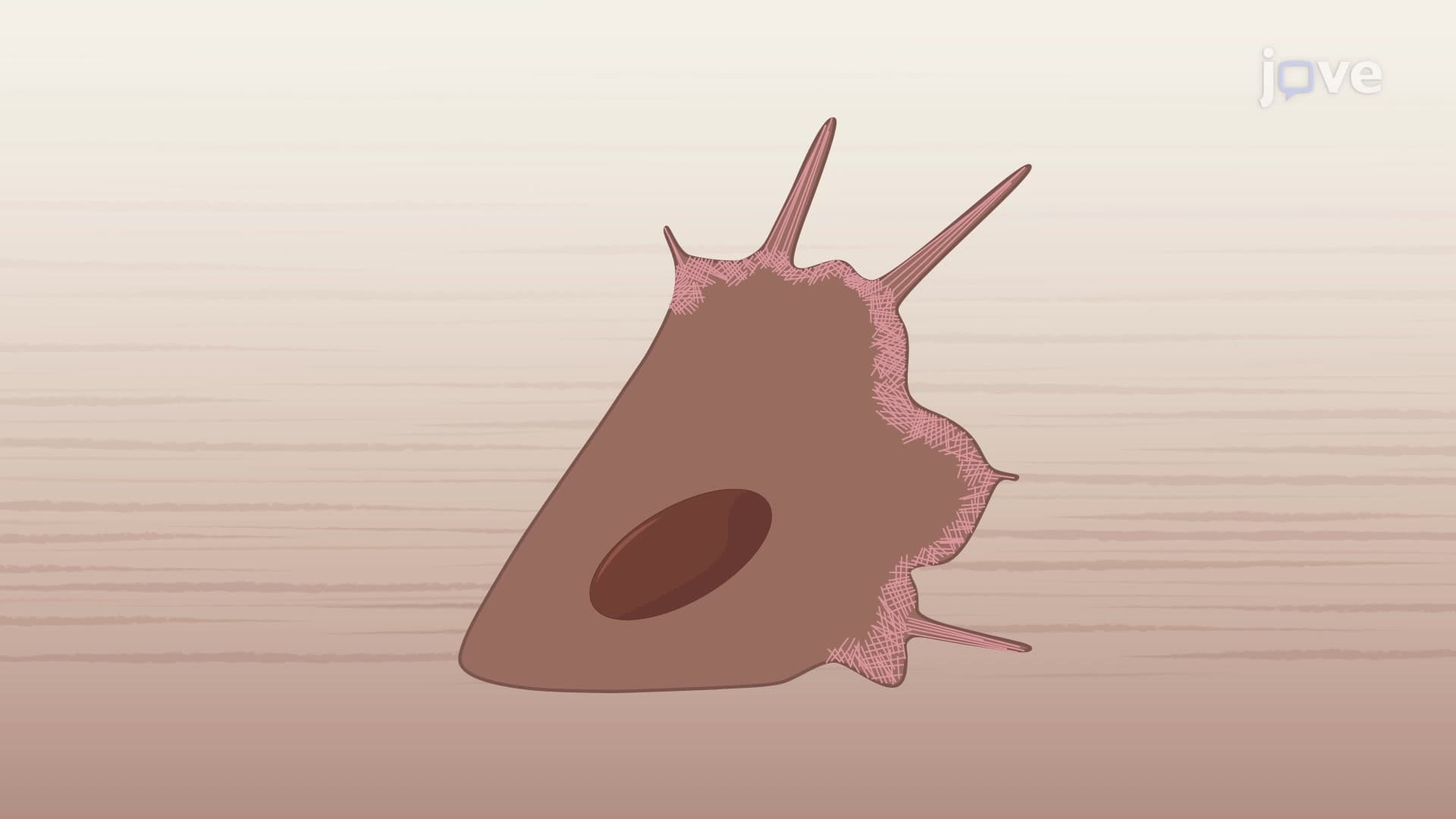
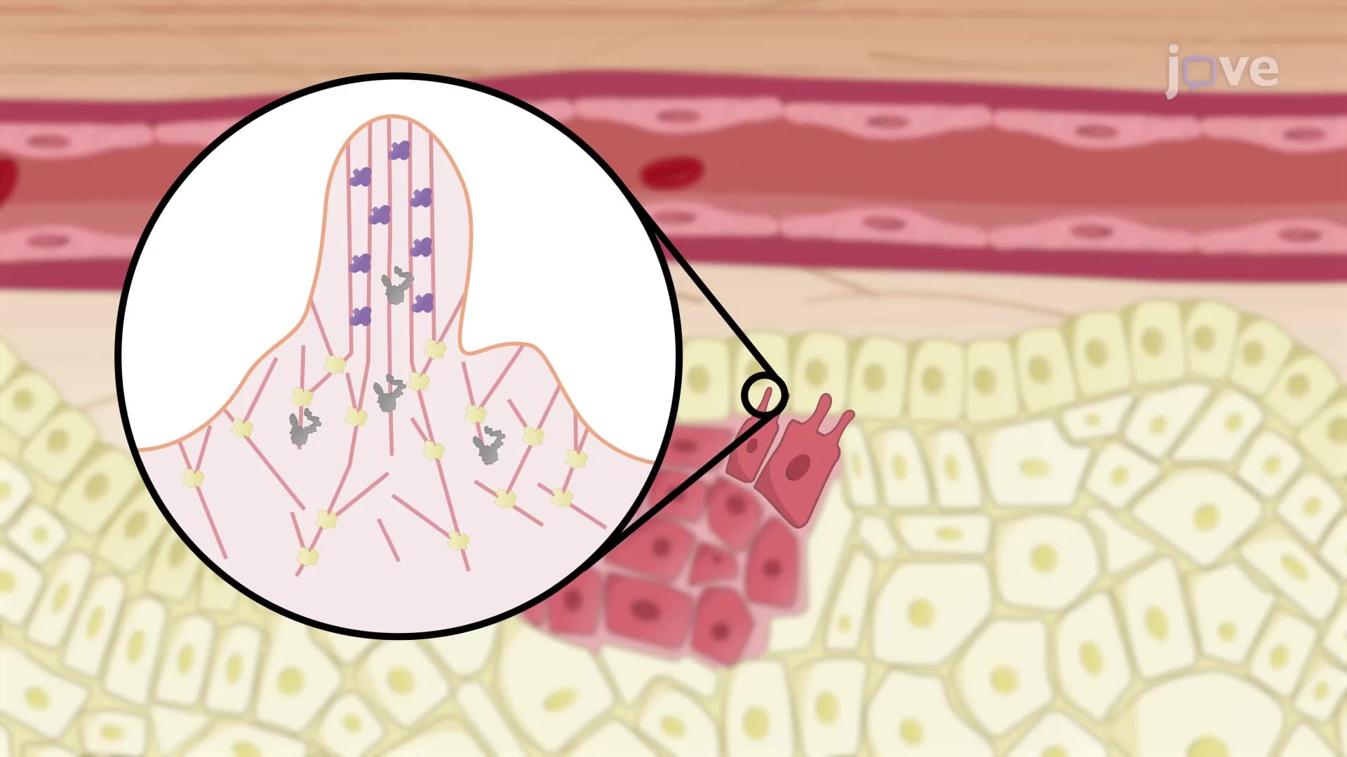
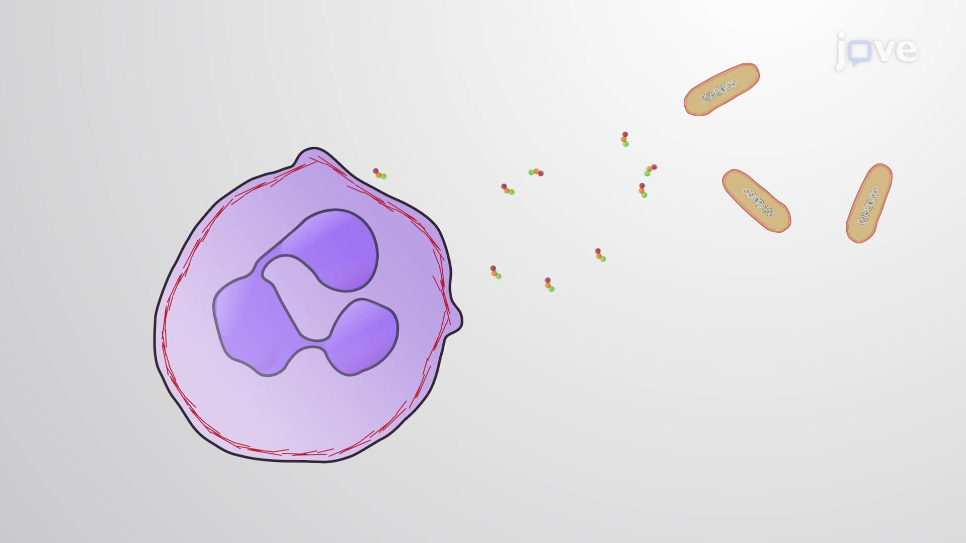
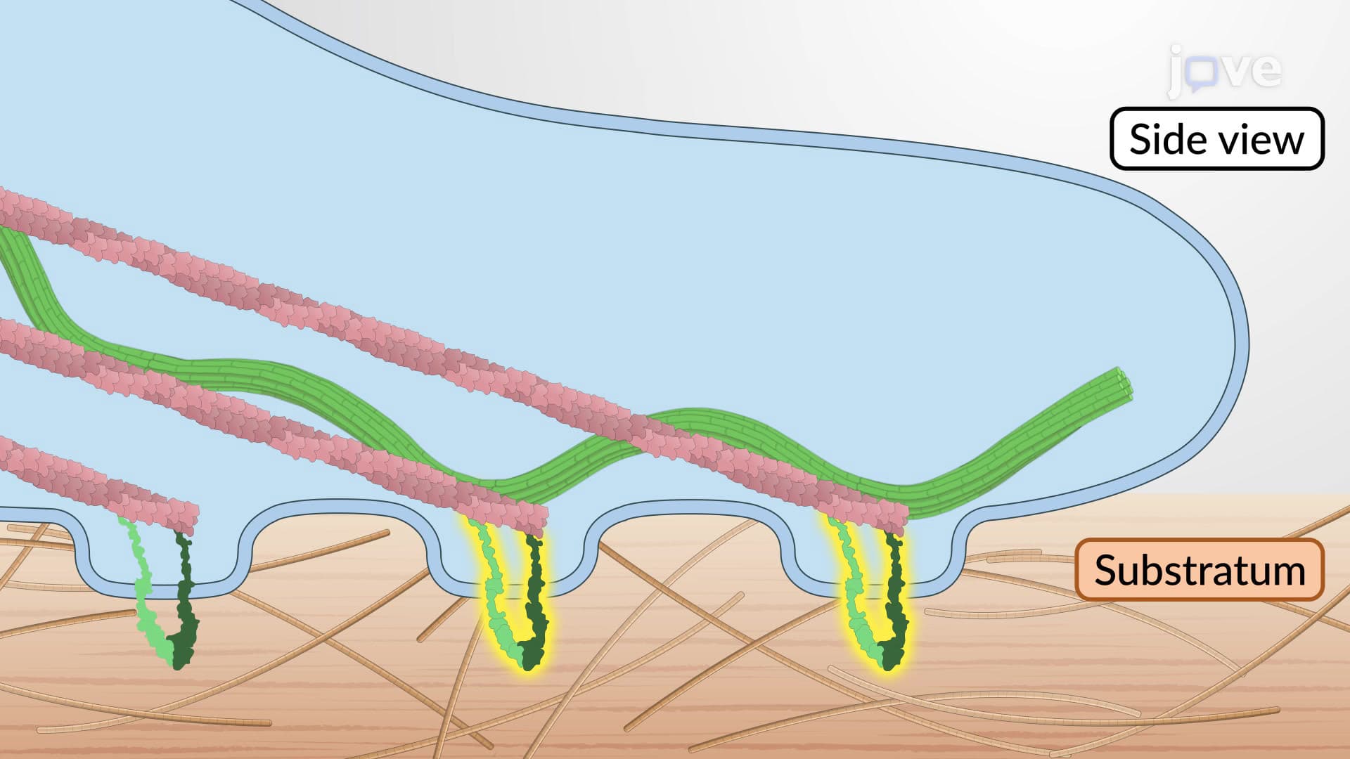
Ever wonder how cancer cells migrate through your body or how immune cells chase down infections? The mechanism of lamellipodia formation explains how cells create their "crawling feet", dynamic membrane protrusions that enable cellular movement. During wound healing at Massachusetts General Hospital, researchers observe these actin-powered structures driving skin cell migration to close injuries. This cellular machinery involves a sophisticated cascade from growth factor binding to membrane extension, orchestrated by proteins like Rac1, WAVE complex, and Arp2/3. Watch the full video on JoVE Coach to master this concept with expert-led visuals and step-by-step explanations.
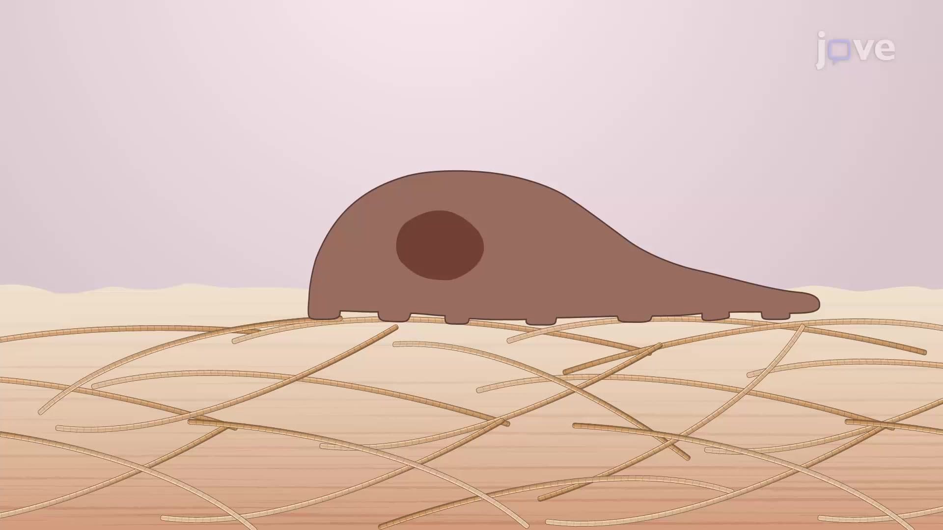
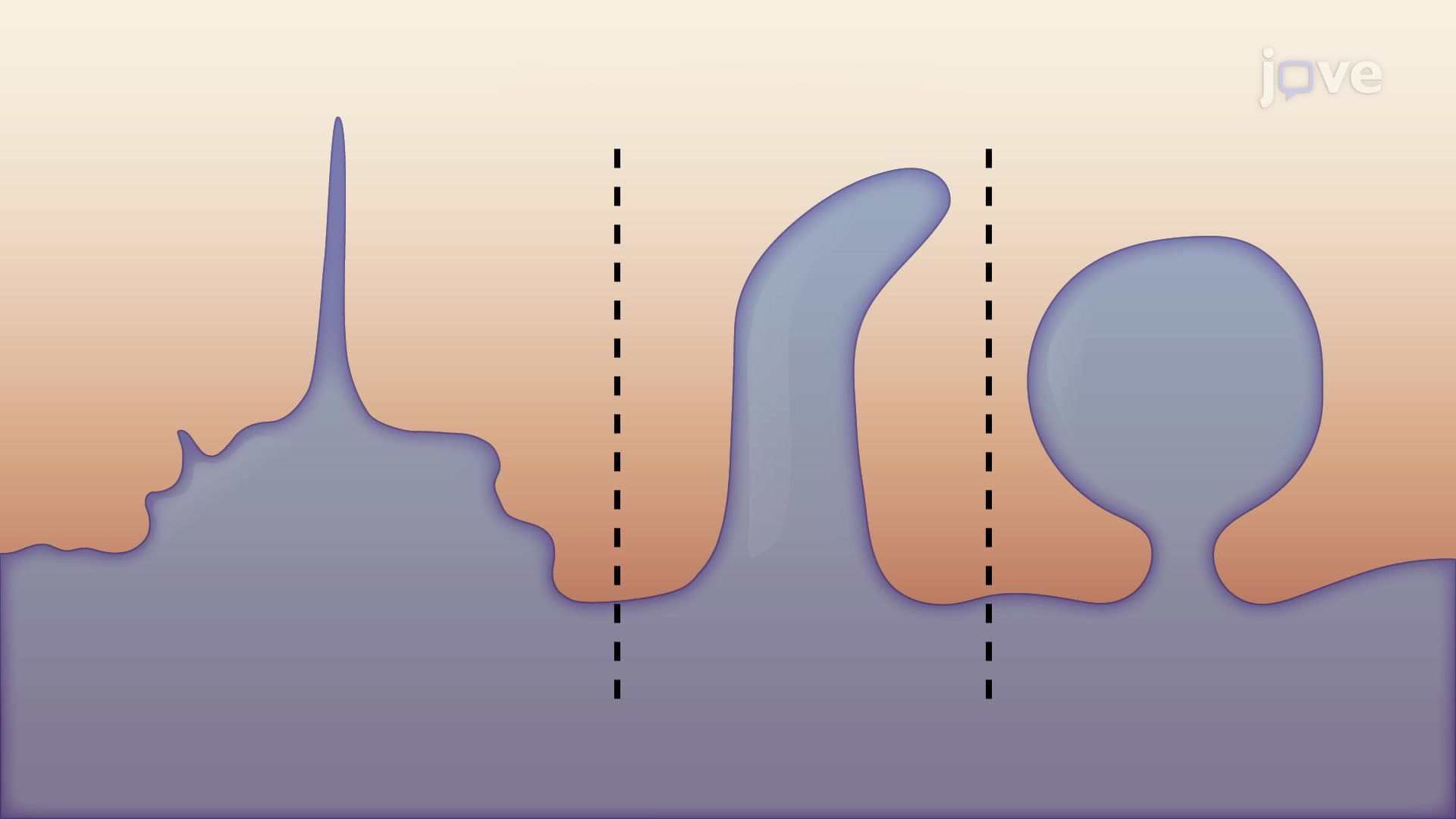
Lamellipodia represent one of biology's most elegant molecular machines, dynamic, sheet-like membrane protrusions that enable cells to crawl, migrate, and explore their environment. Found at the leading edge of motile cells, these structures are essential for processes ranging from embryonic development to immune responses. Students studying for the MCAT or AP Biology will encounter this concept when learning about cell biology and signal transduction.
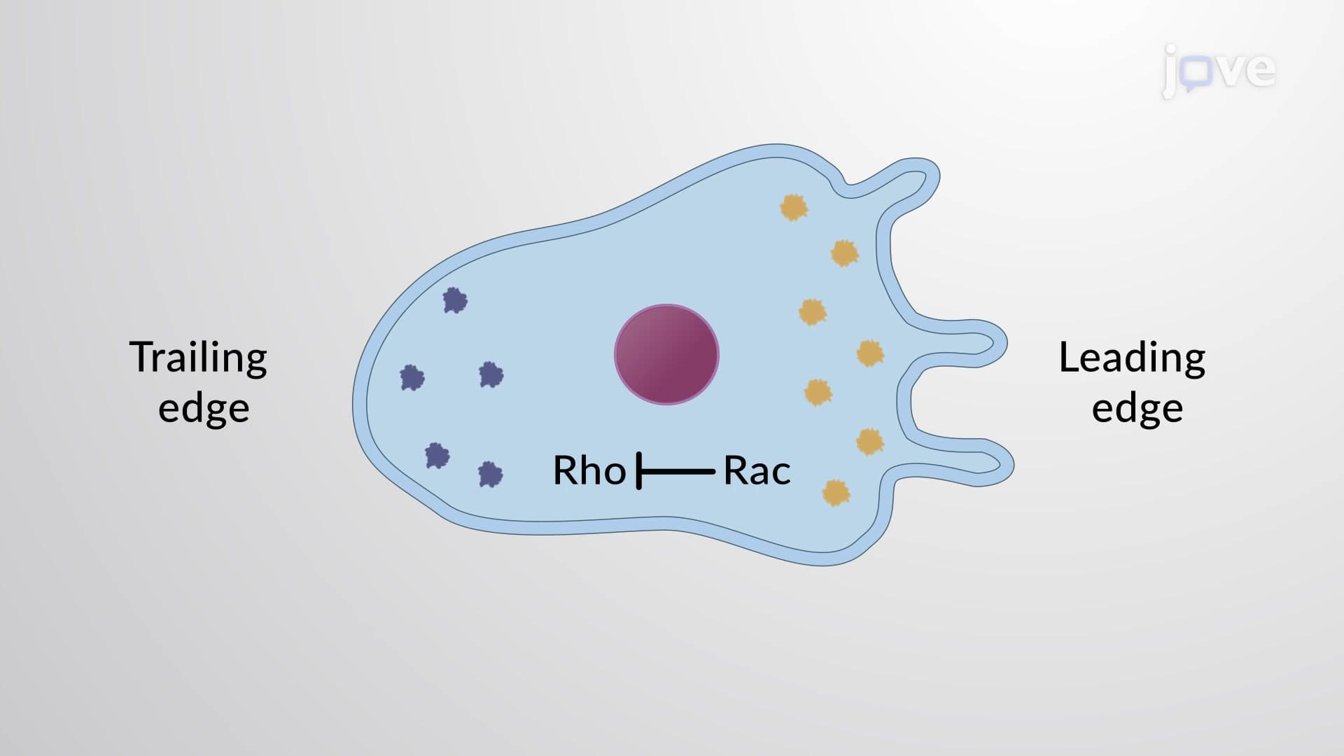
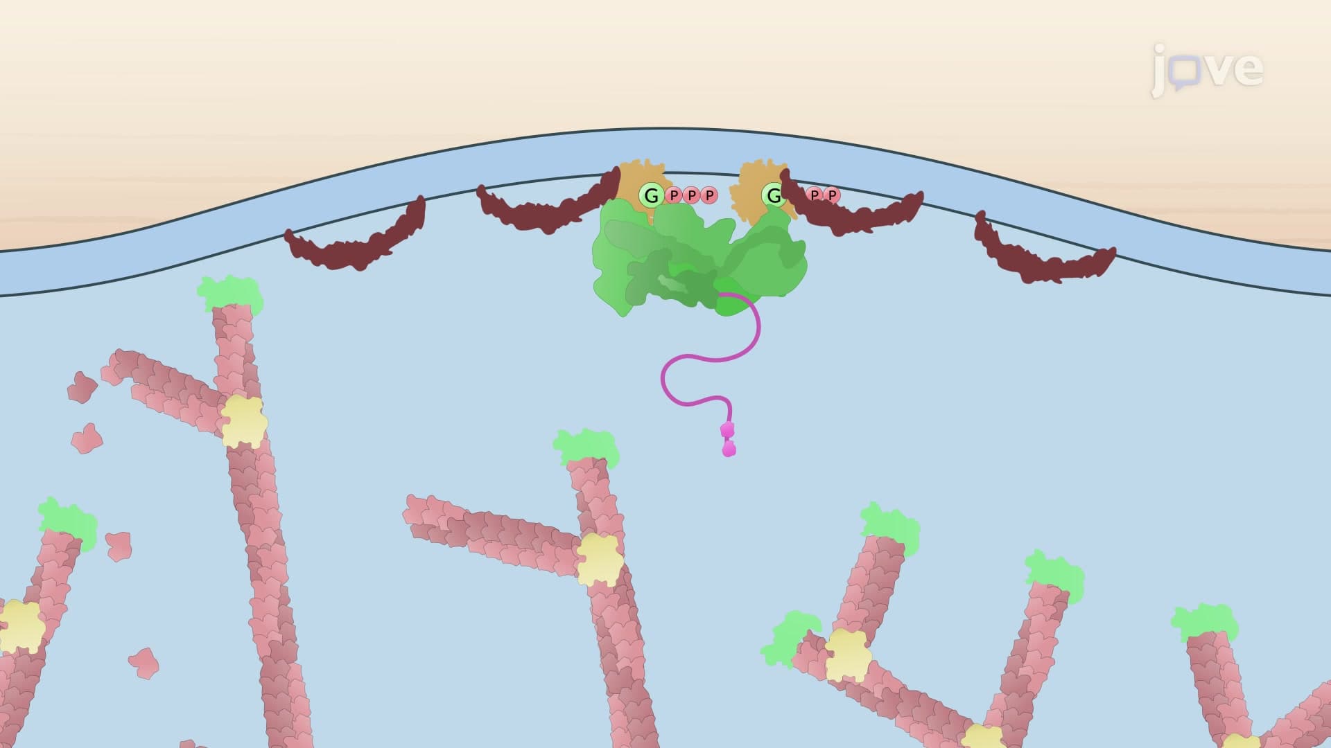
The mechanism begins when extracellular growth factors bind to receptor tyrosine kinases (RTKs) embedded in the cell membrane. This binding event triggers autophosphorylation of the receptor's cytoplasmic domain, creating docking sites for signaling proteins. The activated RTK then stimulates Rac1, a member of the Rho family of small GTPases. When bound to GTP, Rac1 becomes the master regulator of lamellipodia formation, think of it as the "on switch" for cellular crawling. This pathway is frequently tested in college cell biology courses, particularly regarding how external signals translate into cellular responses.
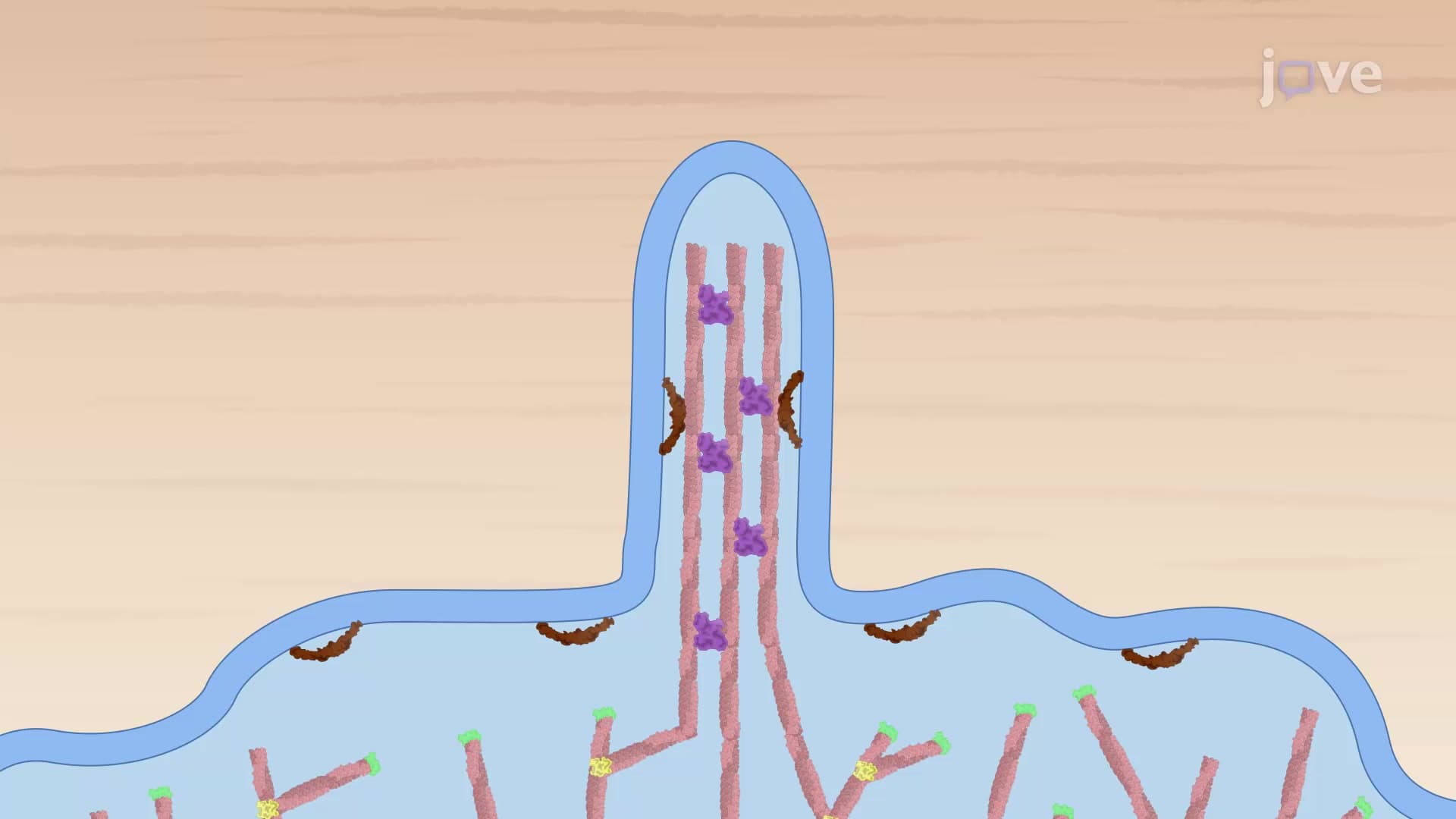
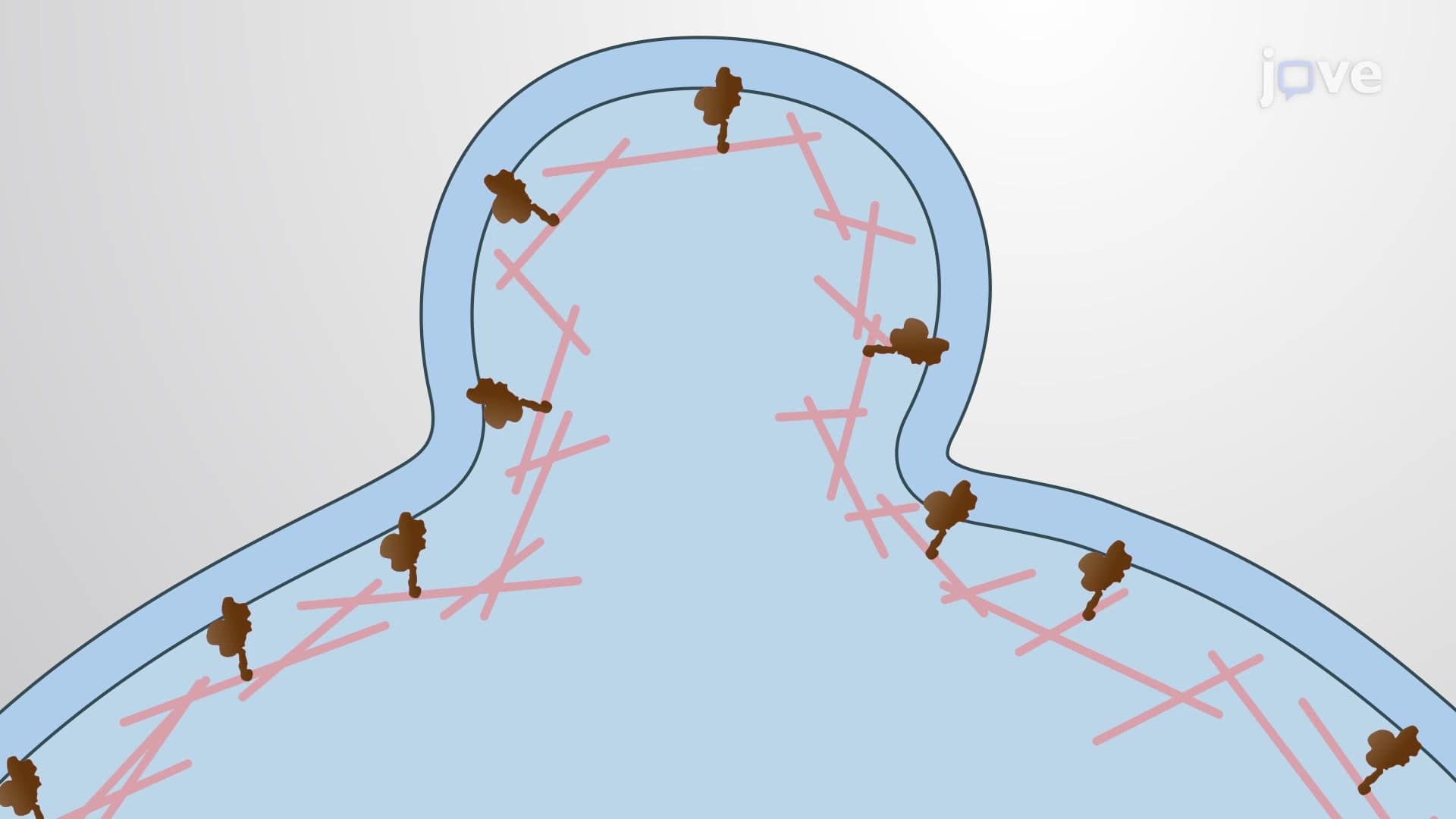
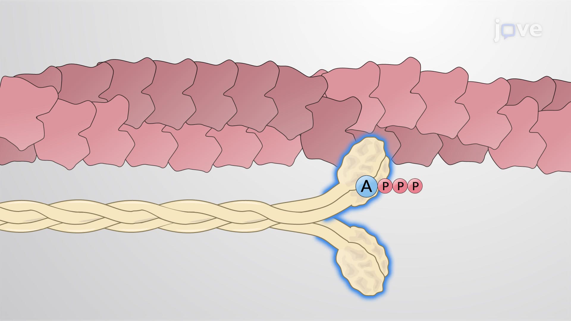
Activated Rac1 recruits the WAVE regulatory complex (WRC) to the plasma membrane, where it becomes a central hub for cytoskeletal reorganization. The WRC then activates two critical components: the Arp2/3 complex and IRSp53 protein. IRSp53 functions as a membrane sculptor, bending the lipid bilayer into the characteristic saddle-shaped curve that defines lamellipodia architecture. Meanwhile, the Arp2/3 complex acts as an actin nucleator, binding to existing actin filaments and creating new branches at 70-degree angles. This branching pattern creates the dense, web-like network that gives lamellipodia their structural integrity.
The beauty of lamellipodia lies in their dynamic nature, powered by actin treadmilling, a process where actin filaments grow at their plus-ends while simultaneously shrinking at their minus-ends. Ena/VASP proteins compete with capping proteins at filament plus-ends, allowing selective elongation that pushes the membrane forward. At the rear of the structure, cofilin severs and depolymerizes older actin filaments, recycling monomers back to the leading edge. This creates a molecular conveyor belt that drives sustained cellular movement. Students preparing for the USMLE Step 1 should understand how disruption of these processes can affect cell migration in cancer metastasis.
Related Micro-courses