Loading video…


 10 Concepts
 76.5K views

Video Summary: Activation and Inactivation of G Proteins Explained
Ever wonder how your morning coffee triggers alertness or why adrenaline makes your heart race during a medical emergency? The activation and inactivation of G proteins serve as molecular switches that convert external signals into cellular responses throughout your body. When a patient receives epinephrine during cardiac arrest at Johns Hopkins Hospital, G protein cycles rapidly switch between "on" and "off" states to amplify life-saving signals. Watch the full video on JoVE Coach to master this concept with expert-led visuals and step-by-step explanations.
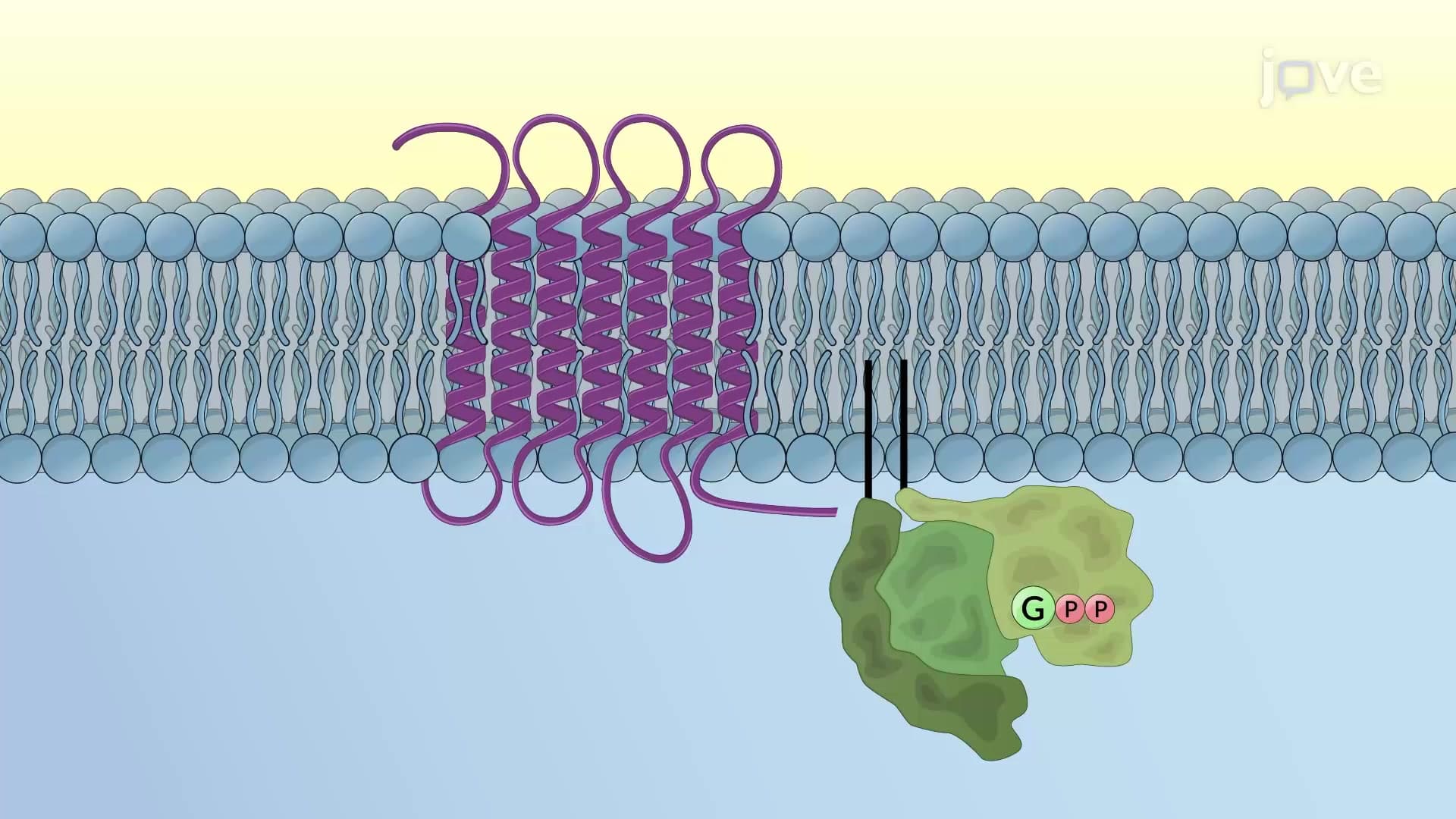
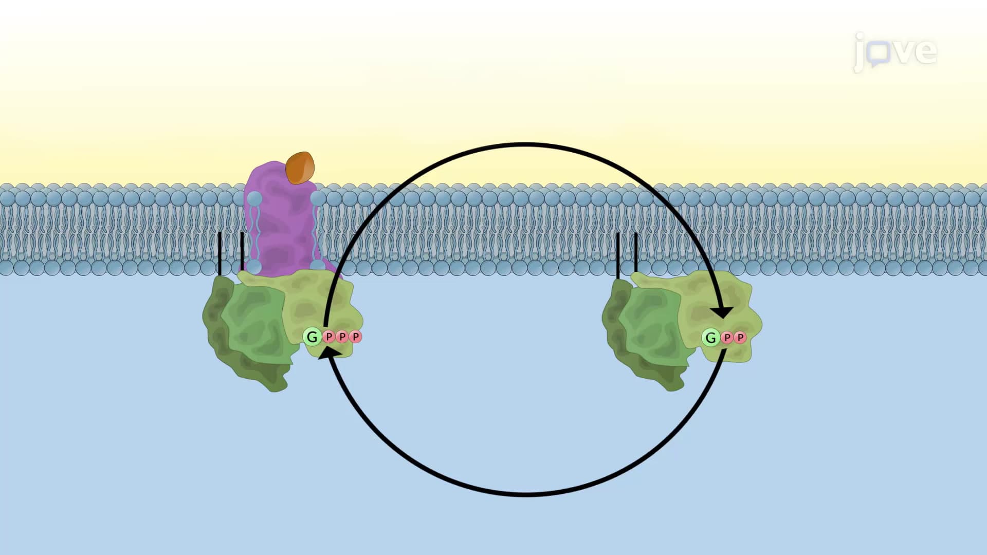
G proteins function as sophisticated molecular switches that translate signals from cell surface receptors into intracellular responses. These heterotrimeric complexes consist of three distinct subunits: the guanosine nucleotide-binding alpha subunit (Gα) and the permanently associated beta-gamma dimer (Gβγ). In their resting state, G proteins remain membrane-bound with GDP occupying the nucleotide-binding pocket of the alpha subunit.
Activation begins when a ligand binds to a G protein-coupled receptor (GPCR), causing conformational changes that expose the G protein's nucleotide-binding site. This exposure facilitates GDP release and GTP binding-a process called nucleotide exchange. The energy released from GTP binding causes the alpha subunit to separate from the beta-gamma dimer, creating two independent signaling units that can simultaneously activate different downstream effectors.
Consider how this works during a stress response: when norepinephrine binds to beta-adrenergic receptors on heart muscle cells, activated Gs-alpha subunits stimulate adenylyl cyclase, while some Gβγ dimers modulate calcium channels. This dual activation amplifies the "fight or flight" response, increasing both heart rate and contractility-mechanisms students encounter in AP Biology and college physiology courses.
Once separated, both G protein subunits can bind and activate specific effector enzymes. Gα-GTP commonly activates adenylyl cyclase (producing cAMP) or phospholipase C-beta (generating IP3 and DAG). Meanwhile, Gβγ dimers can modulate ion channels or activate different enzyme cascades. This parallel processing explains how a single GPCR activation can trigger multiple, simultaneous cellular responses.
G protein inactivation occurs through the alpha subunit's intrinsic GTPase activity, which hydrolyzes bound GTP to GDP and inorganic phosphate. This hydrolysis reduces the alpha subunit's affinity for effector proteins and increases its affinity for the beta-gamma dimer. Reassociation of all three subunits restores the inactive state, terminating the signal. This built-in "timer" mechanism prevents excessive cellular activation and maintains physiological balance-a concept frequently tested on the MCAT and USMLE Step 1.
Related Micro-courses