Loading subject page…


62,212 views

Video Summary: What are Amyloid Fibrils
Did you know that the same protein structures linked to Alzheimer's disease affect over 6.5 million Americans? Amyloid fibrils are highly ordered protein aggregates with distinctive beta-sheet structures that form when proteins misfold and clump together. These rope-like structures can disrupt normal cell function, contributing to diseases like Alzheimer's, Parkinson's, and type 2 diabetes that impact millions of US patients annually. Watch the full video on JoVE Coach to master this concept with expert-led visuals and step-by-step explanations.
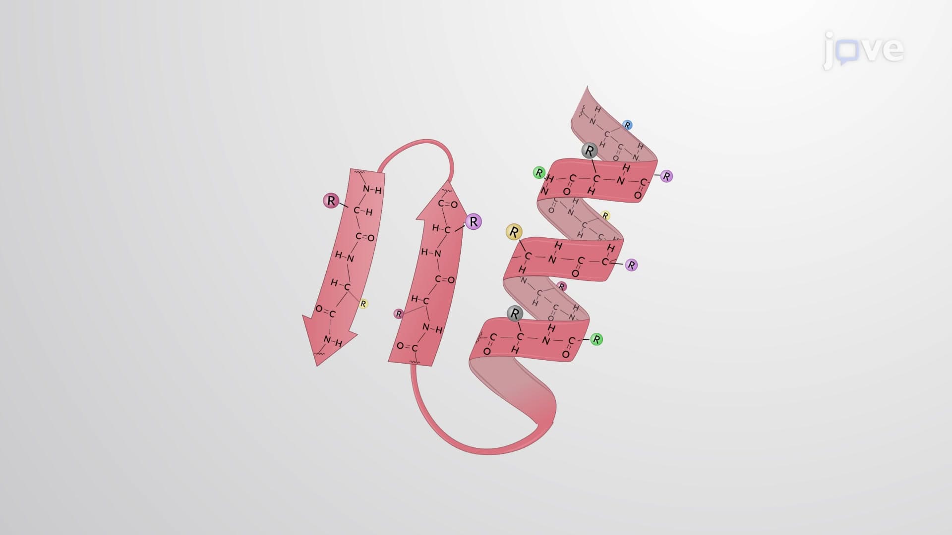
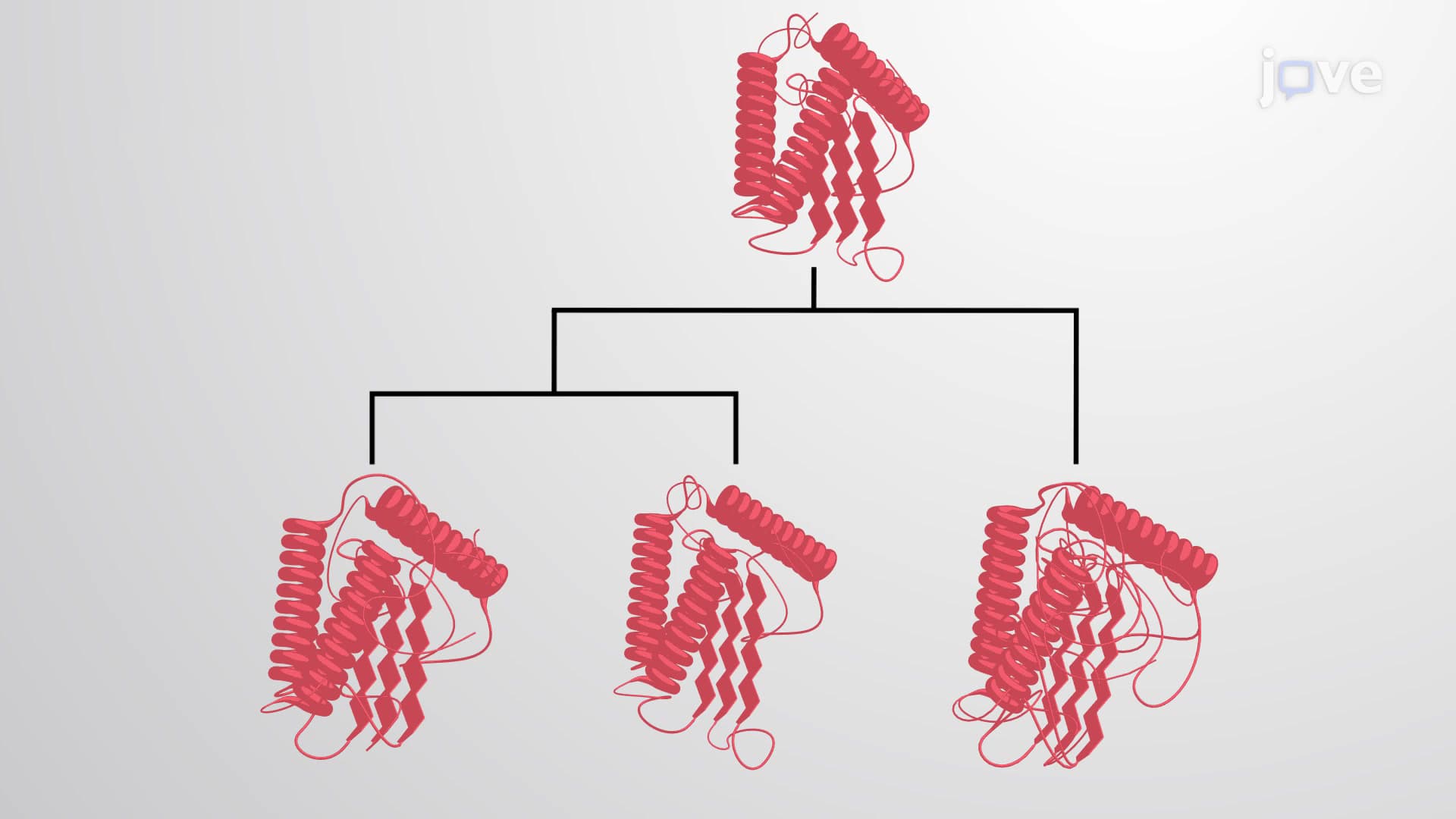
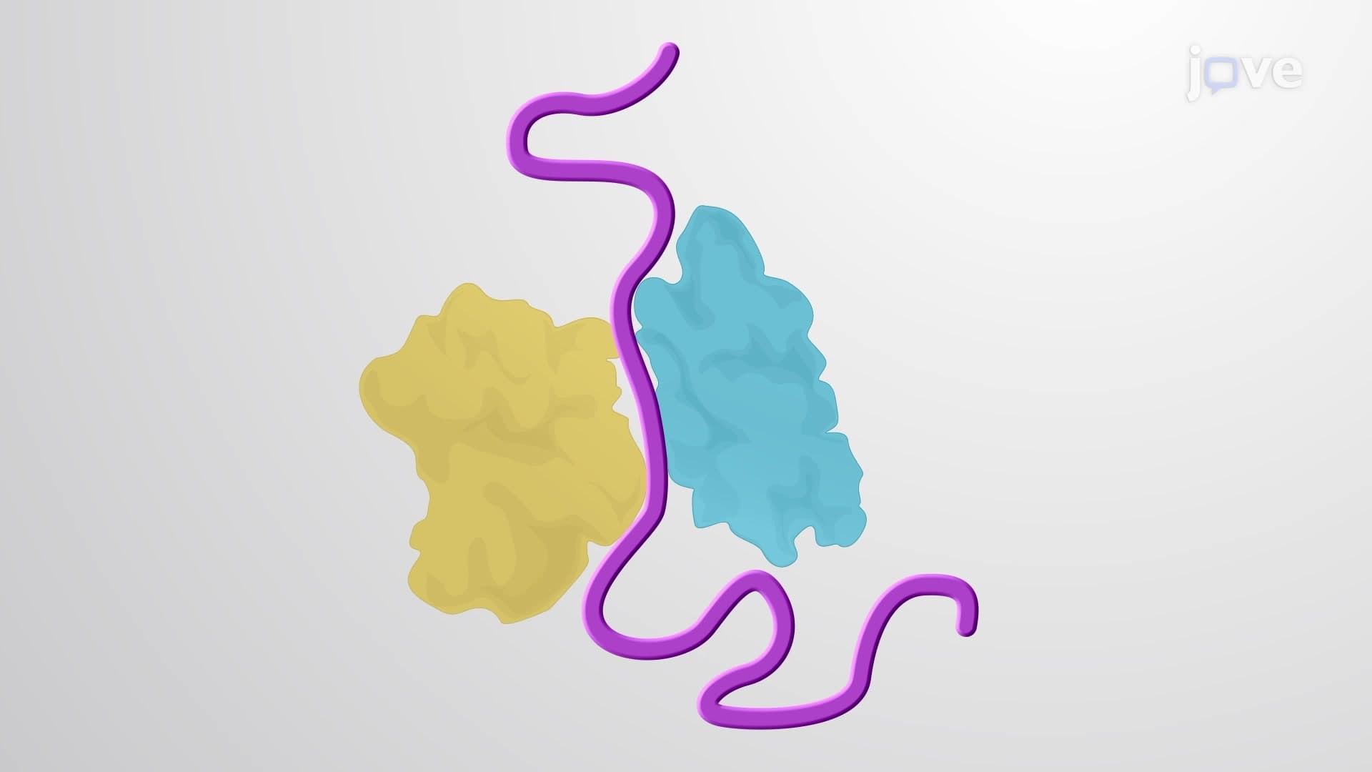
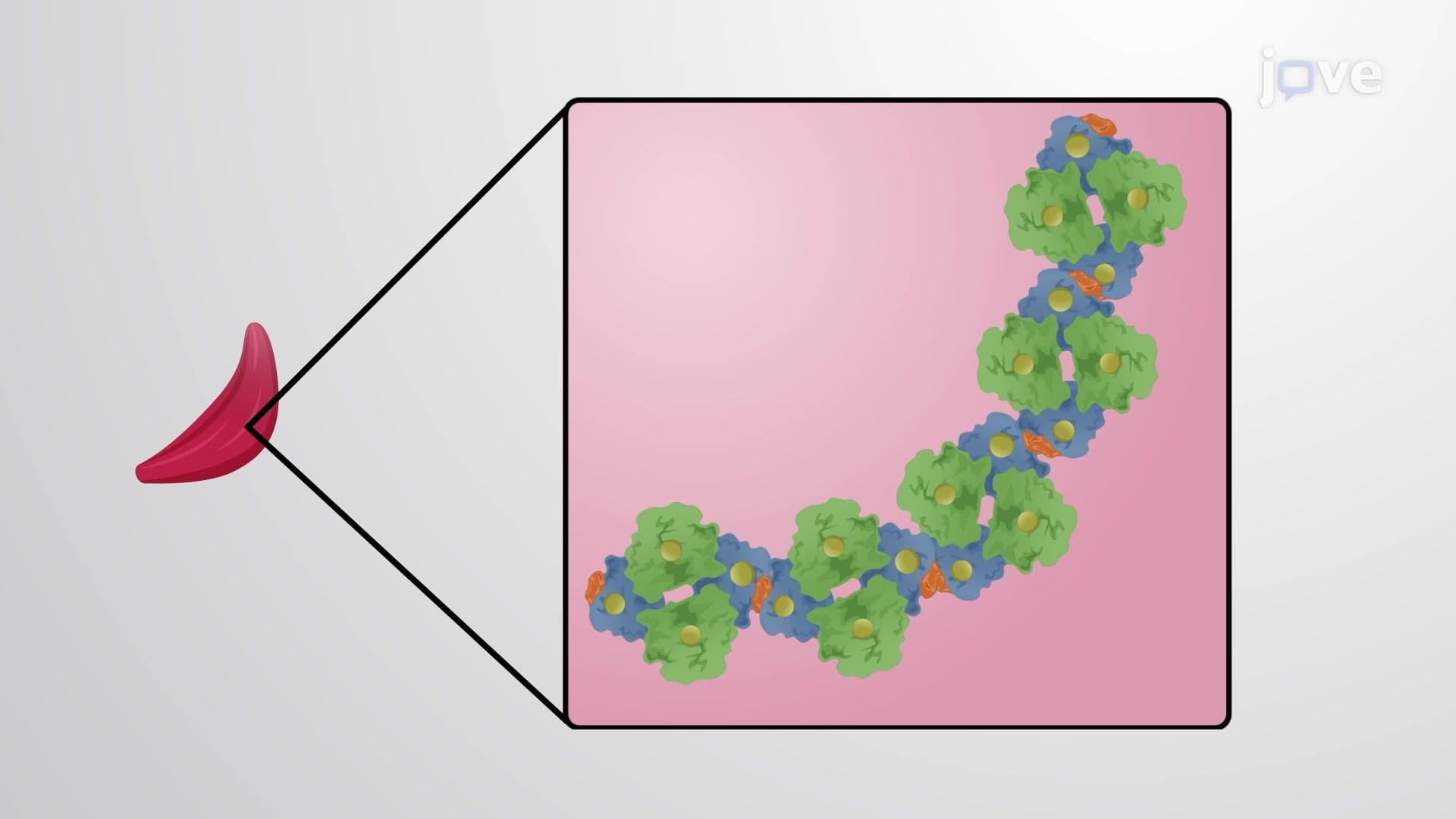
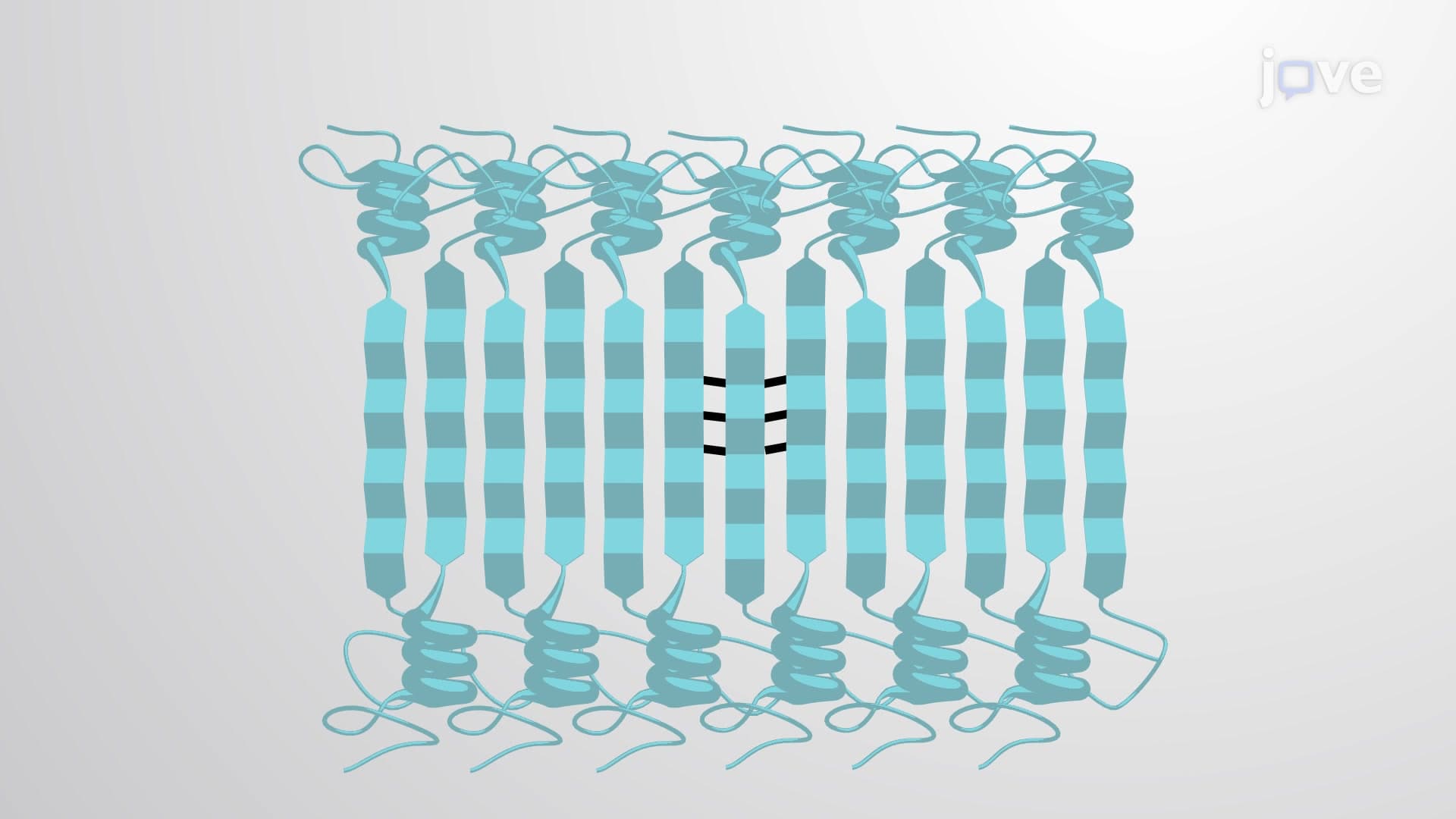
Amyloid fibrils represent one of the most fascinating and medically significant examples of protein misfolding in biology. These highly organized protein aggregates form when normally soluble proteins undergo conformational changes, transforming from their native structures into insoluble, fibrous deposits. The hallmark feature of amyloid fibrils is their cross-beta sheet architecture-a repeating pattern where beta strands run perpendicular to the fibril axis, creating incredibly stable, rope-like structures.
The formation process occurs through three distinct phases. During nucleation, individual misfolded proteins come together to form small, unstable clusters called nuclei. This initial step is typically the slowest and rate-limiting phase. The elongation phase follows, where additional misfolded proteins rapidly add to the growing fibril ends, extending the structure in both directions. Finally, during maturation, the fibrils undergo structural refinement, becoming more organized and resistant to breakdown.
In the United States, amyloid-related diseases affect millions of people, making this topic crucial for students pursuing healthcare careers. Alzheimer's disease, affecting over 6.5 million Americans, involves amyloid-beta plaques in brain tissue. Parkinson's disease features alpha-synuclein fibrils called Lewy bodies. Type 2 diabetes, prevalent in over 37 million US adults, involves islet amyloid polypeptide (IAPP) deposits in pancreatic beta cells.
These diseases share common features: protein misfolding, fibril formation, cellular dysfunction, and progressive tissue damage. Understanding these mechanisms is essential for MCAT preparation and appears frequently in AP Biology exams when covering protein structure and human physiology.
Surprisingly, not all amyloid fibrils cause disease. Functional amyloids serve important biological roles, challenging the traditional view of these structures as purely pathological. For example, certain bacteria use amyloid-like structures in biofilms, and some organisms employ them for hormone storage and release.
Scientists study amyloid fibrils using advanced techniques like transmission electron microscopy, X-ray diffraction, and atomic force microscopy. These methods reveal the intricate details of fibril structure and help researchers at institutions like the National Institutes of Health develop potential therapies. Current research focuses on preventing fibril formation, promoting their clearance, or stabilizing non-toxic protein conformations-knowledge that's increasingly relevant for students considering careers in biochemistry, neuroscience, or pharmaceutical research.
Related Micro-courses