Loading subject page…

- Cell Biology
- Protein Structure
- Protein Organization
Micro-course: Protein Structure
62,212 views
Learn key concepts and earn your certificate.


Protein Organization

00:00
01:24
Video Summary: What Is Protein Organization
Did you know that insulin-the hormone controlling your blood sugar-only works because of its precise protein organization levels? From a simple chain of amino acids emerging from ribosomes, proteins undergo an incredible transformation through primary, secondary, tertiary, and quaternary structures. Consider hemoglobin in your red blood cells: this life-sustaining protein requires all four organizational levels to transport oxygen efficiently throughout your body. Understanding what is protein organization reveals how biological molecules achieve their remarkable functions. Watch the full video on JoVE Coach to master this concept with expert-led visuals and step-by-step explanations.
- Understand what is protein organization and identify the four distinct structural levels that transform amino acid chains into functional proteins
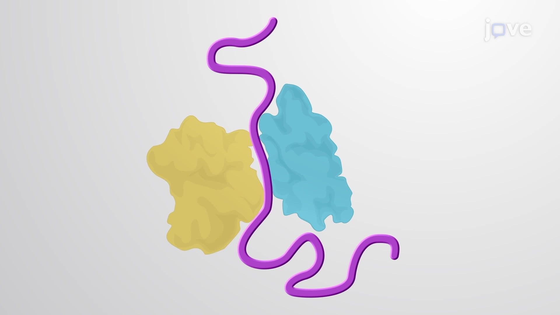
- Recognize how primary structure forms through peptide bonds between amino acids during ribosomal translation
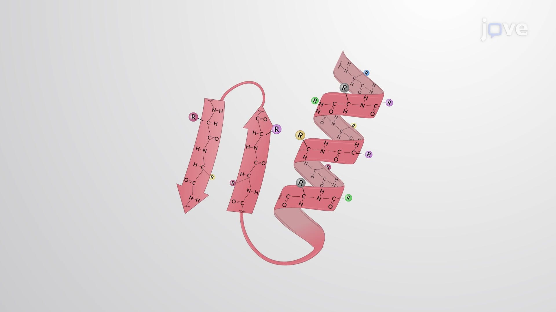
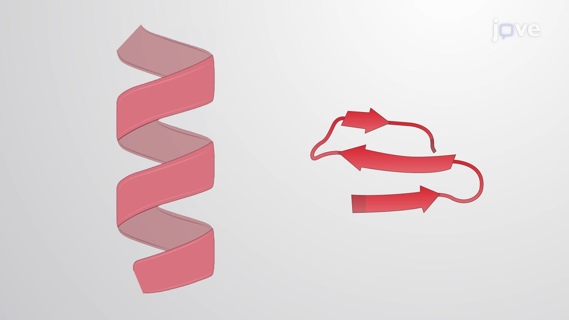
- Analyze secondary structures including alpha helices and beta sheets stabilized by hydrogen bonding patterns
- Compare tertiary structure formation through various molecular interactions including hydrophobic forces, ionic bonds, and disulfide bridges
- Evaluate quaternary structure assembly when multiple polypeptide chains combine into larger protein complexes
- Apply protein structure hierarchy knowledge to predict how mutations might affect protein function
- Identify real-world examples of proteins requiring specific organizational levels for biological activity
- Explore the relationship between protein conformation levels and cellular processes in human physiology
Understanding What is Protein Organization: The Foundation of Life
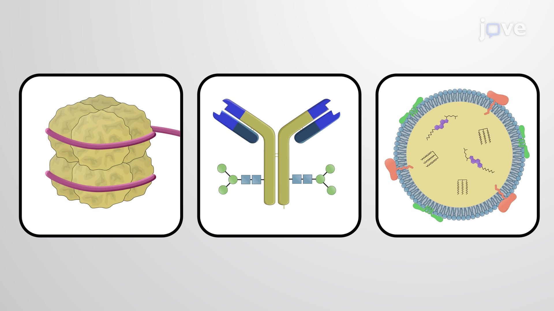
Protein organization represents one of biochemistry's most elegant examples of how simple building blocks create complex, functional molecules. This hierarchical system transforms basic amino acid sequences into the sophisticated proteins that power every cellular process in your body-from the enzymes digesting your lunch to the antibodies protecting you from pathogens.
Primary Structure: The Genetic Blueprint in Action
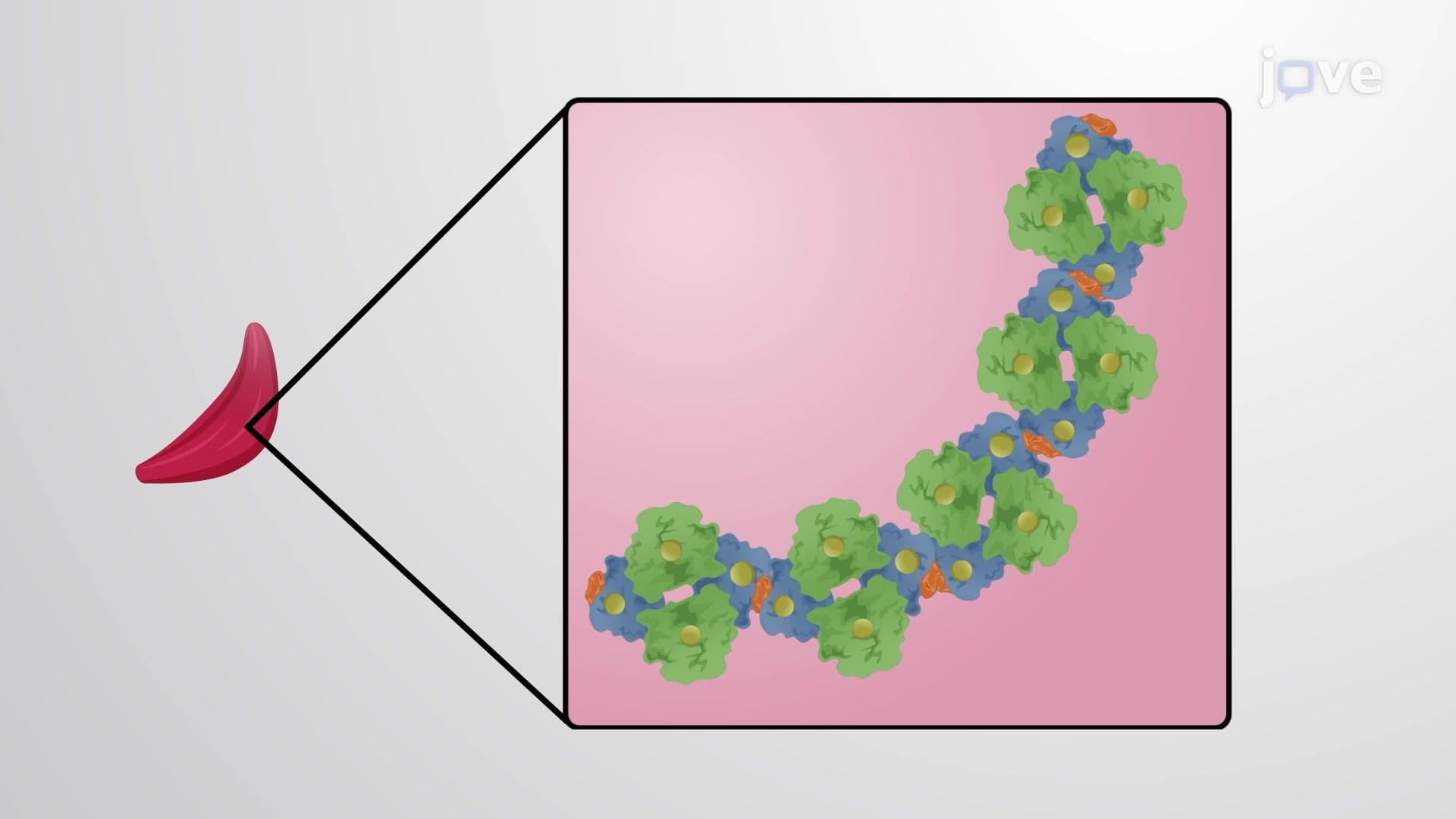
The journey begins with primary structure, where genetic information becomes reality. During translation at ribosomes, amino acids link through peptide bonds between carboxyl and amino groups, creating a linear chain. This sequence, determined by your DNA, serves as the foundation for everything that follows. Consider sickle cell anemia-a single amino acid change in hemoglobin's primary structure (glutamic acid to valine at position 6) demonstrates how critical this level is. Students preparing for the MCAT or AP Biology exams should remember that primary structure directly reflects genetic code, making it testable material for DNA-to-protein synthesis questions.
Secondary Structure: Local Organization Patterns
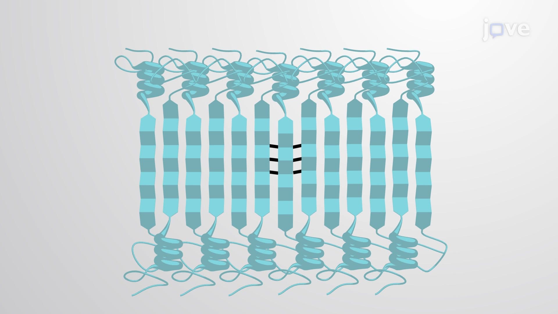
Secondary structures emerge as the polypeptide chain begins folding through hydrogen bonding between backbone atoms. Alpha helices form when every fourth amino acid creates hydrogen bonds, producing characteristic spiral structures found in keratin (your hair protein) and myosin (muscle contraction protein). Beta sheets create zigzag patterns through sideways hydrogen bonding, prominent in silk proteins and amyloid structures. These patterns appear frequently on college biochemistry exams because they represent fundamental ways proteins achieve stability while maintaining flexibility for function.
Tertiary and Quaternary Structure: Functional Architecture
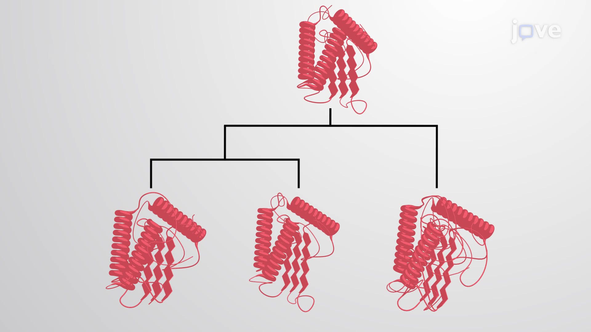
Tertiary structure represents the final 3D shape for single-chain proteins, achieved through interactions between distant amino acid side chains. Hydrophobic forces drive nonpolar residues inward, while ionic bonds, hydrogen bonds, and disulfide bridges provide stability. Enzymes like pepsin (stomach protein digestion) demonstrate how tertiary structure creates active sites with precise molecular recognition.
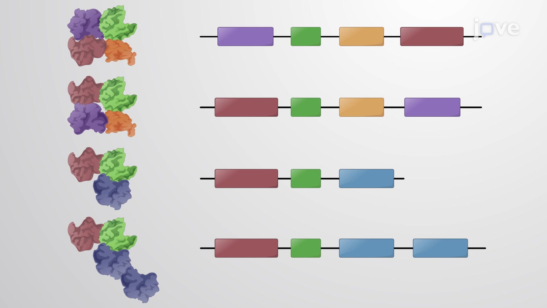
Quaternary structure involves multiple polypeptide chains combining into larger complexes. Hemoglobin exemplifies this beautifully-four separate chains (two alpha, two beta) work together to transport oxygen efficiently. This cooperative binding, where oxygen binding to one subunit increases affinity in others, showcases how quaternary organization enables sophisticated regulatory mechanisms impossible in single-chain proteins.
Clinical and Academic Applications
Understanding polypeptide protein organization proves essential for pre-med students tackling MCAT biochemistry sections and nursing students preparing for NCLEX pharmacology questions. Protein misfolding diseases like Alzheimer's and Parkinson's highlight how disrupted organization leads to cellular dysfunction, while targeted cancer therapies often work by interfering with specific protein conformations.
Frequently Asked Questions
Protein organization refers to the four hierarchical levels (primary, secondary, tertiary, quaternary) that transform linear amino acid chains into functional 3D proteins. This organization determines every protein's specific biological role, from enzyme catalysis to immune system recognition. Understanding these levels helps explain how genetic mutations affect protein function and why certain diseases occur when protein folding goes wrong.
The MCAT heavily tests all four protein organization levels, particularly focusing on structure-function relationships and how disruptions affect biological processes. Expect questions about alpha helices in membrane proteins, beta sheets in amyloid diseases, tertiary structure in enzyme active sites, and quaternary structure in allosteric regulation. Practice identifying these structures and predicting functional consequences of structural changes.
AP Biology frequently tests protein structure through free-response questions connecting DNA mutations to protein dysfunction. Students must explain how primary structure changes affect secondary and tertiary folding, ultimately impacting biological function. Expect questions about sickle cell anemia, enzyme inhibition, and how environmental factors like pH affect protein conformation levels.
Introductory biochemistry, organic chemistry II, molecular biology, and cell biology courses extensively cover protein folding organization. Pre-med students encounter this material in both general chemistry and advanced biochemistry, while nursing programs address protein structure in pharmacology and pathophysiology courses. Graduate programs in biotechnology and pharmaceutical sciences require deep understanding for drug design applications.
Protein misfolding causes numerous diseases affecting millions of Americans, including Alzheimer's (affecting 6 million), Parkinson's (1 million patients), and cystic fibrosis (30,000 cases). These conditions result from disrupted tertiary or quaternary structure, demonstrating why understanding normal protein conformation levels is crucial for developing treatments and managing patient care.
Not at all! While detailed biochemical mechanisms require advanced study, high school students can easily grasp the basic hierarchy and real-world connections. Start with familiar examples like hair structure (keratin) or blood oxygen transport (hemoglobin), then build understanding of how shape determines function. Most students find protein organization intuitive once they see concrete examples.
Create visual flowcharts connecting each organizational level to specific examples and functions. Use mnemonics like "Pretty Students Take Quality notes" for Primary, Secondary, Tertiary, Quaternary structures. Practice drawing protein structures and explaining how changes at each level affect function. Focus on understanding cause-and-effect relationships rather than memorizing isolated facts.
Explore protein folding kinetics, chaperone mechanisms, and post-translational modifications that fine-tune protein function. Advanced students can investigate computational protein modeling, drug design targeting specific conformations, and emerging therapies for protein misfolding diseases. These topics connect protein organization to cutting-edge biotechnology and pharmaceutical research.
Related Micro-courses

12 Concepts
10 Concepts

17 Concepts