Loading subject page…

1,694 views

Video Summary: Cardiopulmonary Resuscitation Iii Aed Use Explained
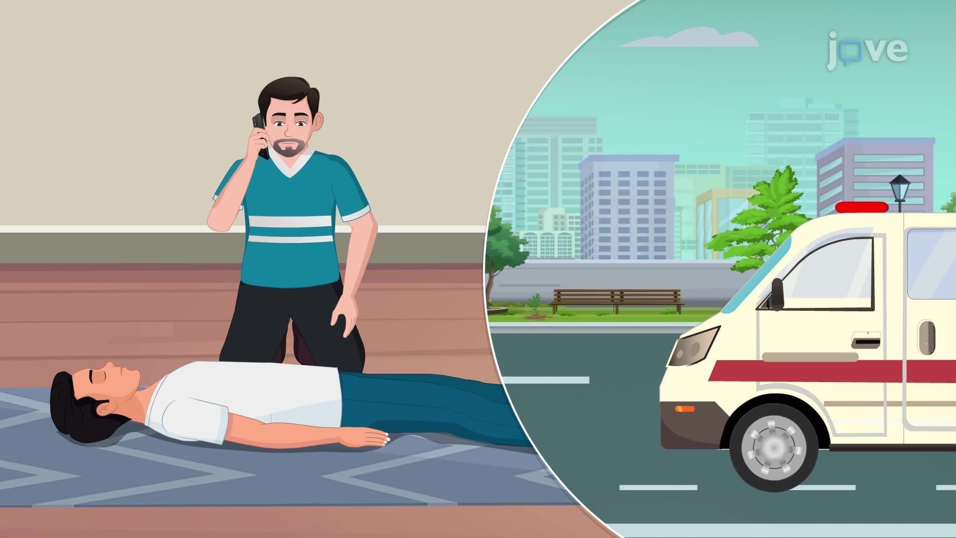
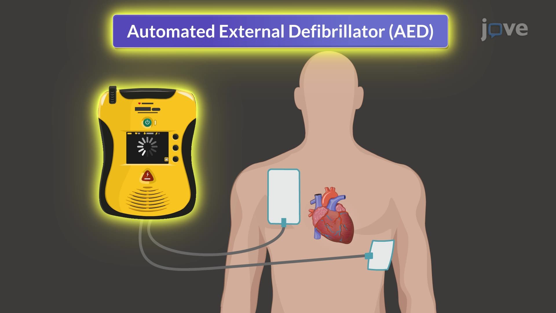
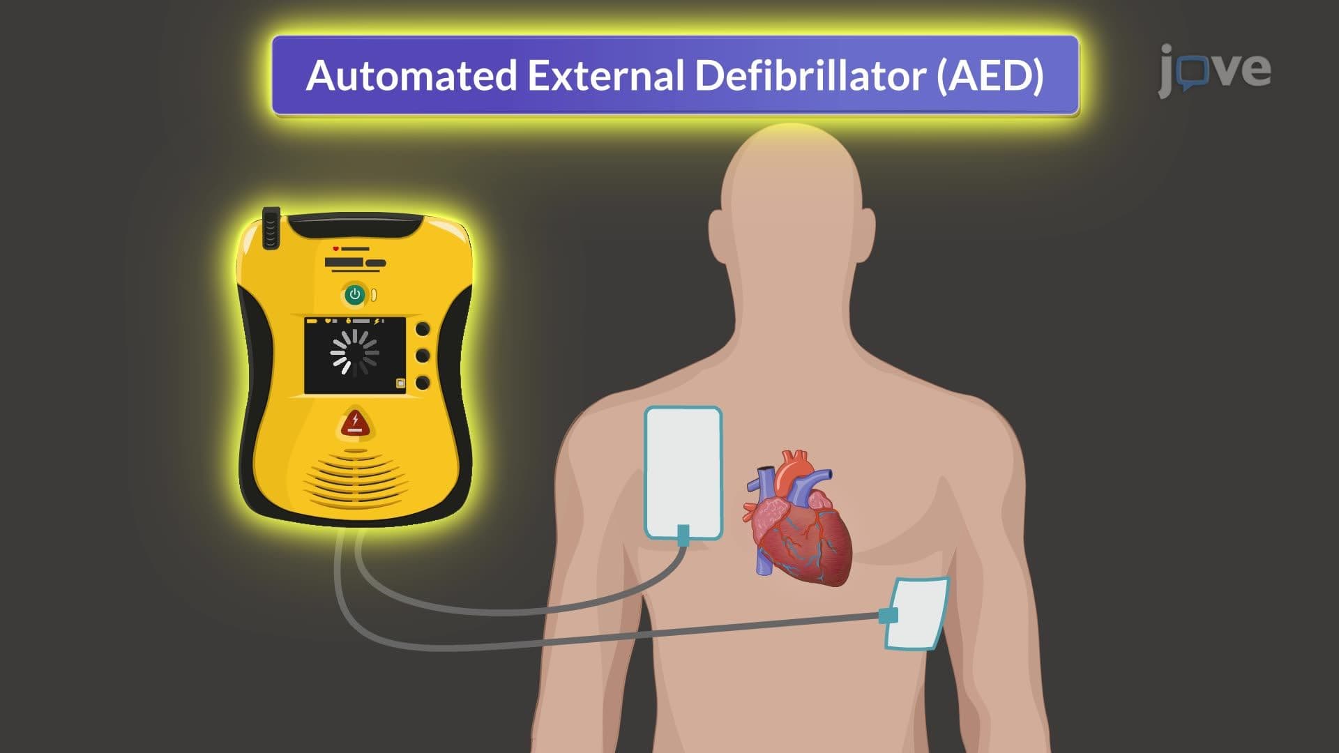
Every minute without defibrillation during sudden cardiac arrest reduces survival chances by 10%. Cardiopulmonary resuscitation III AED use combines chest compressions with automated external defibrillator technology to treat life-threatening arrhythmias like ventricular fibrillation. When a high school athlete collapses during basketball practice at Chicago's Lincoln Park High School, proper Cardiopulmonary Resuscitation III AED Use protocol can mean the difference between life and death. Watch the full video on JoVE Coach to master this concept with expert-led visuals and step-by-step explanations.
Cardiopulmonary resuscitation III AED use represents the advanced integration of mechanical chest compressions with electrical cardioversion therapy. While basic CPR maintains minimal blood circulation, certain lethal arrhythmias require electrical intervention to restore coordinated cardiac muscle contractions. The American Heart Association emphasizes that defibrillation within the first three minutes of ventricular fibrillation onset can achieve survival rates exceeding 70%.
Ventricular fibrillation (VF) and pulseless ventricular tachycardia (pVT) are the primary rhythms responsive to defibrillation. In VF, cardiac muscle fibers contract chaotically, producing no effective blood pumping. The heart essentially "quivers" like a bag of worms. Pulseless VT involves rapid, organized ventricular contractions that fail to generate adequate cardiac output. Both conditions prove fatal within minutes without electrical intervention.
Medical students preparing for USMLE Step 1 must understand that asystole (flatline) and pulseless electrical activity (PEA) are non-shockable rhythms. These require continued high-quality CPR and identification of reversible causes using the "H's and T's" mnemonic: hypoxia, hypovolemia, hydrogen ion (acidosis), hypo/hyperkalemia, hypothermia, toxins, tamponade, tension pneumothorax, thrombosis (coronary and pulmonary), and trauma.
Modern AEDs utilize sophisticated algorithms analyzing multiple ECG parameters including QRS morphology, heart rate, and amplitude. The device samples the cardiac rhythm for 5-15 seconds, filtering out chest compression artifacts and patient movement. Nursing students studying for NCLEX-RN should remember that AEDs are designed for lay rescuer use, featuring automated rhythm interpretation that eliminates manual ECG analysis requirements.
Emergency medical technicians responding to cardiac arrests in Chicago's Millennium Park or nurses working Cleveland Clinic's cardiac units regularly implement these protocols. The 2020 American Heart Association guidelines emphasize minimizing pre-shock pauses to less than 10 seconds, as even brief interruptions reduce defibrillation success rates. High school students taking AP Biology will encounter this concept when studying cardiovascular physiology and electrical conduction systems.
The two-minute cycle timing correlates with optimal CPR effectiveness before rescuer fatigue impacts compression quality. Research conducted at Johns Hopkins Hospital demonstrates that continuous chest compressions between AED analyses maintain coronary perfusion pressure, increasing the likelihood of return of spontaneous circulation (ROSC).
Related Micro-courses