Loading subject page…

- Medical Surgical Nursing
- Chest Injuries
- Pleural Disorders: Types and Brief Description
Micro-course: Chest Injuries
5,442 views
Learn key concepts and earn your certificate.



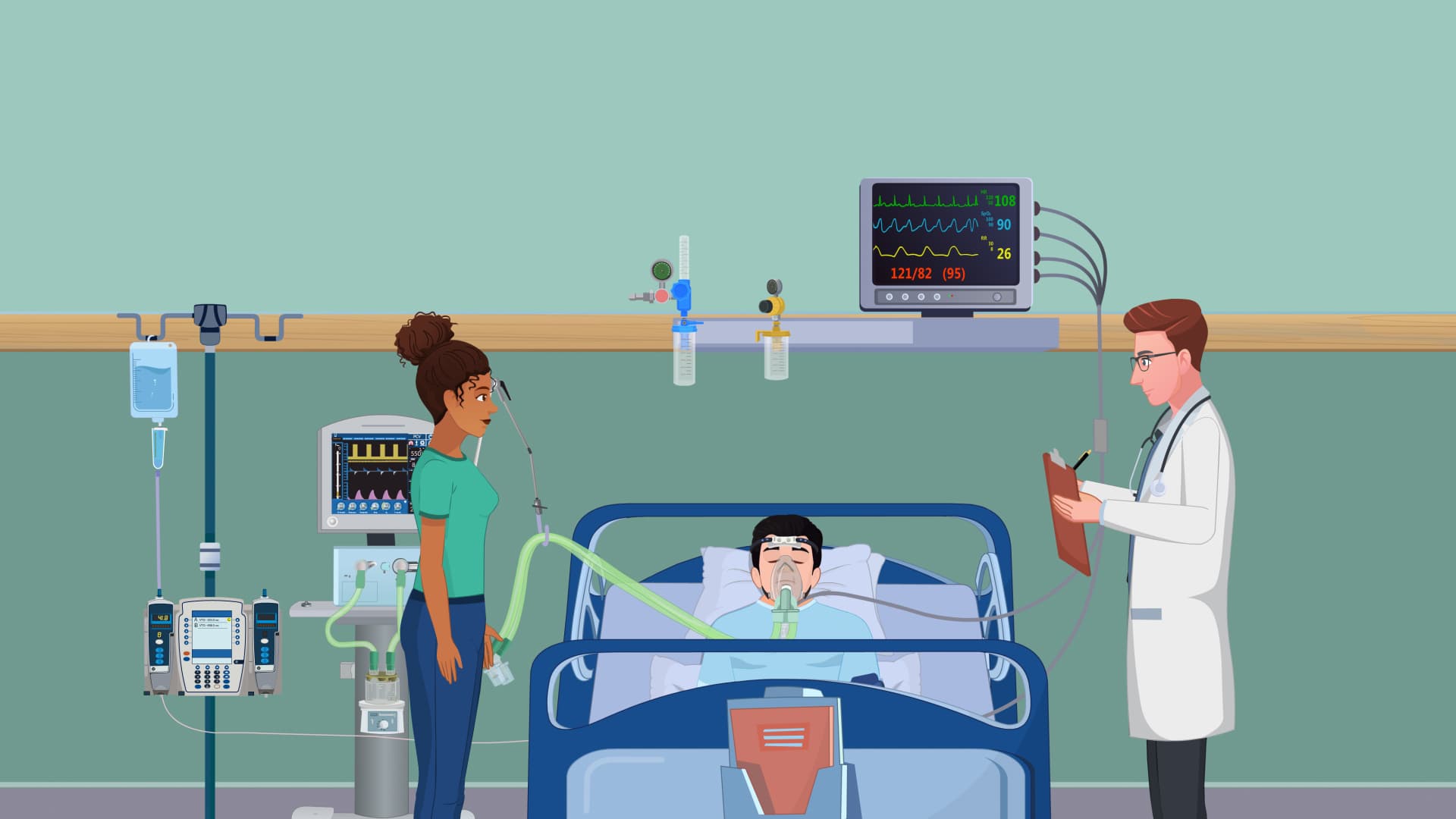
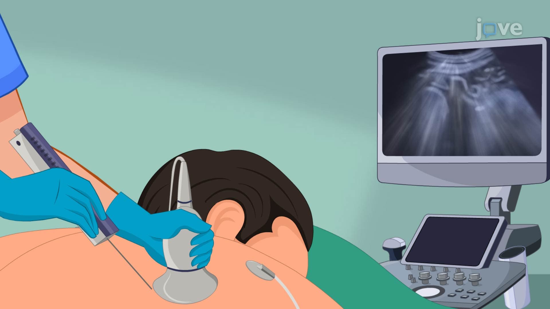
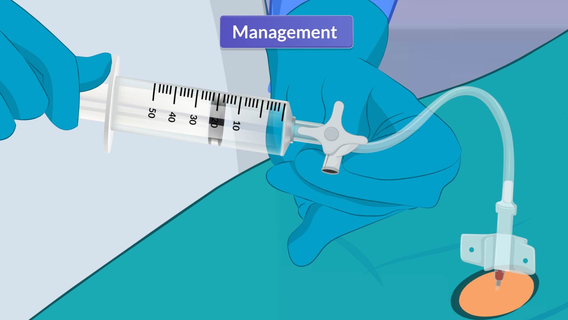
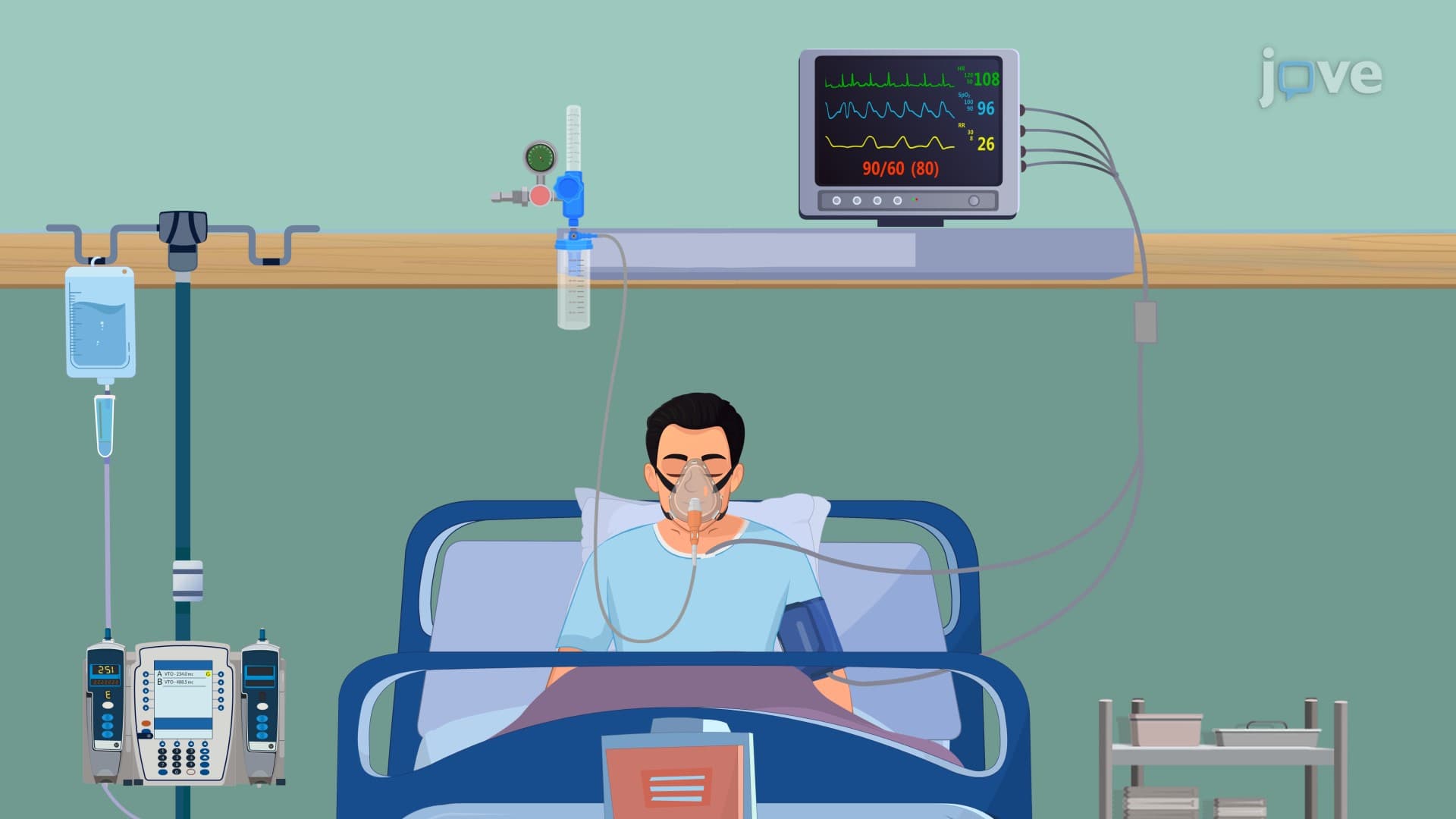
Pleural Disorders: Types and Brief Description

00:00
01:30
Video Summary: Pleural Disorders Types and Brief Description Explained
Did you know that over 1.5 million Americans develop pleural effusion annually, making pleural disorders types and brief description one of the most critical topics in respiratory medicine? These conditions affect the thin membrane surrounding your lungs, potentially causing severe breathing difficulties seen in emergency rooms across hospitals like Mayo Clinic and Johns Hopkins. Understanding pleural disorders types and brief description explained helps students recognize symptoms from chest pain to lung collapse. Watch the full video on JoVE Coach to master this concept with expert-led visuals and step-by-step explanations.
- Understand the fundamental anatomy of pleural disorders types and brief description explained, including the structure and function of pleural membranes
- Identify the five major categories of pleural disorders: pleurisy, pleural effusion, pneumothorax, hemothorax, and empyema
- Analyze the distinct symptoms and causes associated with each pleural disorder type
- Compare inflammatory versus mechanical pleural conditions and their different treatment approaches
- Evaluate clinical presentations to distinguish between life-threatening and manageable pleural disorders
- Apply knowledge of pleural anatomy to predict how different disorders affect breathing mechanics
- Recognize emergency warning signs that require immediate medical intervention
- Explore the relationship between underlying diseases and secondary pleural complications
Understanding Pleural Disorders Types And Brief Description Fundamentals
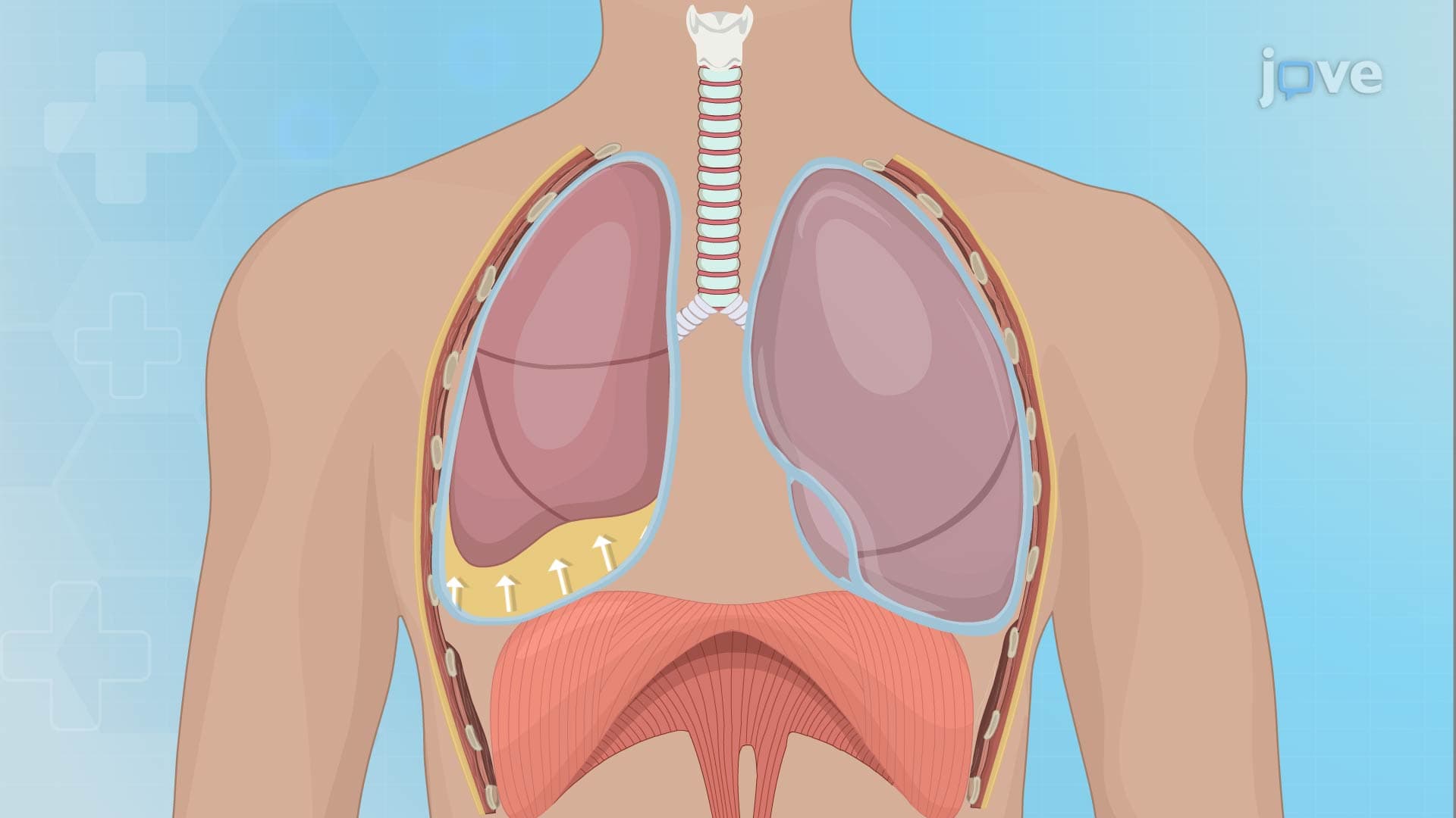
The pleural system represents one of medicine's most elegant protective mechanisms. This double-layered membrane system-consisting of visceral pleura (covering the lungs) and parietal pleura (lining the chest wall)-creates a sealed cavity filled with approximately 10-20 mL of lubricating fluid. When this delicate balance becomes disrupted, various pleural disorders emerge, each with distinct pathophysiology and clinical implications that medical students encounter on the MCAT and healthcare professionals see daily in clinical practice.
Inflammatory Pleural Conditions: Pleurisy and Empyema
Pleurisy (pleuritis) represents the most common inflammatory pleural disorder, affecting over 200,000 Americans annually. This condition involves inflammation of pleural surfaces, typically triggered by pneumonia, autoimmune diseases like lupus, or viral infections. Patients experience characteristic sharp, stabbing chest pain that worsens with breathing-a symptom pattern frequently tested on AP Biology exams and nursing school assessments like the NCLEX.
Empyema represents a more severe inflammatory condition where bacterial infections, particularly from Streptococcus pneumoniae or Staphylococcus aureus, cause pus accumulation in the pleural space. This condition requires aggressive antibiotic therapy and often surgical drainage, making it a critical topic in medical education curricula nationwide.
Fluid and Air Accumulation Disorders
Pleural effusion involves abnormal fluid accumulation exceeding the normal 10-20 mL capacity. Transudative effusions result from pressure imbalances due to heart failure or kidney disease, while exudative effusions stem from inflammatory processes or malignancy. This distinction proves crucial for USMLE Step 1 preparation, as students must differentiate between these types using Light's criteria-a diagnostic framework developed at American medical institutions.
Pneumothorax occurs when air enters the pleural cavity, disrupting the negative pressure essential for lung expansion. Primary spontaneous pneumothorax typically affects tall, thin young males, while secondary pneumothorax complicates existing lung diseases like COPD. This condition frequently appears in emergency medicine scenarios on medical licensing exams.
Trauma-Related Pleural Disorders
Hemothorax involves blood accumulation in the pleural space, usually following chest trauma from motor vehicle accidents-a leading cause of emergency department visits in the United States. This condition can rapidly progress to hemorrhagic shock, making early recognition vital for healthcare providers. The combination of pneumothorax and hemothorax, called hemopneumothorax, represents a medical emergency requiring immediate intervention.
Understanding these pleural disorders proves essential for students pursuing healthcare careers, as these conditions commonly appear on standardized exams and clinical rotations throughout American medical education systems.
Frequently Asked Questions
Pleural disorders are medical conditions affecting the thin membrane surrounding your lungs and lining your chest cavity. These disorders include five main types: pleurisy (inflammation), pleural effusion (excess fluid), pneumothorax (air accumulation), hemothorax (blood accumulation), and empyema (pus collection). Each type has distinct causes, symptoms, and treatment approaches that are fundamental to understanding respiratory medicine.
Medical exams like the MCAT, USMLE, and NCLEX frequently test pleural disorders through clinical scenarios requiring symptom recognition and diagnostic reasoning. Expect questions about distinguishing chest pain patterns, interpreting chest X-rays showing fluid levels, and understanding pressure changes in pneumothorax. The MCAT particularly emphasizes the physics of pleural pressure and gas exchange disruption.
Concentrate on the structural anatomy of pleural layers, the normal function of pleural fluid, and how each disorder disrupts normal breathing mechanics. Practice identifying symptoms like pleuritic chest pain versus dyspnea, and understand the pathophysiology behind fluid accumulation. Many exams include diagram labeling and cause-effect relationships between underlying diseases and pleural complications.
Emergency departments across major medical centers like Cleveland Clinic and Massachusetts General Hospital regularly treat pleural disorders, particularly pneumothorax in young athletes and pleural effusions in elderly patients with heart failure. Nursing students often encounter these conditions during clinical rotations, where they learn to assess breathing patterns, monitor oxygen saturation, and recognize when patients need immediate physician evaluation.
Basic high school biology and introductory anatomy provide sufficient background for understanding pleural disorders. Focus on fundamental concepts like membrane structure, fluid dynamics, and basic respiratory physiology. Most students find pleural disorders accessible once they grasp the simple concept of two membrane layers with fluid between them-similar to a water balloon inside another balloon.
Create memory associations linking each disorder to its key feature: pleurisy = pain (both start with 'p'), effusion = excess fluid (both start with 'e'), pneumothorax = air (pneumo = air), hemothorax = blood (hemo = blood), empyema = pus/infection (think "yucky empyema"). Practice with clinical scenarios rather than isolated definitions to improve retention and application on exams.
After mastering basic pleural disorders, explore chest tube placement procedures, thoracentesis techniques, and advanced diagnostic methods like pleural biopsy. Study the relationship between pleural disorders and systemic diseases like cancer metastasis, tuberculosis, and autoimmune conditions. This knowledge proves valuable for advanced placement courses and pre-med preparation.
Pleural disorders integrate closely with lung compliance, ventilation-perfusion relationships, and respiratory failure mechanisms commonly covered in physiology courses. Understanding pleural pressure changes helps explain concepts like atelectasis and mechanical ventilation that appear on nursing exams like HESI A2 and TEAS. These connections frequently appear as integrated questions on comprehensive exams.
Related Micro-courses