Loading subject page…


5,442 views


Video Summary: Pleural Effusion Ii Symptoms and Management Explained
Did you know that over 1.5 million Americans develop pleural effusion annually, with symptoms ranging from barely noticeable to life-threatening? Pleural effusion II symptoms and management involves recognizing critical signs like progressive shortness of breath and chest pain, then implementing targeted treatments from simple thoracentesis to complex pleurodesis procedures. Consider a patient at Johns Hopkins Hospital presenting with persistent cough and breathing difficulties-proper symptom assessment and management protocols can mean the difference between rapid recovery and serious complications. Watch the full video on JoVE Coach to master this concept with expert-led visuals and step-by-step explanations.
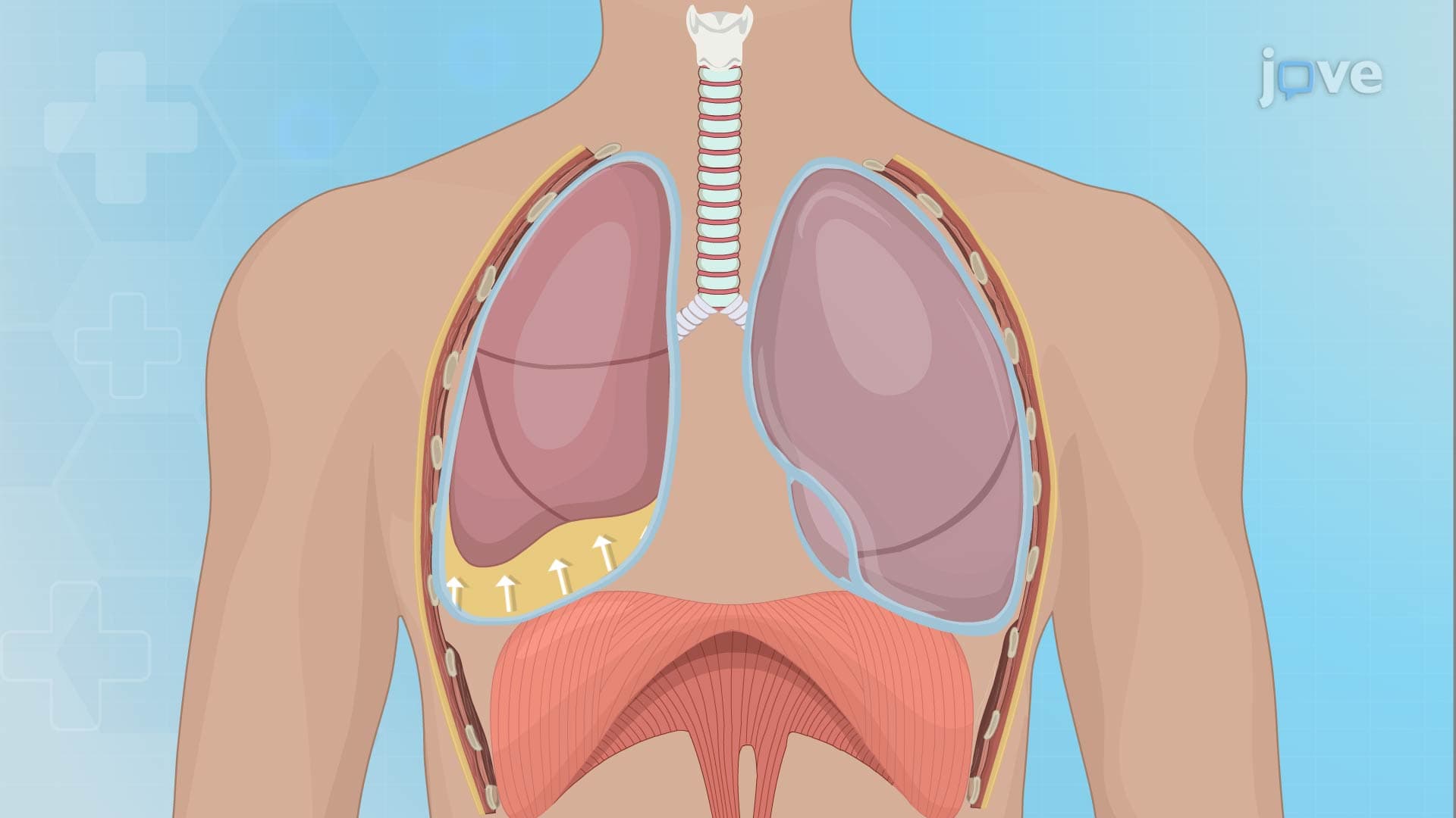
Pleural effusion represents abnormal fluid accumulation in the pleural space, creating a complex clinical scenario requiring systematic symptom recognition and management. The condition affects hundreds of thousands of Americans annually, with presentations ranging from asymptomatic small effusions discovered incidentally on chest X-rays to massive effusions causing respiratory failure. Understanding the symptom-management relationship becomes crucial for healthcare providers working in emergency departments, internal medicine units, and pulmonology clinics across the United States.
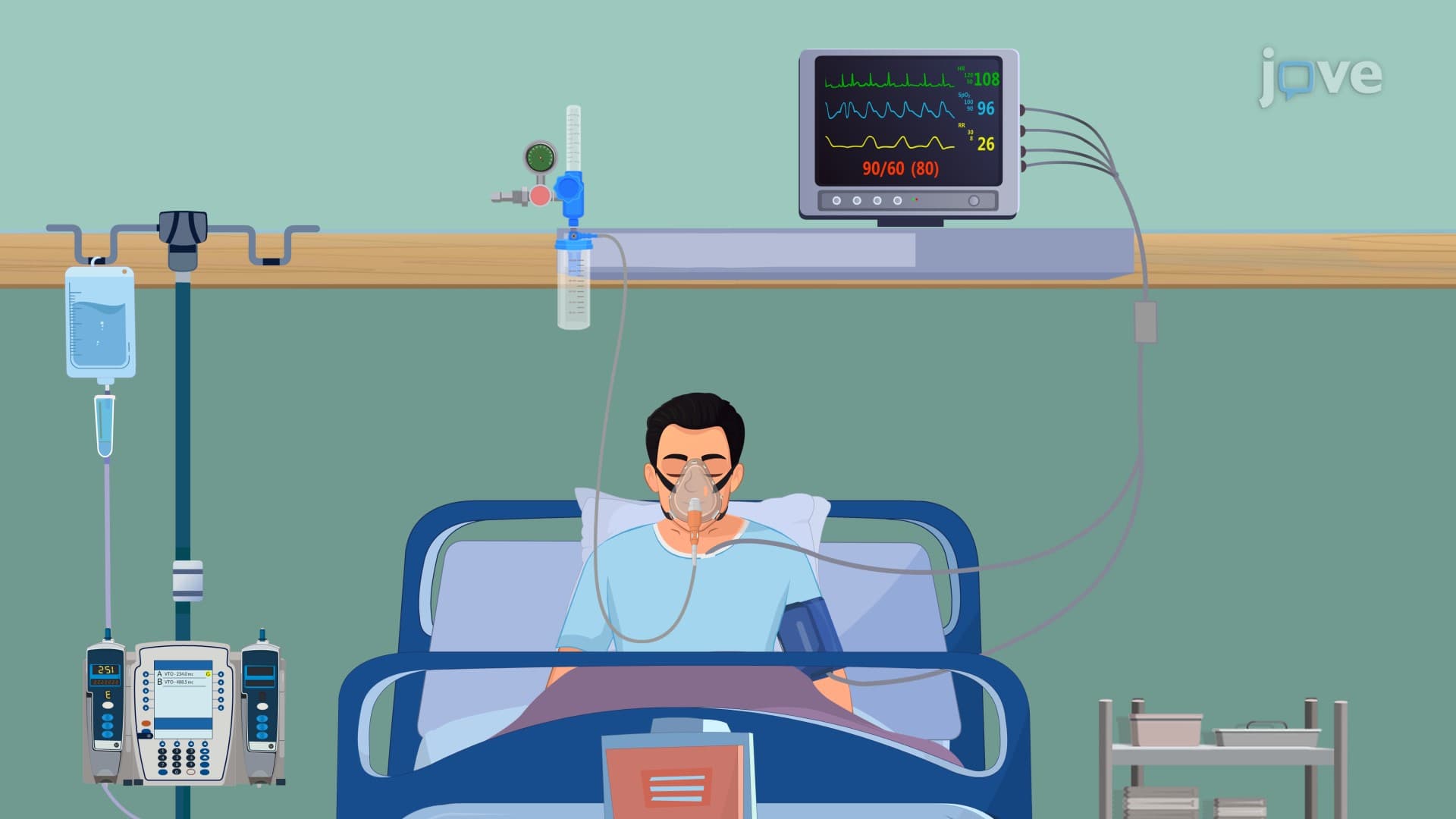
The symptomatic presentation of pleural effusion correlates directly with the volume of accumulated fluid and the underlying etiology. Small effusions (less than 500 mL) often remain asymptomatic, while larger accumulations progressively impair respiratory mechanics. Pleuritic chest pain-sharp, stabbing pain that worsens with inspiration-commonly accompanies inflammatory conditions like pneumonia or pulmonary embolism. Progressive dyspnea represents the hallmark symptom, developing as fluid compresses lung tissue and reduces functional respiratory capacity.
Malignant effusions, frequently seen in patients with lung, breast, or lymphatic cancers treated at major cancer centers like MD Anderson or Memorial Sloan Kettering, typically present with insidious onset dyspnea and persistent non-productive cough. These patients often experience constitutional symptoms including weight loss and fatigue. Conversely, parapneumonic effusions associated with bacterial pneumonia present acutely with fever, chills, and productive cough alongside respiratory symptoms.
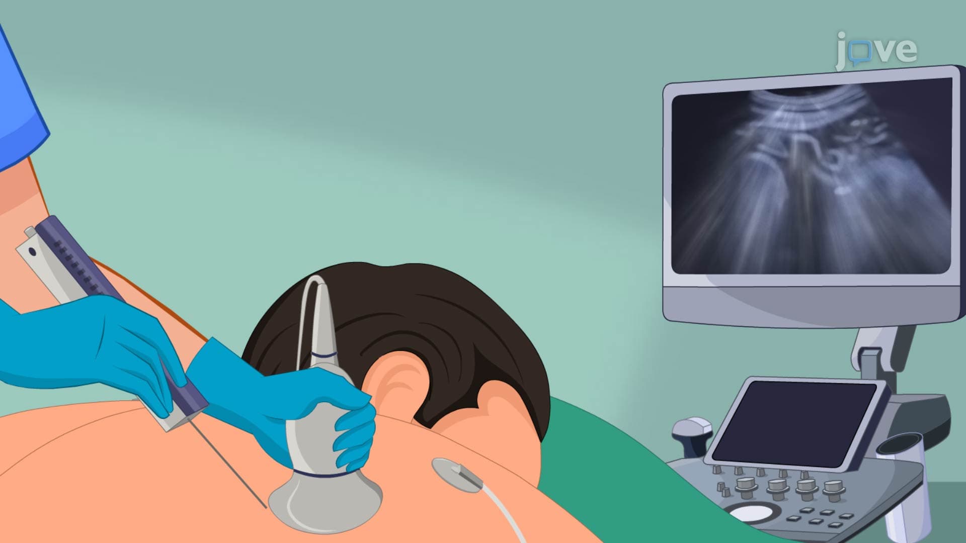
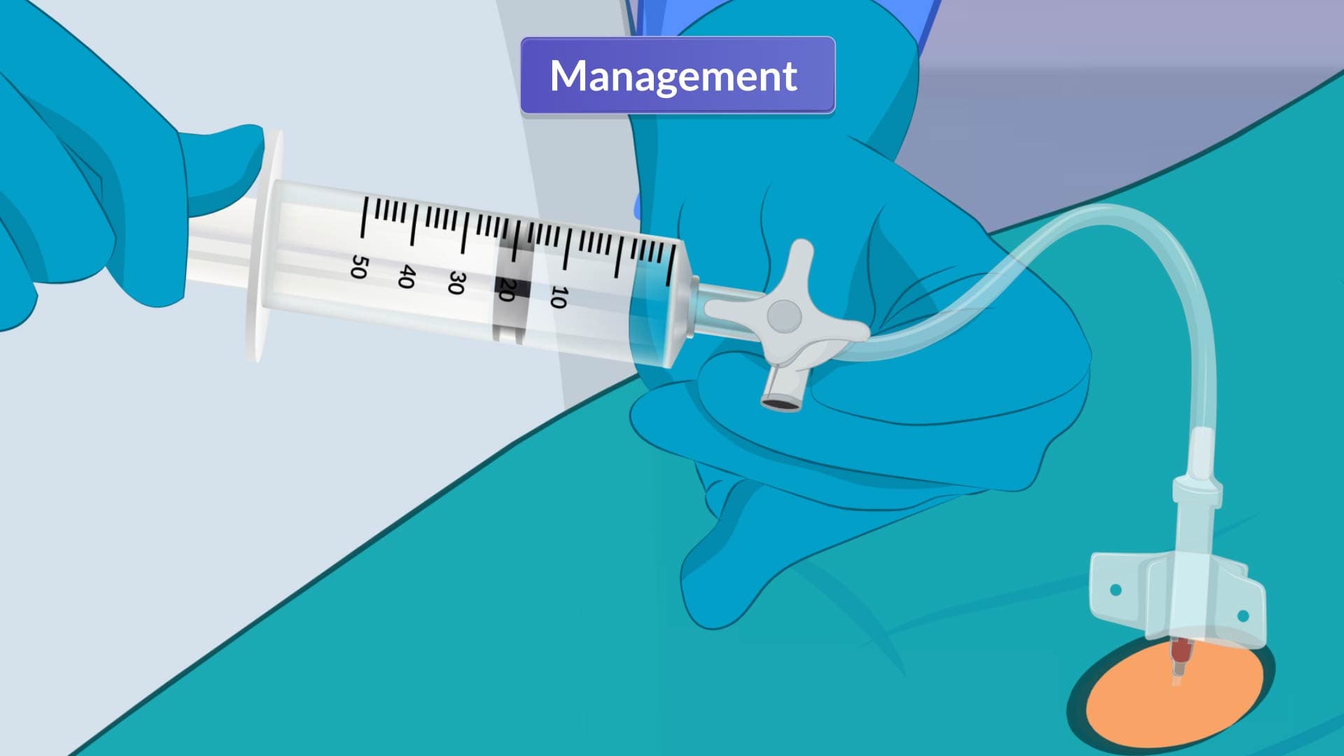
Management protocols focus on two primary objectives: identifying and treating the underlying cause while providing symptomatic relief. Thoracentesis serves dual diagnostic and therapeutic purposes, allowing fluid analysis for cellular composition, protein levels, and microbiological studies while simultaneously relieving respiratory distress. The procedure, commonly performed in hospital settings from Cleveland Clinic to UCLA Medical Center, can safely remove up to 1.5 liters of fluid per session.
For recurrent or large-volume effusions, particularly those associated with malignancy, more aggressive interventions become necessary. Chest tube thoracostomy enables continuous drainage, while chemical pleurodesis using talc or doxycycline creates permanent adhesions between pleural surfaces. This prevents future fluid reaccumulation, though the procedure requires careful patient selection and monitoring.
These concepts frequently appear on standardized examinations including the MCAT's biological sciences section, NCLEX-RN respiratory disorders questions, and internal medicine board certifications. Students preparing for these assessments should focus on correlating symptom severity with management intensity-understanding when conservative observation suffices versus when immediate intervention prevents respiratory compromise. Clinical rotations in pulmonology and internal medicine provide hands-on experience with these decision-making processes in real healthcare settings.
Related Micro-courses