Loading subject page…


5,442 views

Video Summary: What Is Flail Chest Ii
Did you know that flail chest injuries occur in nearly 10% of all serious chest trauma cases treated in US emergency departments? Understanding flail chest ii becomes crucial when examining how multiple rib fractures create a paradoxical breathing pattern that can be life-threatening. Consider a motorcyclist involved in a high-speed collision on Interstate 95, the impact force could fracture three or more adjacent ribs in two places, creating the unstable chest segment that defines What is Flail Chest II. Watch the full video on JoVE Coach to master this concept with expert-led visuals and step-by-step explanations.
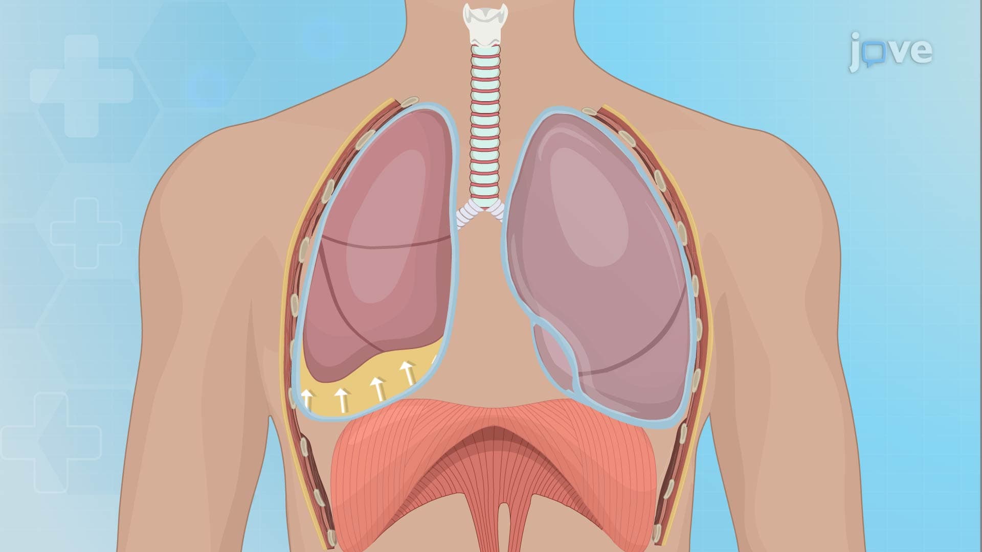
What is Flail Chest II represents a critical thoracic injury where three or more adjacent ribs are fractured in at least two locations, creating a free-floating segment of the chest wall. This unstable segment moves paradoxically during respiration, moving inward during inspiration when the rest of the chest expands outward, and vice versa during expiration. The condition significantly compromises respiratory efficiency and can lead to life-threatening complications if not properly managed.
The mechanism typically involves high-energy trauma such as motor vehicle accidents, falls from significant heights, or direct blunt force trauma to the chest. In the United States, flail chest accounts for approximately 5-15% of all rib fractures and carries a mortality rate of 10-15% when associated with underlying pulmonary contusion.
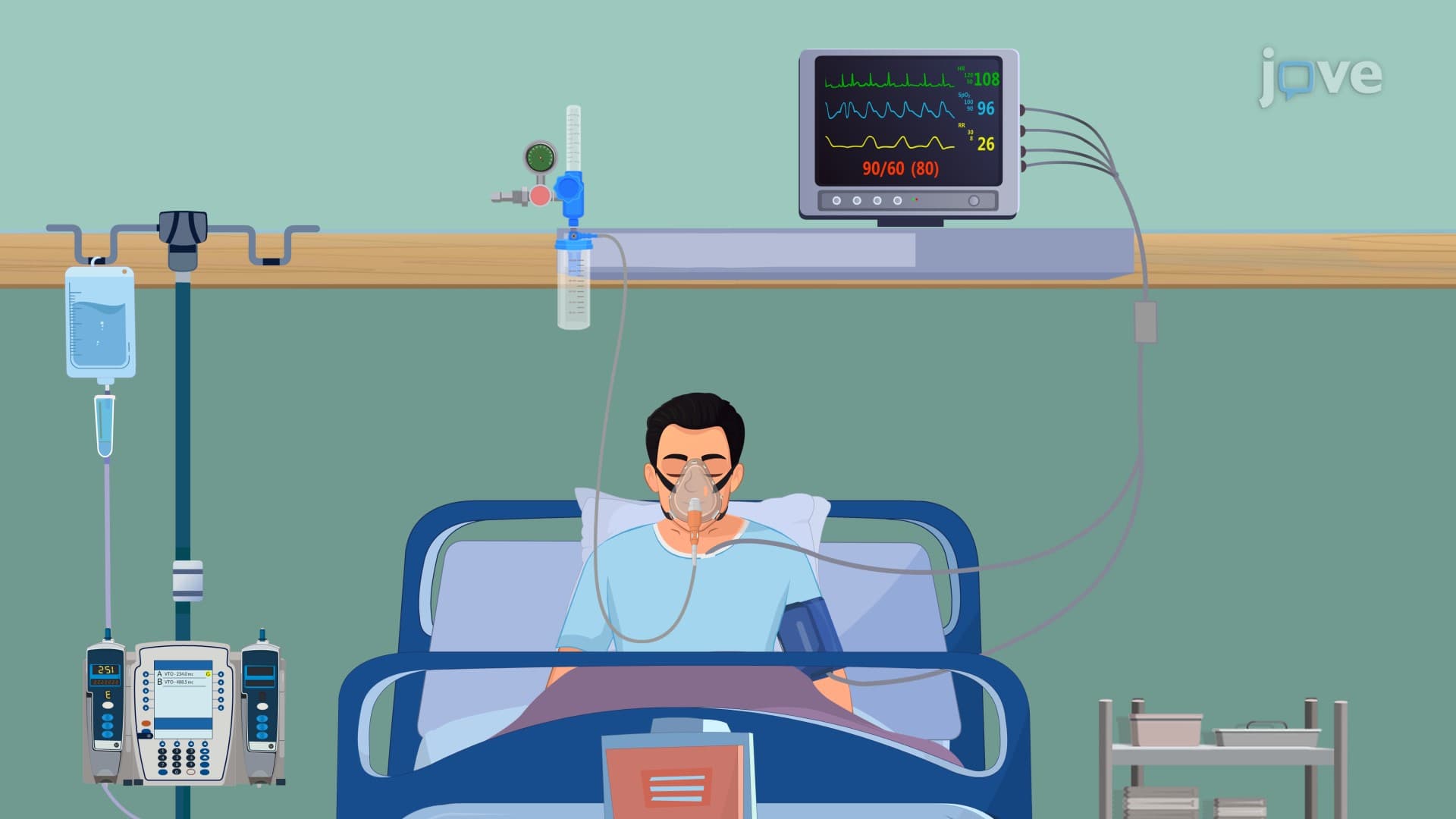
Healthcare providers must conduct comprehensive trauma assessments focusing on the mechanism of injury. Physical examination reveals abnormal chest wall movement, crepitus (crackling sounds from bone fragments), and signs of respiratory distress including tachypnea, shallow breathing, and accessory muscle use. Patients often present with severe chest pain that worsens with breathing or movement.
Imaging studies play crucial roles in diagnosis and management planning. Chest X-rays provide initial assessment of rib fractures and associated injuries, while CT scans offer detailed visualization of the extent of chest wall damage, underlying lung contusions, and potential complications like pneumothorax or hemothorax. These diagnostic tools are essential for determining appropriate treatment strategies.
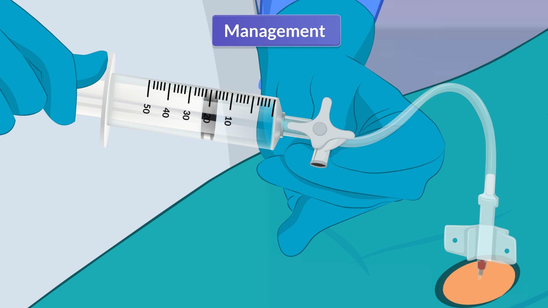
Treatment approaches vary significantly based on injury severity and patient stability. For mild-to-moderate cases, conservative management focuses on maintaining airway clearance through positioning techniques, encouraging deep breathing exercises, and implementing effective coughing strategies to prevent pneumonia. Pain control becomes paramount, as inadequate analgesia leads to shallow breathing and respiratory complications.
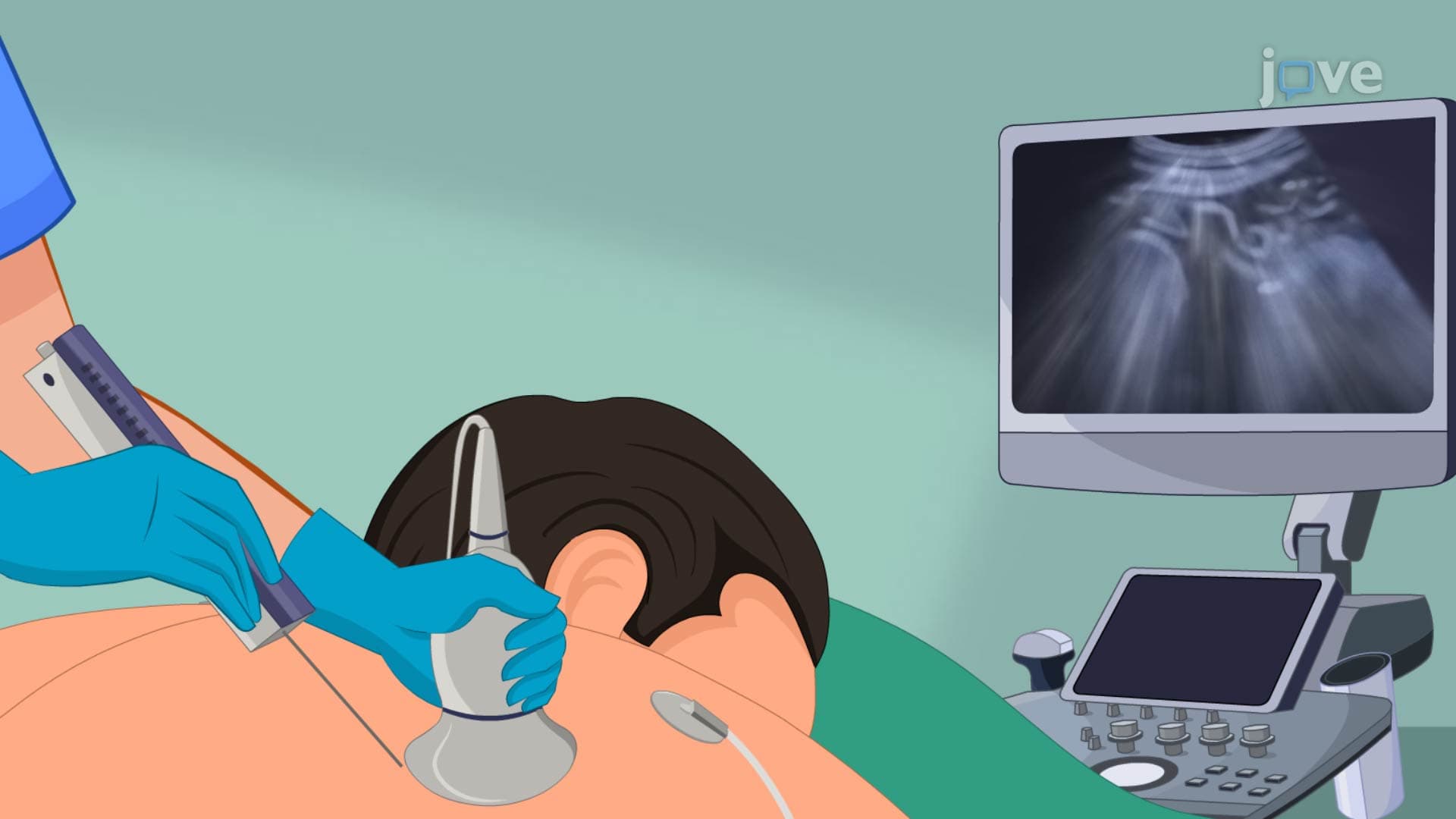
Advanced pain management options include patient-controlled analgesia (PCA) systems, intercostal nerve blocks targeting specific rib segments, epidural analgesia for comprehensive pain relief, and intrapleural opioid administration. These techniques allow patients to breathe more effectively while promoting healing.
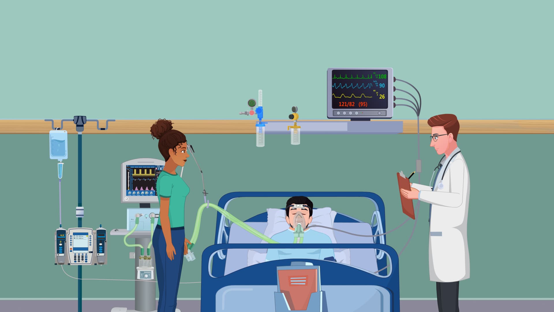
Severe flail chest injuries often require mechanical ventilation with positive end-expiratory pressure (PEEP) to stabilize the chest wall internally and improve gas exchange. Modern approaches may include surgical fixation using titanium plates and screws to restore chest wall stability, particularly in cases with significant displacement or when conservative management fails. The decision for surgical intervention considers factors such as patient age, comorbidities, and overall trauma severity.
Related Micro-courses