Loading subject page…


5,442 views

Video Summary: What Is Pneumothorax I
Did you know that a simple sneeze or cough can cause your lung to collapse in certain individuals? Pneumothorax I represents one of the most critical respiratory emergencies seen in US emergency departments, affecting over 20,000 Americans annually. This condition occurs when air abnormally accumulates between the chest wall and lungs, disrupting the delicate pressure balance that keeps our lungs inflated. Watch the full video on JoVE Coach to master this concept with expert-led visuals and step-by-step explanations.
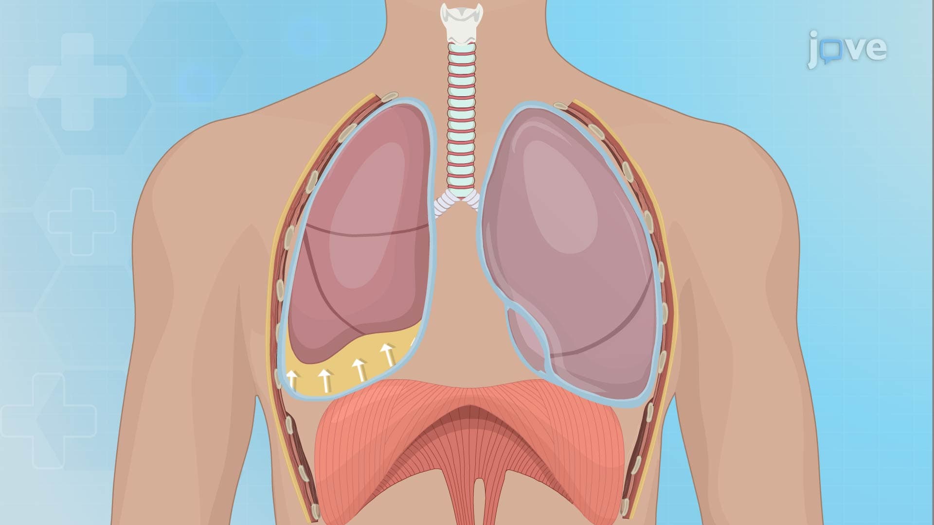
Pneumothorax I fundamentally represents a disruption of the respiratory system's most critical pressure relationship. Under normal circumstances, the pleural space-the thin cavity between your chest wall and lungs-maintains a negative pressure of approximately -5 mmHg. This negative pressure acts like a vacuum, keeping your lungs expanded against the chest wall. When air enters this space, it eliminates this negative pressure, causing the elastic lung tissue to recoil and collapse.
This concept frequently appears on the MCAT and in college-level anatomy and physiology courses, where students must understand the relationship between Boyle's Law and respiratory mechanics. The pleural pressure gradient is essential for ventilation, making pneumothorax a perfect example of how physics principles apply to human biology.
Healthcare professionals classify pneumothorax into three distinct categories, each with unique underlying mechanisms. Spontaneous pneumothorax typically affects tall, thin young adults, particularly males aged 15-30. Primary spontaneous pneumothorax occurs in healthy individuals when small air-filled sacs called blebs rupture on the lung surface. Secondary spontaneous pneumothorax develops in patients with existing lung disease, most commonly chronic obstructive pulmonary disease (COPD) or emphysema.
Traumatic pneumothorax results from external forces or medical interventions. Motor vehicle accidents, sports injuries, and penetrating chest wounds represent common injury-related causes seen in US trauma centers. Iatrogenic pneumothorax occurs during medical procedures-central line placement, thoracentesis, or mechanical ventilation can inadvertently puncture lung tissue.
Tension pneumothorax represents the most dangerous variant, creating a life-threatening emergency requiring immediate intervention. As air continues entering the pleural space without escape, pressure builds progressively, shifting the mediastinum and compressing the opposite lung and major blood vessels. This compression reduces venous return to the heart, potentially causing cardiovascular collapse.
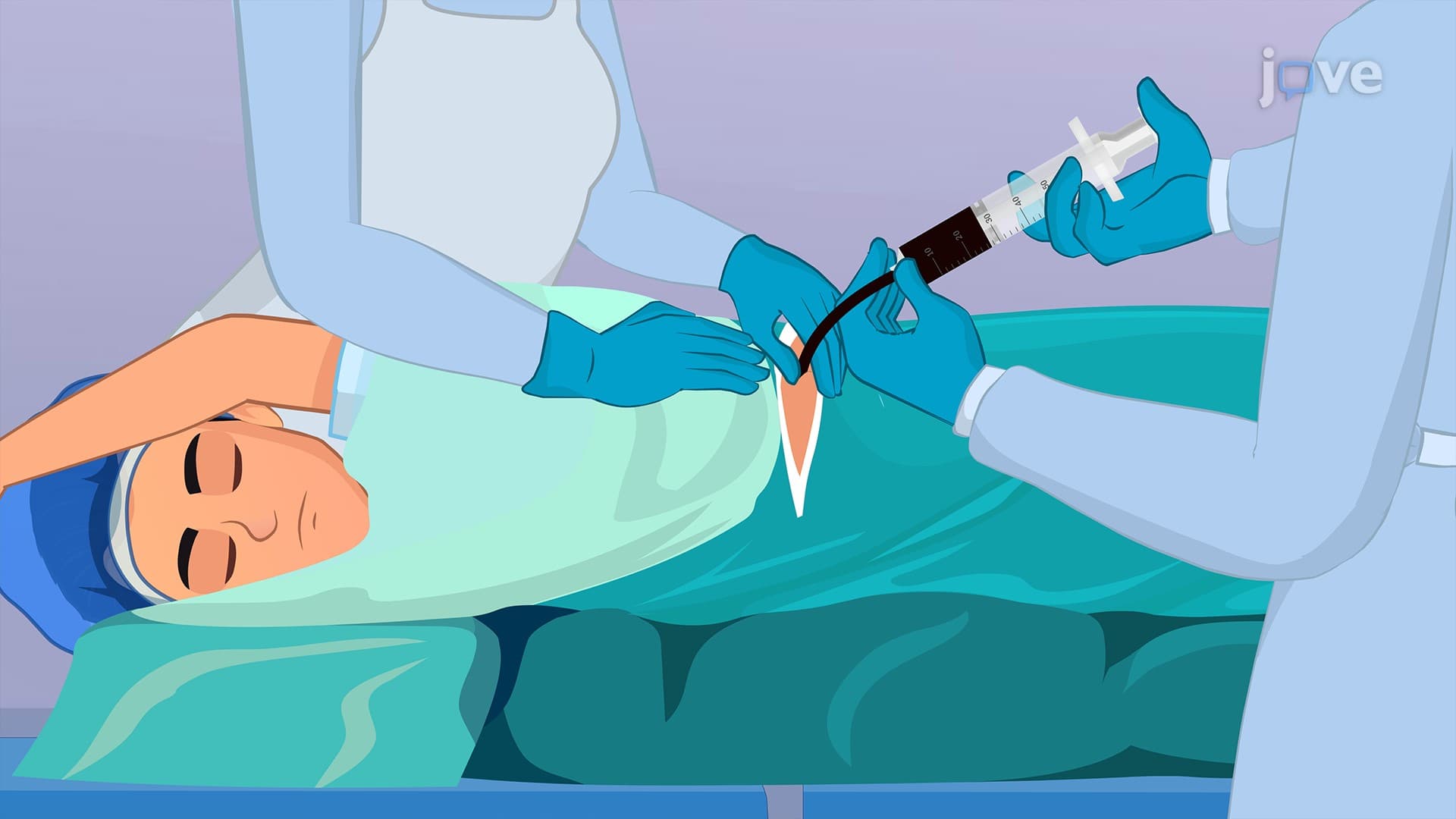
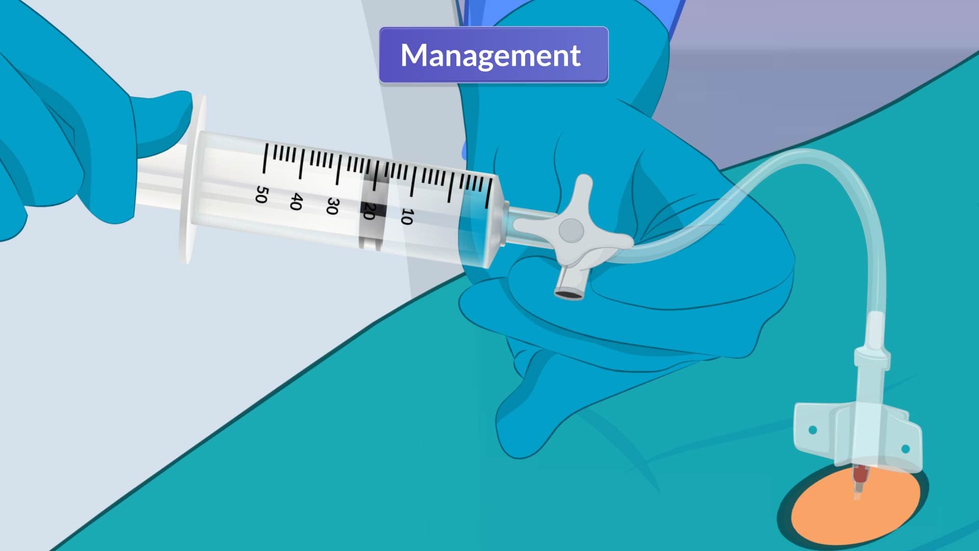
Emergency physicians in US hospitals must recognize tension pneumothorax signs rapidly: severe respiratory distress, absent breath sounds, hyperresonance to percussion, and tracheal deviation away from the affected side. Treatment requires immediate needle decompression followed by chest tube placement-procedures covered extensively in emergency medicine residency training and tested on the USMLE Step 2 Clinical Knowledge examination.
Related Micro-courses