Loading subject page…


5,442 views

Video Summary: What Is Pneumothorax Ii
Did you know that a collapsed lung can occur spontaneously in seemingly healthy teenagers, often during routine activities like playing basketball? Pneumothorax II represents the advanced understanding of how air trapped between lung layers creates life-threatening breathing emergencies. Emergency departments across the US see pneumothorax cases daily, from spontaneous collapses in tall, thin young adults to trauma-induced injuries in car accidents. Understanding What is Pneumothorax II means grasping both the pathophysiology and critical interventions that save lives. Watch the full video on JoVE Coach to master this concept with expert-led visuals and step-by-step explanations.
What is Pneumothorax II encompasses the comprehensive study of pleural space disorders where air accumulates between the visceral and parietal pleura, disrupting normal respiratory mechanics. This advanced concept builds upon basic respiratory anatomy to explore the complex interplay between intrapleural pressure, lung compliance, and cardiovascular function. In US medical education, this topic appears prominently in pathophysiology courses, MCAT preparation, and clinical rotations.
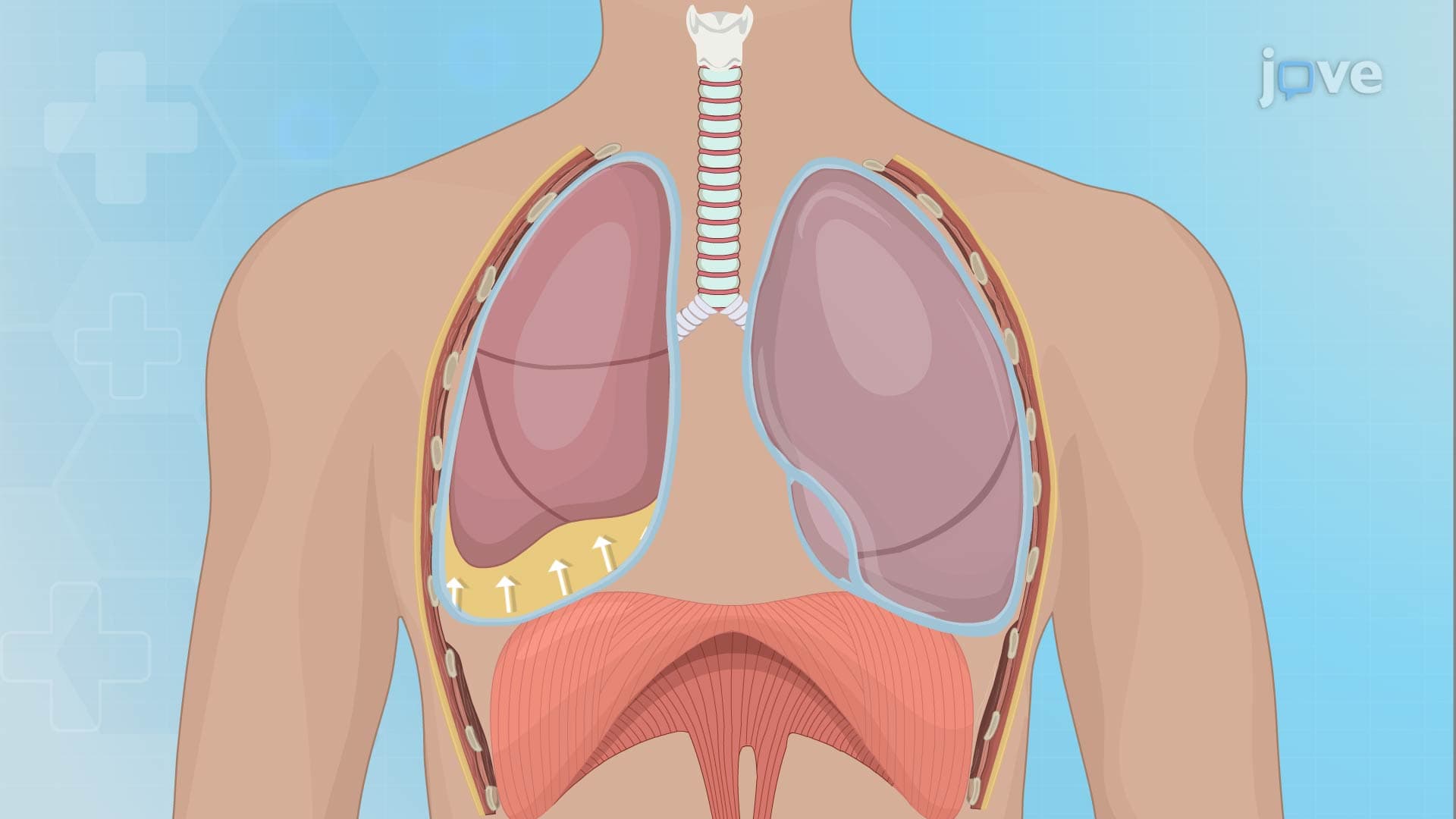
The pathophysiology involves disruption of the normally negative intrapleural pressure (-4 to -8 mmHg), which maintains lung expansion against the chest wall. When air enters this space through pleural rupture or external trauma, the pressure gradient equalizes, causing lung collapse proportional to the air volume accumulated.
Pneumothorax II presentation varies dramatically based on underlying lung health and pneumothorax size. Primary spontaneous pneumothorax typically affects healthy individuals aged 15-30, particularly tall, thin males with a 6:1 male-to-female ratio. These patients often experience sudden onset during minimal exertion-studies from US emergency departments show peak incidence during activities like weightlifting or playing wind instruments.
Secondary pneumothorax occurs in patients with underlying pulmonary pathology such as COPD, cystic fibrosis, or Pneumocystis pneumonia, common in immunocompromised patients. The clinical severity correlates with reduced pulmonary reserve, making even small pneumothoraces potentially life-threatening.
Physical examination reveals classic findings: hyperresonance to percussion, diminished tactile fremitus, and absent breath sounds on the affected side. Advanced practitioners recognize subtle signs like tracheal deviation (in tension pneumothorax) and paradoxical chest wall movement.
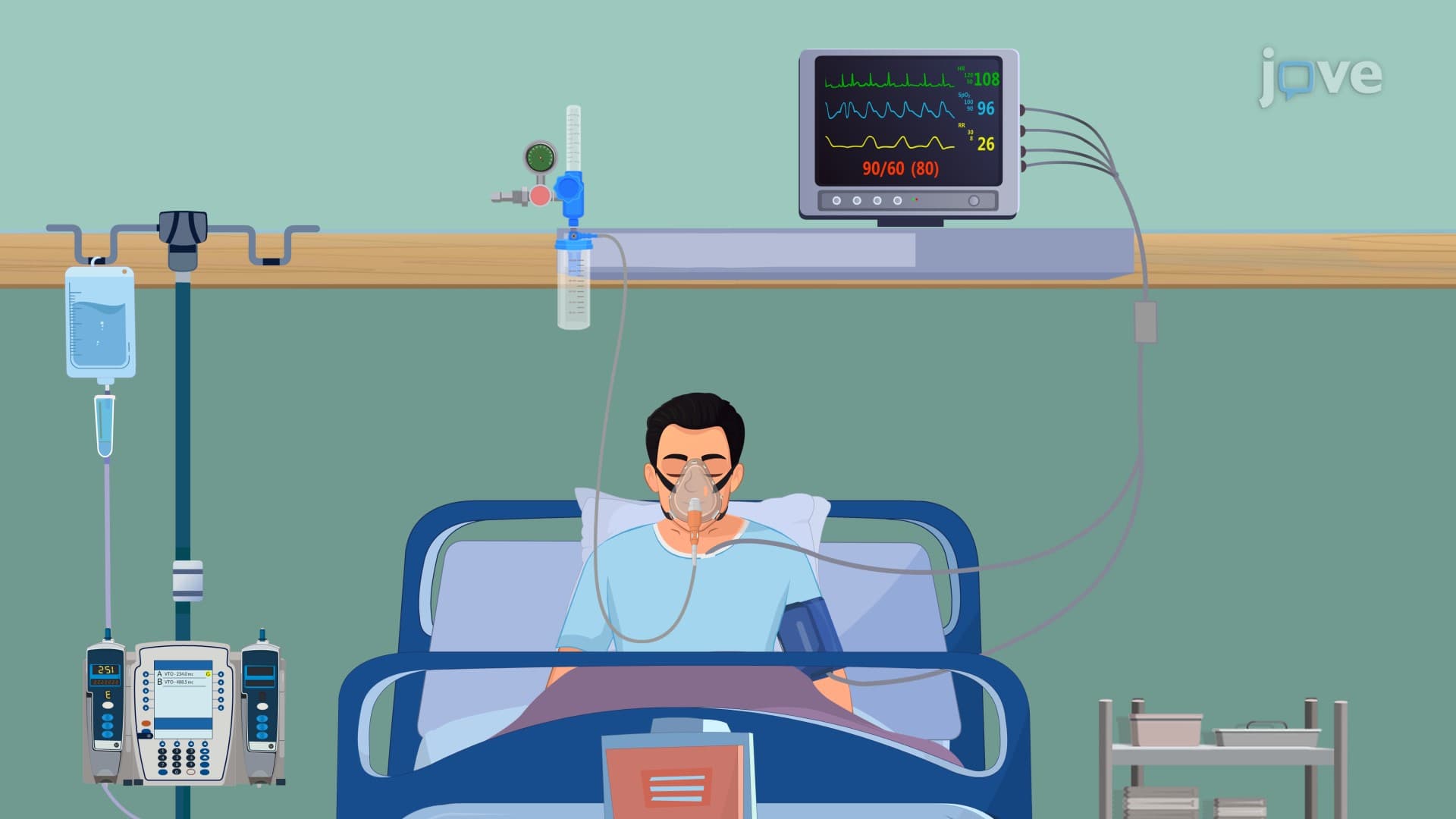
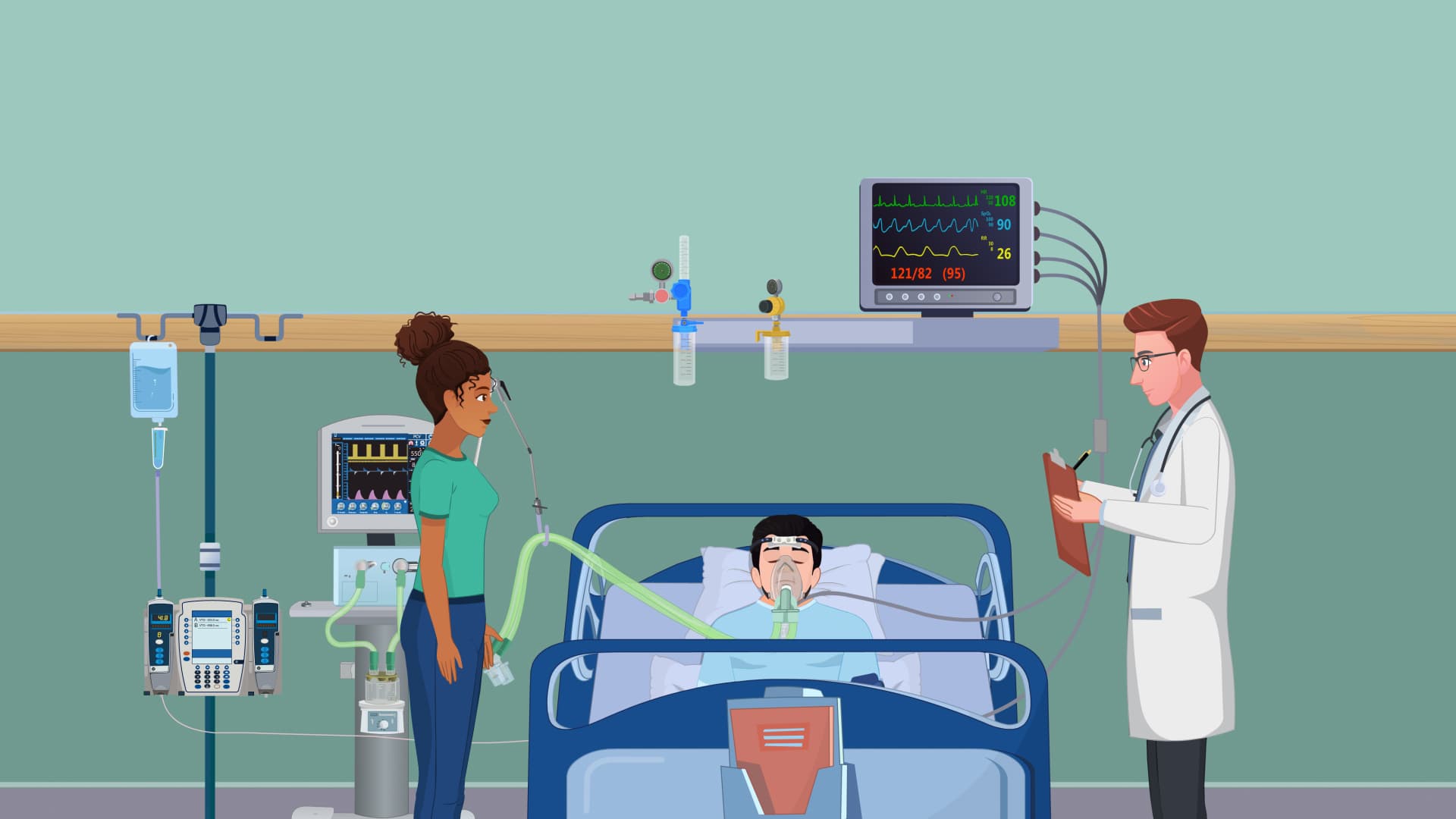
Treatment protocols follow evidence-based guidelines established by the American College of Emergency Physicians. Pneumothorax II management stratifies by size and clinical stability. Small pneumothoraces (<20% lung collapse) in stable patients may resolve with observation and supplemental oxygen, which increases nitrogen absorption from the pleural space fivefold.
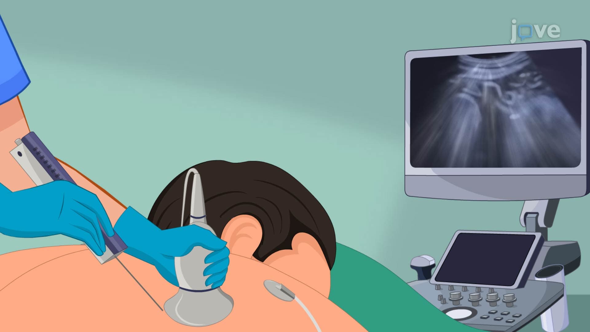
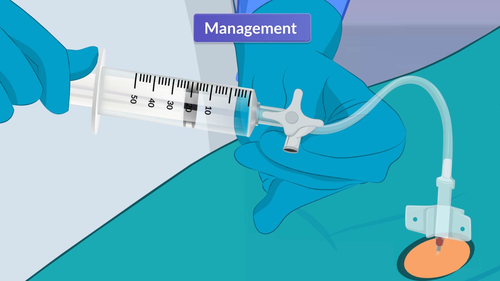
Moderate to large pneumothoraces require active intervention. Needle thoracostomy-performed at the second intercostal space, midclavicular line-provides immediate decompression in tension pneumothorax. This technique, taught in Advanced Trauma Life Support (ATLS) courses across US medical schools, can be lifesaving within minutes.
Chest tube thoracostomy remains the definitive treatment for most cases. The tube, typically placed at the fourth or fifth intercostal space in the mid-axillary line, connects to a water-seal drainage system maintaining negative pressure while preventing air re-entry.
Recurrent pneumothorax affects 20-30% of patients after initial episodes, necessitating surgical intervention. Video-assisted thoracoscopic surgery (VATS) has become the gold standard at major US medical centers, offering minimal invasive access for bleb resection and pleurodesis. This procedure reduces recurrence rates to less than 5% while maintaining excellent cosmetic outcomes important to young patients.
Chemical pleurodesis using talc or doxycycline creates pleural adhesions preventing future air accumulation. These procedures require careful patient selection and informed consent regarding potential complications like chronic pain or reduced pulmonary function.
For MCAT and USMLE preparation, students should understand the physiological rationale behind each intervention and recognize scenarios requiring emergent versus elective surgical management. Case-based questions often test the ability to prioritize treatments based on clinical presentation and underlying pathology.
Related Micro-courses