Loading video…

 15 Concepts
 2.5K views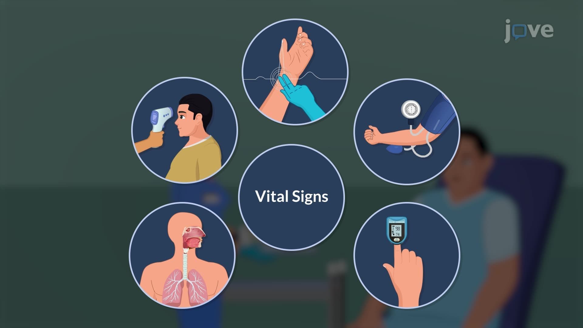

Video Summary: Acute Coronary Syndrome Iii Diagnostic Studies Explained
Every 40 seconds, someone in the United States suffers a heart attack, making rapid diagnosis critical for patient survival. Acute coronary syndrome III diagnostic studies provide healthcare professionals with essential tools to identify and assess heart attacks through systematic evaluation methods. At Cleveland Clinic, emergency physicians use these diagnostic protocols to differentiate between STEMI and NSTEMI cases within minutes of patient arrival. Watch the full video on JoVE Coach to master this concept with expert-led visuals and step-by-step explanations.
The systematic evaluation of suspected acute coronary syndrome represents one of the most critical decision-making processes in emergency medicine. This diagnostic framework combines multiple assessment tools to rapidly identify patients experiencing heart attacks, ensuring appropriate treatment within the crucial "golden hour" window.
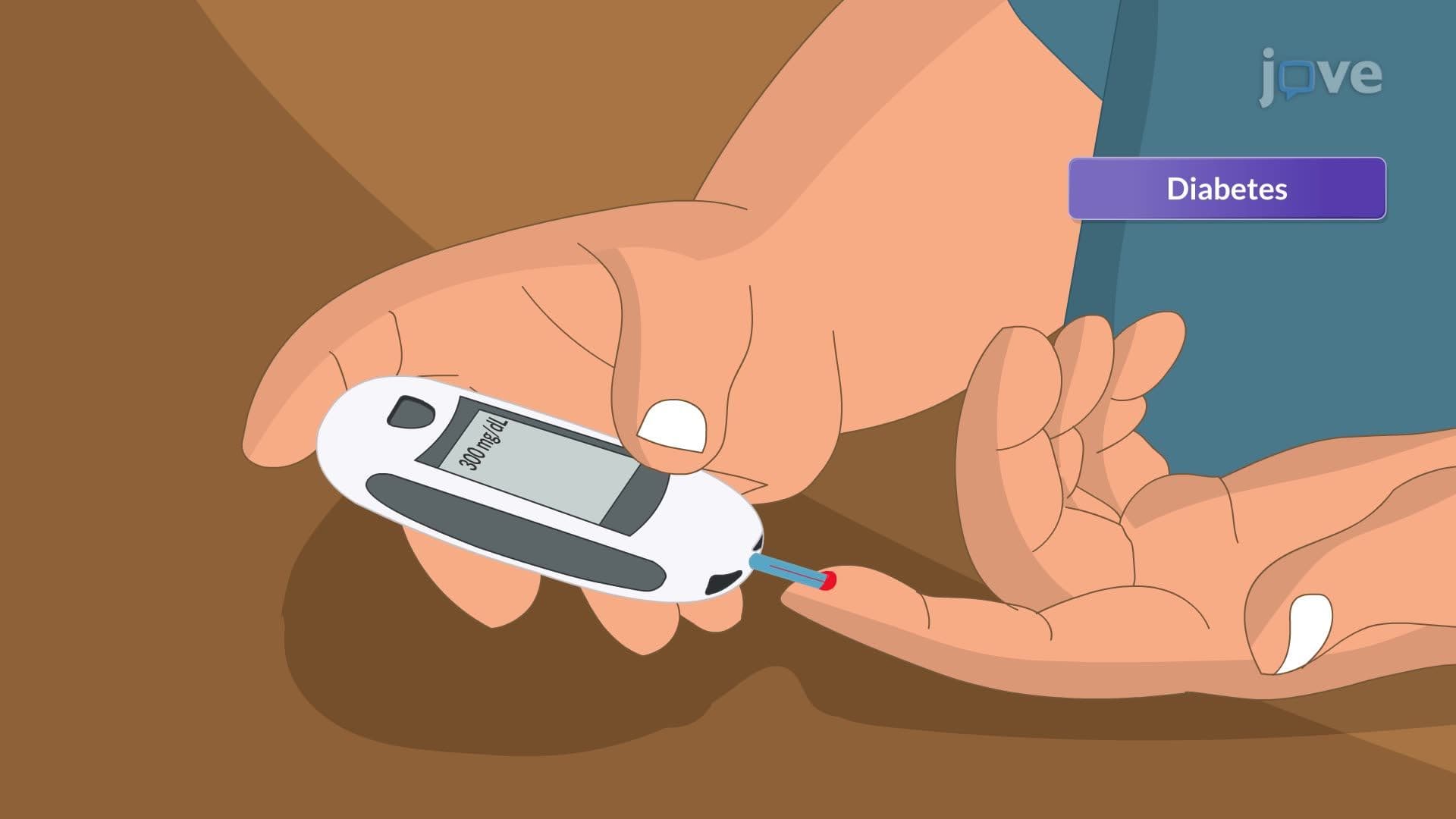
Comprehensive patient evaluation begins with detailed history-taking focused on chest pain characteristics. Healthcare providers assess pain quality, radiation patterns, duration, and associated symptoms like shortness of breath or diaphoresis. Previous cardiac history, including prior heart attacks, coronary artery disease, or cardiac procedures, significantly influences diagnostic probability. Family history analysis examines first-degree relatives with early-onset coronary disease, particularly fathers under 55 or mothers under 65. Risk factor evaluation encompasses modifiable factors like smoking history, diabetes mellitus, hyperlipidemia, and hypertension, alongside non-modifiable factors including age and gender.
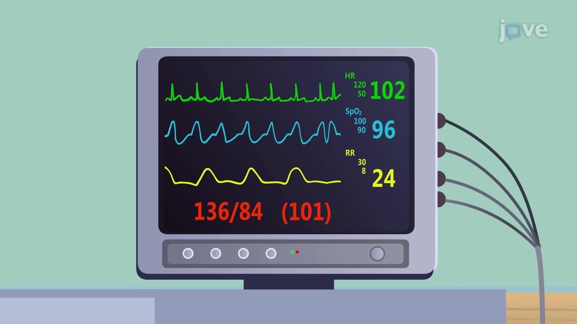
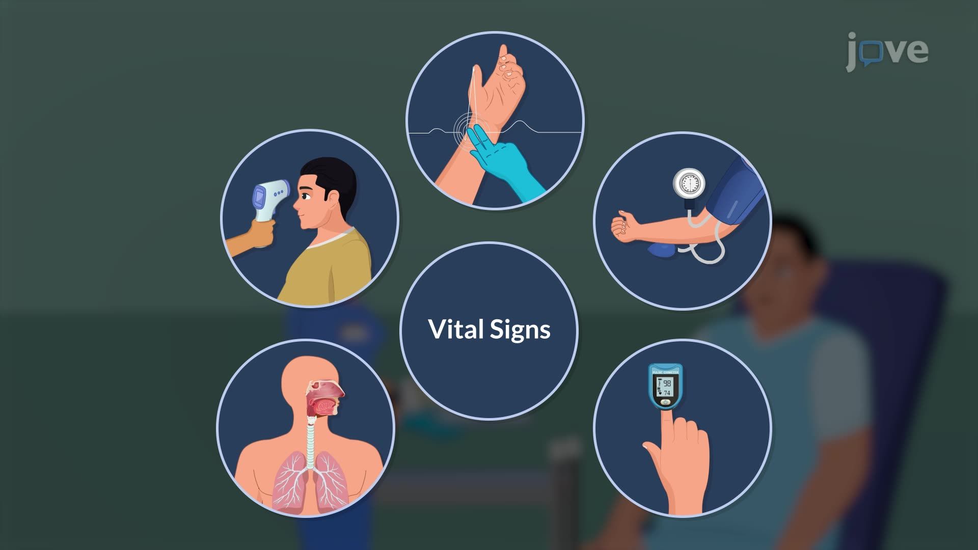
The focused cardiovascular examination seeks evidence of acute heart failure and hemodynamic compromise. Vital sign abnormalities may include hypotension, tachycardia, or irregular rhythms suggesting arrhythmias. Jugular venous distention indicates elevated right heart pressures, while peripheral edema suggests fluid retention from decreased cardiac output. Cardiac auscultation identifies new murmurs, particularly mitral regurgitation from papillary muscle dysfunction or ventricular septal defects from myocardial rupture. Pulmonary examination detects rales or crackles indicating left heart failure and pulmonary edema development.
The 12-lead ECG serves as the cornerstone diagnostic tool, with ST-elevation myocardial infarction (STEMI) showing characteristic ST-segment elevation in anatomically corresponding leads. Non-ST-elevation myocardial infarction (NSTEMI) typically presents with ST-depression and T-wave inversions, while established infarcts demonstrate pathological Q-waves indicating myocardial necrosis.
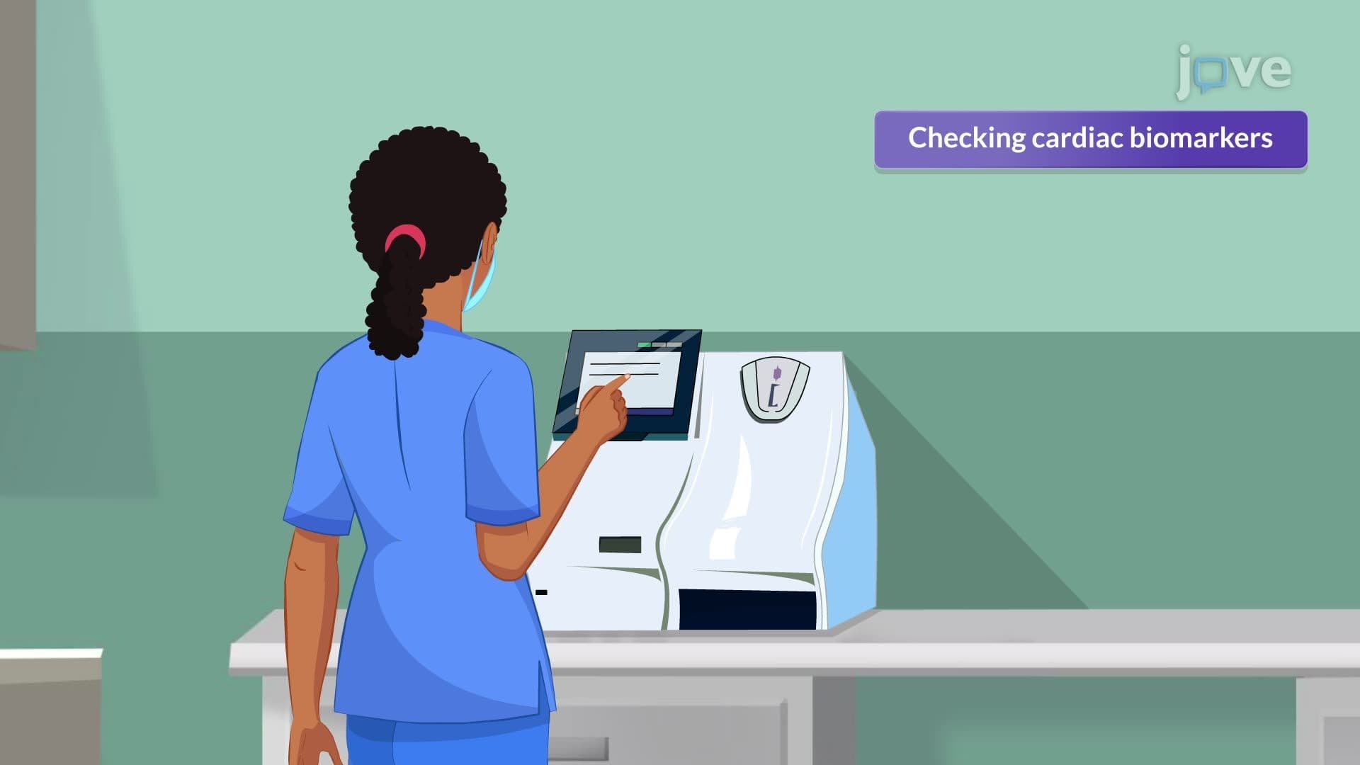
Cardiac biomarker interpretation follows specific temporal patterns crucial for MCAT and USMLE preparation. Troponin I and T, the most specific cardiac markers, become detectable 3-4 hours post-injury and remain elevated for days. Creatine kinase-MB rises within 3-6 hours and normalizes within 24-48 hours, making it useful for detecting reinfarction. Myoglobin, though less specific, appears earliest at 1-3 hours, serving as an early exclusion marker.
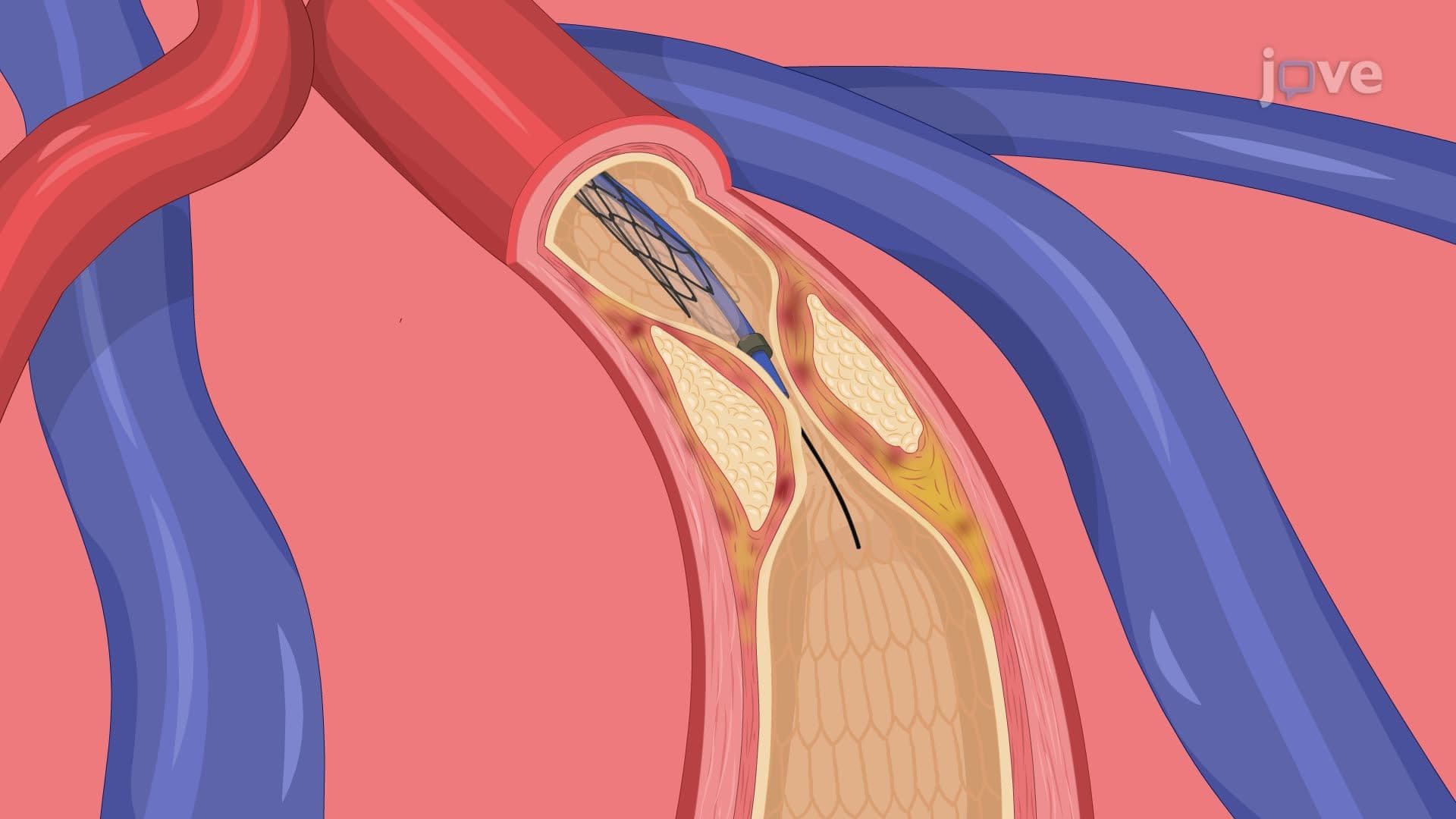
Advanced imaging includes echocardiography for wall motion assessment and ejection fraction calculation, while coronary angiography provides definitive anatomical evaluation of coronary artery patency and guides percutaneous coronary intervention decisions.
Related Micro-courses