Loading subject page…

2,474 views

Video Summary: Coronary Artery Disease Ii Pathophysiology Explained
Did you know that heart disease kills one American every 34 seconds, making coronary artery disease ii pathophysiology crucial for understanding our nation's leading killer? This complex process transforms healthy arteries into dangerous, plaque-filled vessels through a cascade of cellular events involving oxidized LDL cholesterol, inflammatory responses, and foam cell formation. Consider how a simple cholesterol particle can trigger the life-threatening blockages seen in over 18 million Americans with coronary artery disease. Watch the full video on JoVE Coach to master this concept with expert-led visuals and step-by-step explanations.
Coronary Artery Disease II Pathophysiology represents one of the most clinically significant disease processes in modern medicine, affecting over 20 million Americans annually. This complex cascade begins at the microscopic level with endothelial dysfunction-the critical first step that transforms healthy arterial walls into sites of chronic inflammation and eventual obstruction.
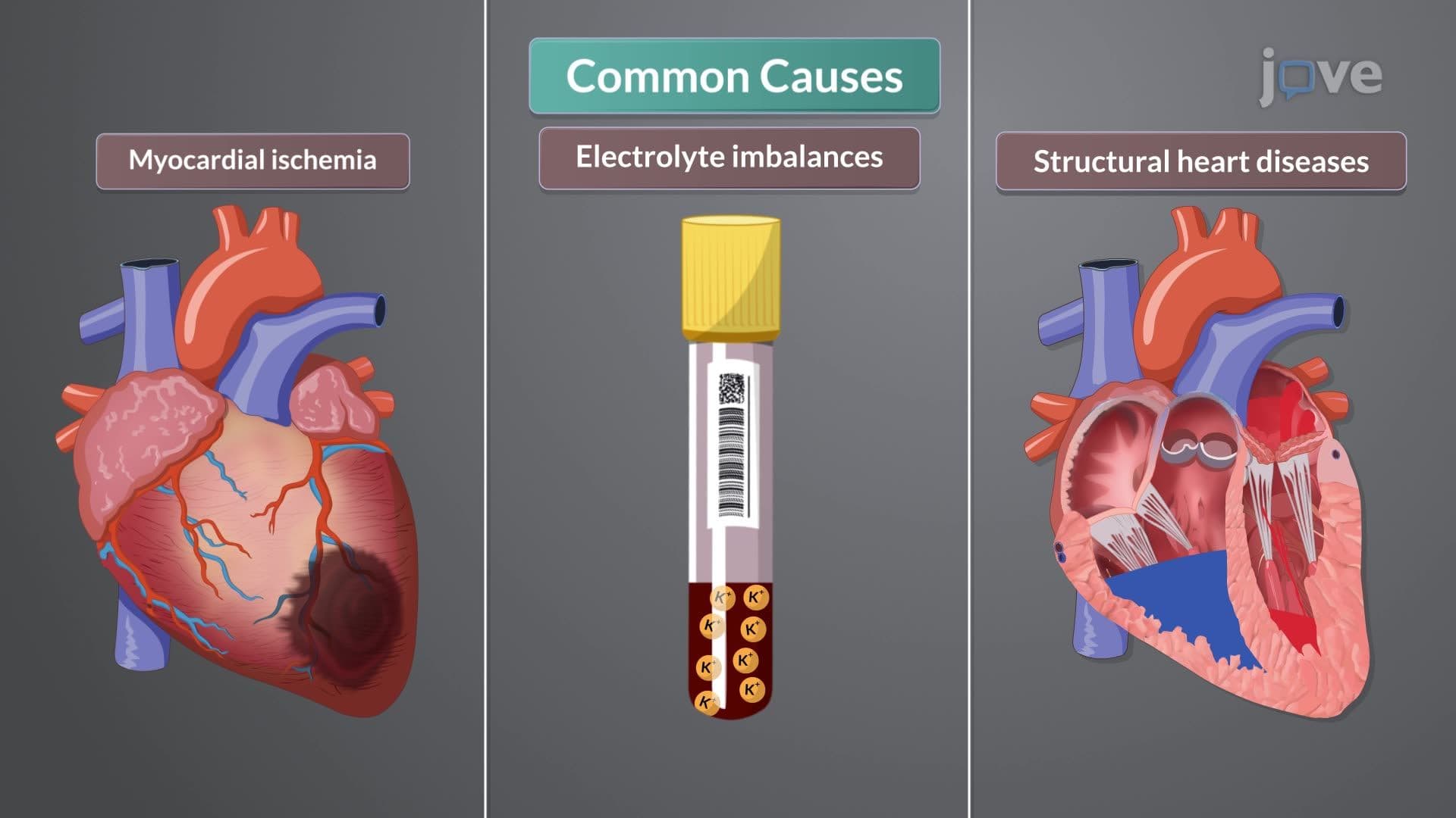
The pathophysiological process initiates when various risk factors-including hypertension, diabetes, smoking, and hyperlipidemia-damage the delicate endothelial lining of coronary arteries. This damage triggers immediate oxidative stress responses, compromising the endothelium's normal protective functions. In healthy individuals, endothelial cells regulate vasodilation, prevent thrombosis, and maintain arterial wall integrity. However, once dysfunction occurs, these protective mechanisms fail, creating an environment conducive to atherosclerotic development.
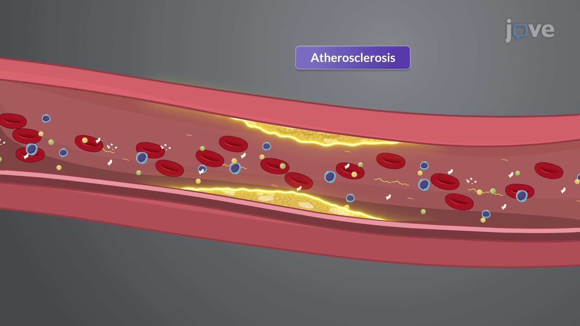
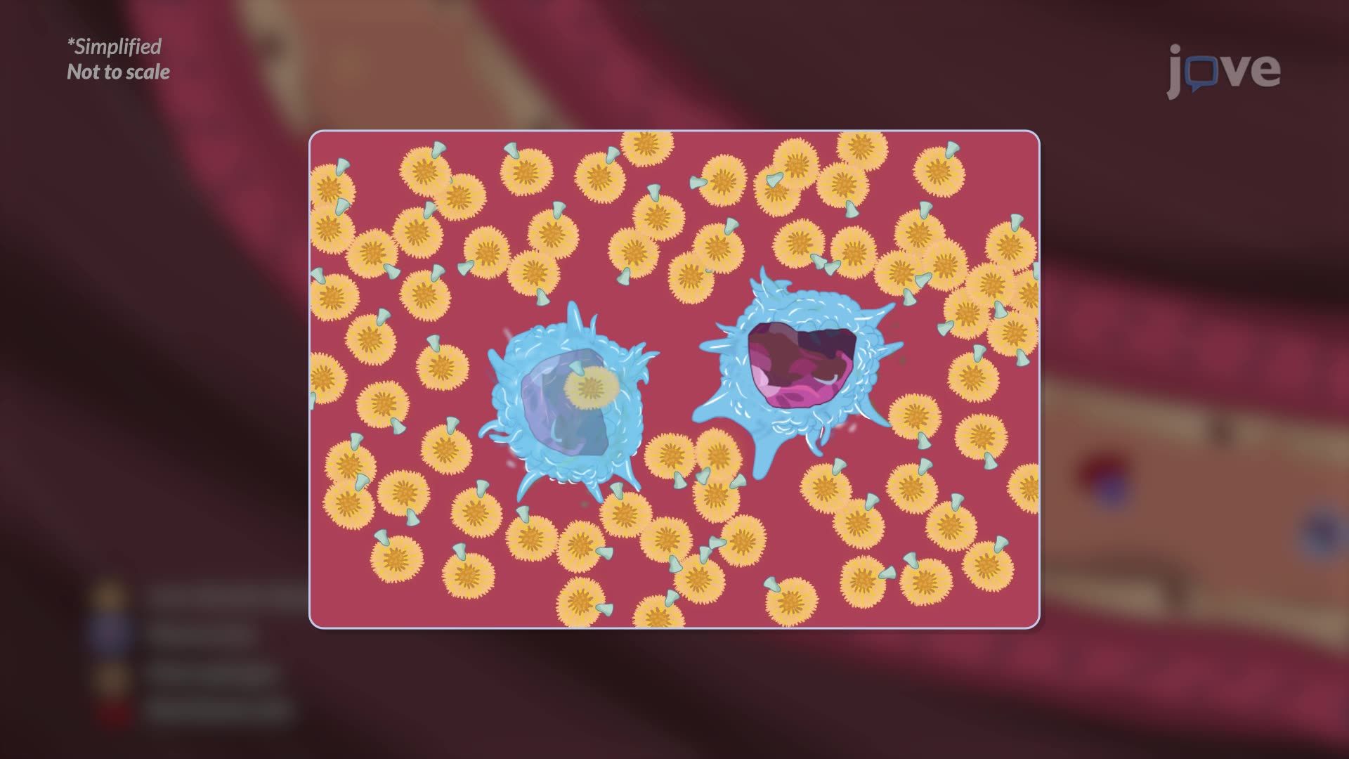
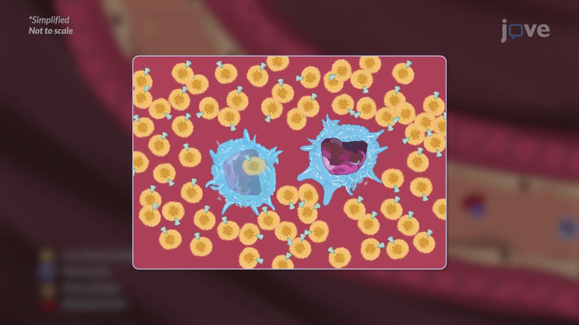
Low-density lipoproteins (LDL) exploit this compromised endothelial barrier, penetrating into the arterial wall's intimal layer. Within this pro-inflammatory environment, LDL particles undergo oxidative modification, transforming from relatively benign cholesterol carriers into highly inflammatory molecules. These oxidized LDL particles act as danger signals, recruiting circulating monocytes to the site of injury through chemotactic gradients.
The recruited monocytes undergo a remarkable transformation upon entering the arterial wall. These immune cells differentiate into macrophages-specialized phagocytic cells designed to consume foreign materials and cellular debris. However, in the context of atherosclerosis, this protective mechanism becomes pathological. Macrophages aggressively uptake oxidized LDL through scavenger receptors, accumulating lipid droplets within their cytoplasm until they resemble foam-hence the term "foam cells."
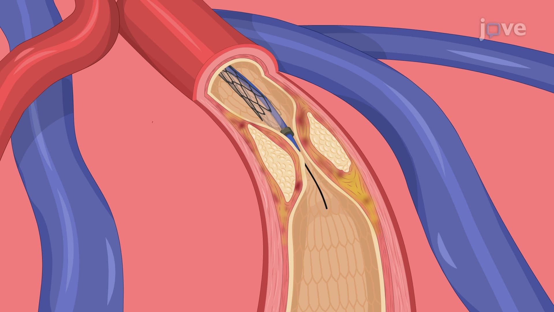
These foam cells represent the building blocks of atherosclerotic plaques, clustering together to form fatty streaks visible even in young adults during autopsy studies. As the process continues, vascular smooth muscle cells migrate from the arterial media to the intima, where they proliferate and synthesize extracellular matrix proteins including collagen and elastin. This fibrous tissue forms a protective cap over the underlying lipid-rich core, creating the characteristic atherosclerotic plaque structure.
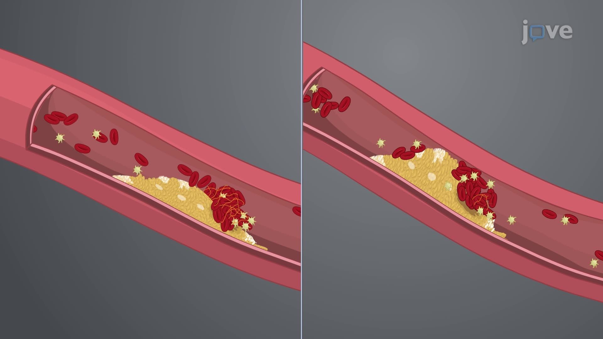
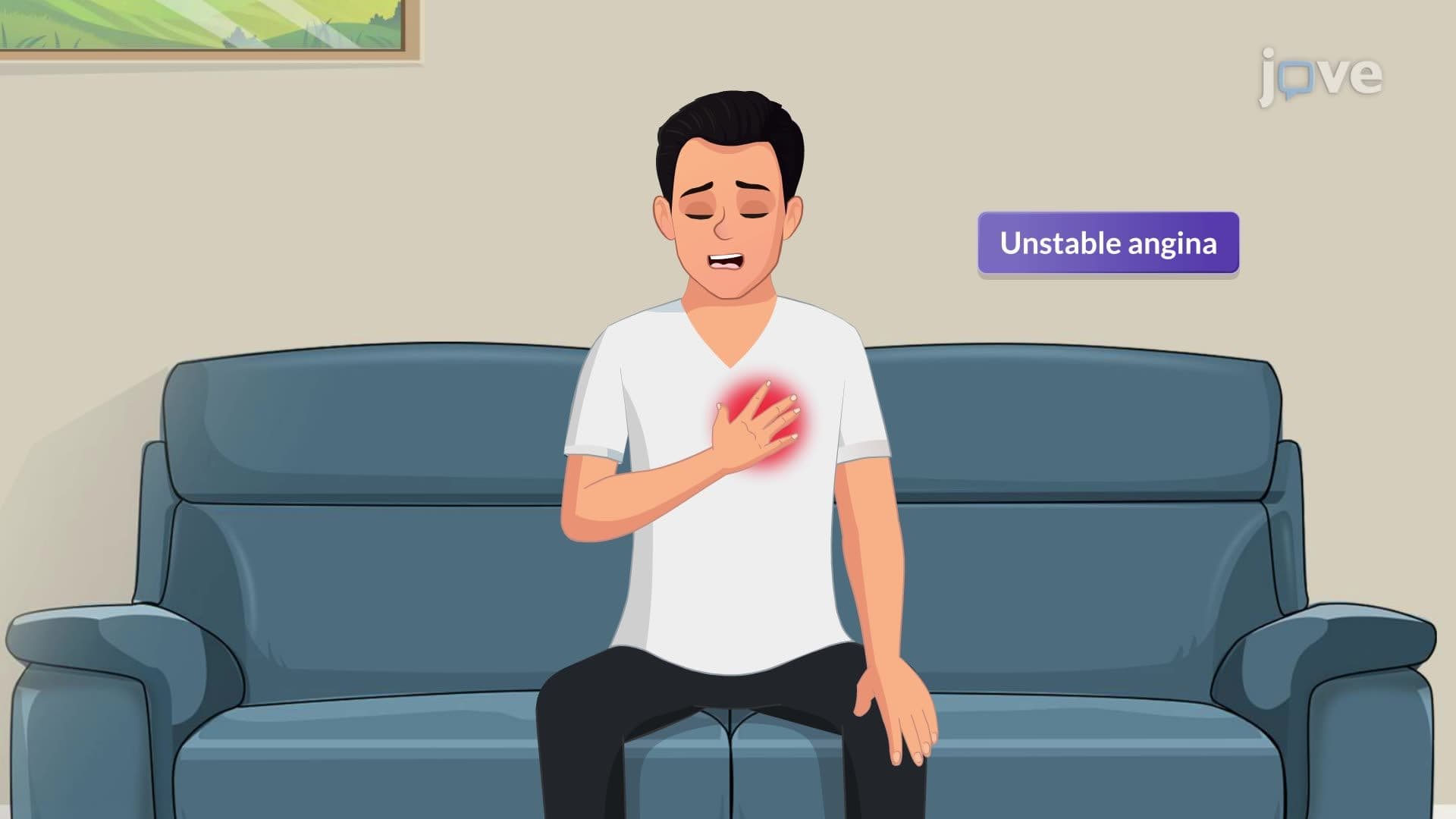
Understanding plaque morphology proves crucial for predicting clinical outcomes. Stable plaques feature thick fibrous caps with relatively small lipid cores, typically causing gradual luminal narrowing and stable angina symptoms. Conversely, unstable plaques contain large lipid cores covered by thin, vulnerable fibrous caps susceptible to rupture. When unstable plaques rupture, they expose highly thrombogenic material to the bloodstream, triggering rapid thrombus formation and potential complete vessel occlusion-the mechanism underlying most myocardial infarctions.
This pathophysiological knowledge directly applies to clinical scenarios tested on the MCAT, USMLE Step 1, and advanced placement biology exams, where students must connect molecular mechanisms to clinical presentations and therapeutic interventions.
Related Micro-courses