Loading subject page…

1,657 views

Video Summary: Myocarditis Ii Clinical Features and Diagnostic Evaluation
Did you know that myocarditis can silently damage your heart without any symptoms? Myocarditis II: clinical features and diagnostic evaluation reveals how this inflammatory heart condition progresses from subtle flu-like symptoms to serious cardiac complications. Consider a college athlete who develops chest pain after a viral infection-early recognition could prevent sudden cardiac death during sports participation. This comprehensive overview covers the spectrum from asymptomatic cases to acute presentations, plus the diagnostic tools physicians use to confirm myocarditis in US medical centers. Watch the full video on JoVE Coach to master this concept with expert-led visuals and step-by-step explanations.
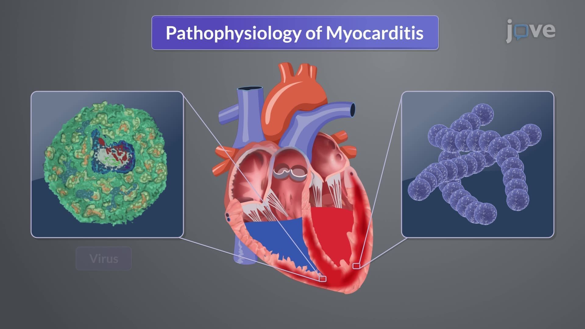
Myocarditis represents one of cardiology's most challenging diagnostic puzzles. This inflammatory heart condition affects thousands of Americans annually, with cases ranging from completely silent infections to rapidly fatal cardiac collapse. The clinical presentation exists on a broad spectrum, making early recognition crucial for optimal patient outcomes.
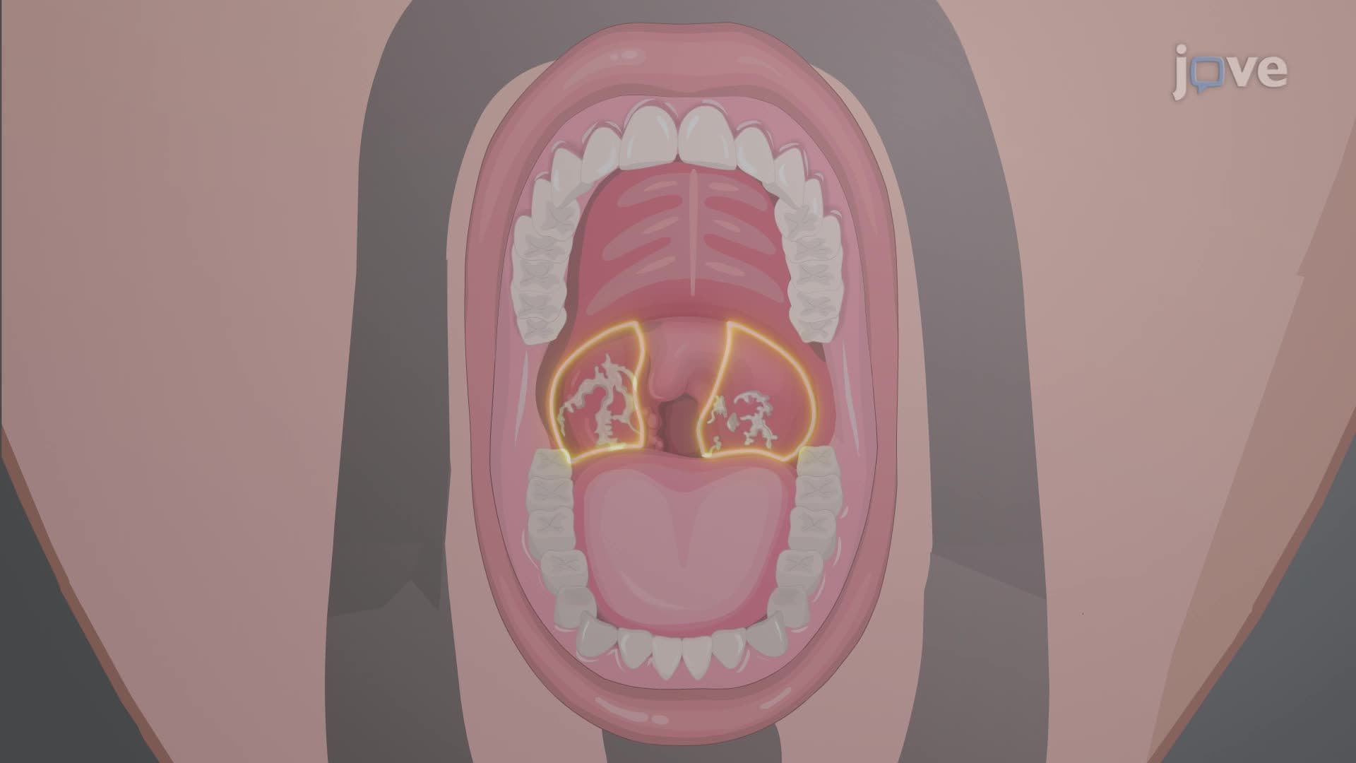
The myocarditis II: clinical features and diagnostic evaluation overview begins with understanding symptom progression. Many patients experience a biphasic illness pattern-initial viral symptoms followed by cardiac manifestations days to weeks later. Early systemic symptoms mirror common viral infections: fever, muscle aches, sore throat, and fatigue. These nonspecific presentations often lead patients to dismiss symptoms as "just the flu."
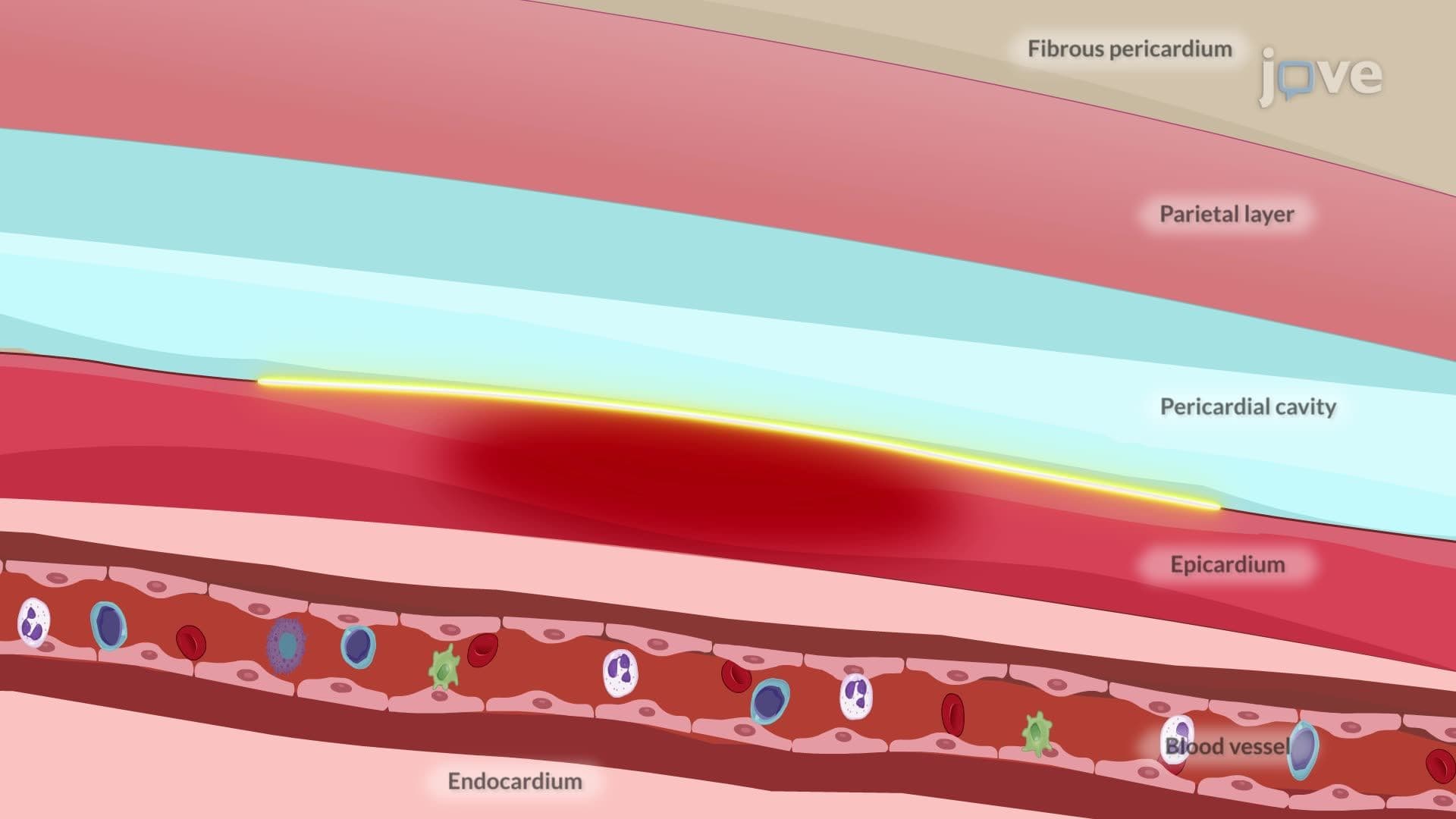
However, as inflammation spreads to cardiac tissue, more concerning signs emerge. Pleuritic chest pain-sharp, positional discomfort worsened by breathing-signals pericardial involvement. The classic pericardial friction rub, a scratchy sound heard through a stethoscope, indicates inflammation of the heart's outer lining. At major medical centers like Johns Hopkins or Mayo Clinic, emergency physicians remain alert for these subtle cardiac clues in patients with recent viral illnesses.
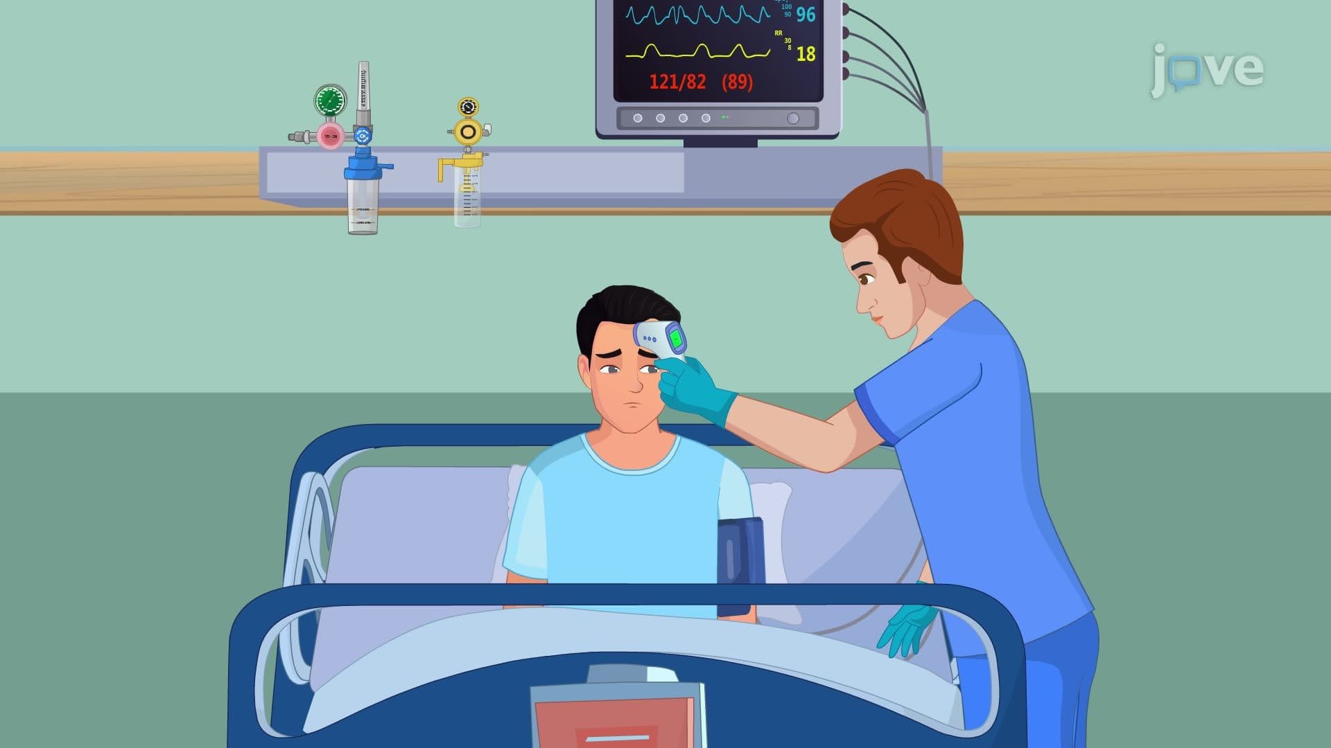
The myocarditis II: clinical features and diagnostic evaluation study guide emphasizes systematic evaluation. Healthcare providers begin with detailed history-taking, focusing on recent infections, travel patterns, and autoimmune disease history. Physical examination targets cardiovascular signs: irregular heartbeats, lung congestion, neck vein distention, and ankle swelling-all indicators of heart failure.
Laboratory testing provides crucial supporting evidence. Elevated troponin levels-proteins released during heart muscle damage-serve as sensitive markers of myocardial injury. Inflammatory markers like C-reactive protein and erythrocyte sedimentation rate typically rise significantly. Complete blood counts often reveal mild increases in white blood cells, reflecting the body's immune response.
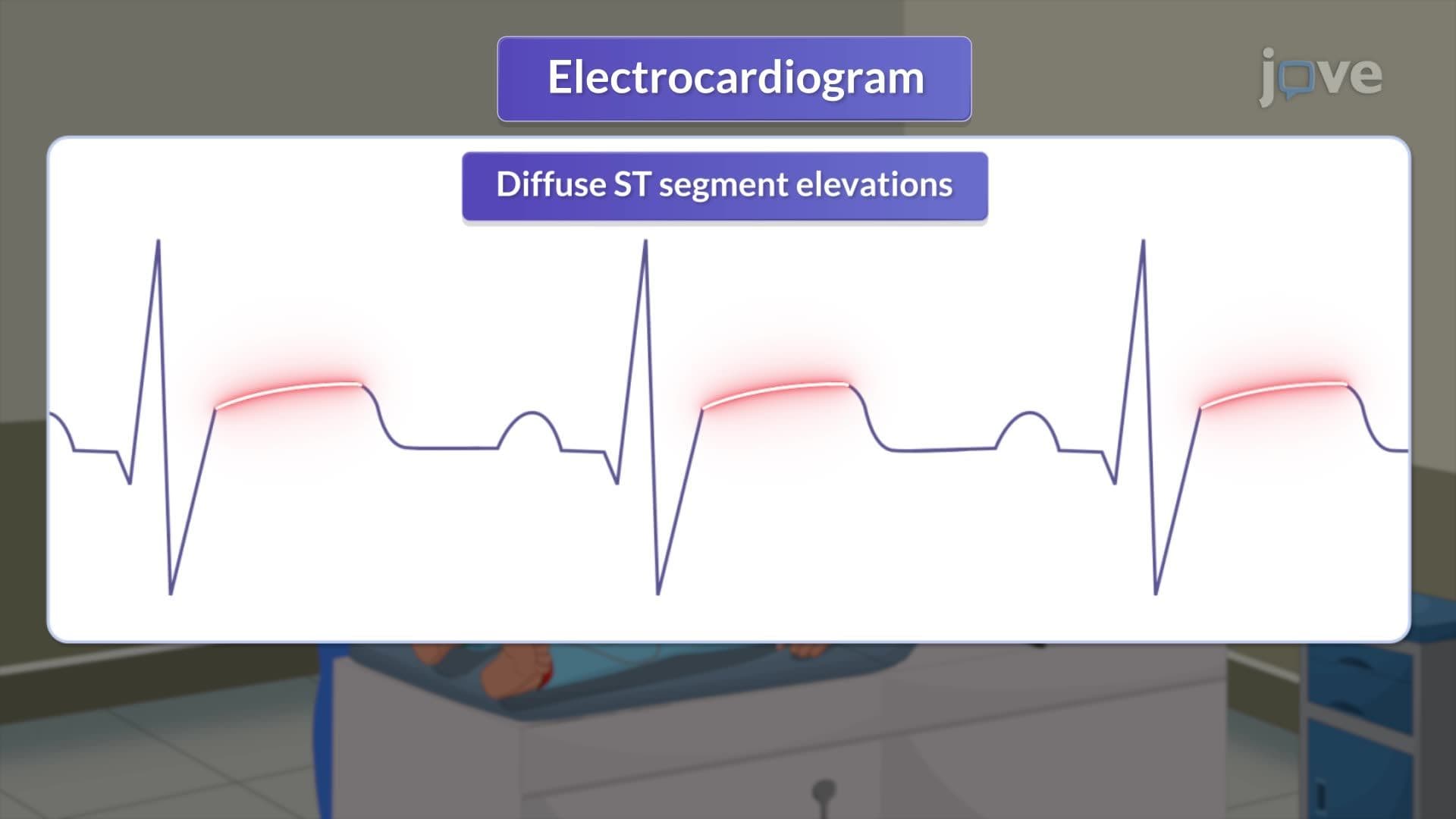
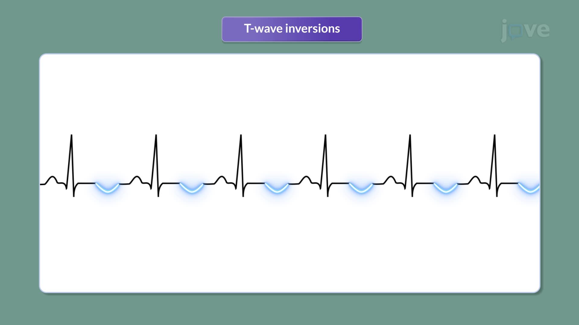
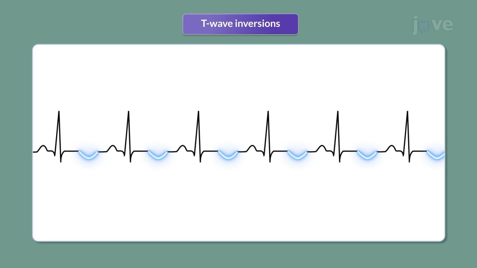
Electrocardiograms (ECGs) demonstrate characteristic patterns in myocarditis cases. Unlike heart attacks that show localized changes, myocarditis typically produces diffuse ST-segment elevations and T-wave inversions across multiple leads. These patterns help distinguish myocarditis from coronary artery disease-a critical differentiation for treatment planning.
Endomyocardial biopsy remains the definitive diagnostic test, though it's reserved for severe cases due to procedural risks. This technique involves threading a catheter through blood vessels to obtain tiny heart tissue samples. Microscopic examination reveals the hallmark features: lymphocytes infiltrating heart muscle and damaged myocytes.
For students preparing for the MCAT or AP Biology exams, understanding this diagnostic progression illustrates how physicians combine clinical reasoning with technological tools. College-level pathophysiology courses frequently test students on distinguishing myocarditis from other cardiac conditions based on presentation patterns and diagnostic findings.
Related Micro-courses