Loading subject page…

1,657 views

Video Summary: Pericarditis Ii Clinical Features and Diagnostic Evaluation
Did you know that pericarditis can mimic a heart attack on an EKG, leading to misdiagnosis in emergency rooms across the United States? Pericarditis II: clinical features and diagnostic evaluation reveals how this inflammatory condition presents with distinctive chest pain that worsens when lying flat and creates a characteristic "scratchy" heart sound. At Johns Hopkins Hospital, emergency physicians use specific diagnostic criteria including EKG changes and echocardiograms to differentiate pericarditis from myocardial infarction. Watch the full video on JoVE Coach to master this concept with expert-led visuals and step-by-step explanations.
Pericarditis represents one of the most clinically challenging cardiac conditions due to its variable presentation and potential to mimic life-threatening conditions like myocardial infarction. The clinical features form a constellation of symptoms that, when properly recognized, lead to accurate diagnosis and appropriate treatment.
The hallmark symptom of pericarditis is sharp, pleuritic chest pain with unique positional characteristics. Unlike the crushing substernal pain of myocardial infarction, pericarditic pain typically improves when patients sit upright and lean forward-a finding so characteristic that emergency physicians at major medical centers like Mayo Clinic use it as a key diagnostic criterion. The pain often radiates to the trapezius ridge, a pattern rarely seen in other cardiac conditions.
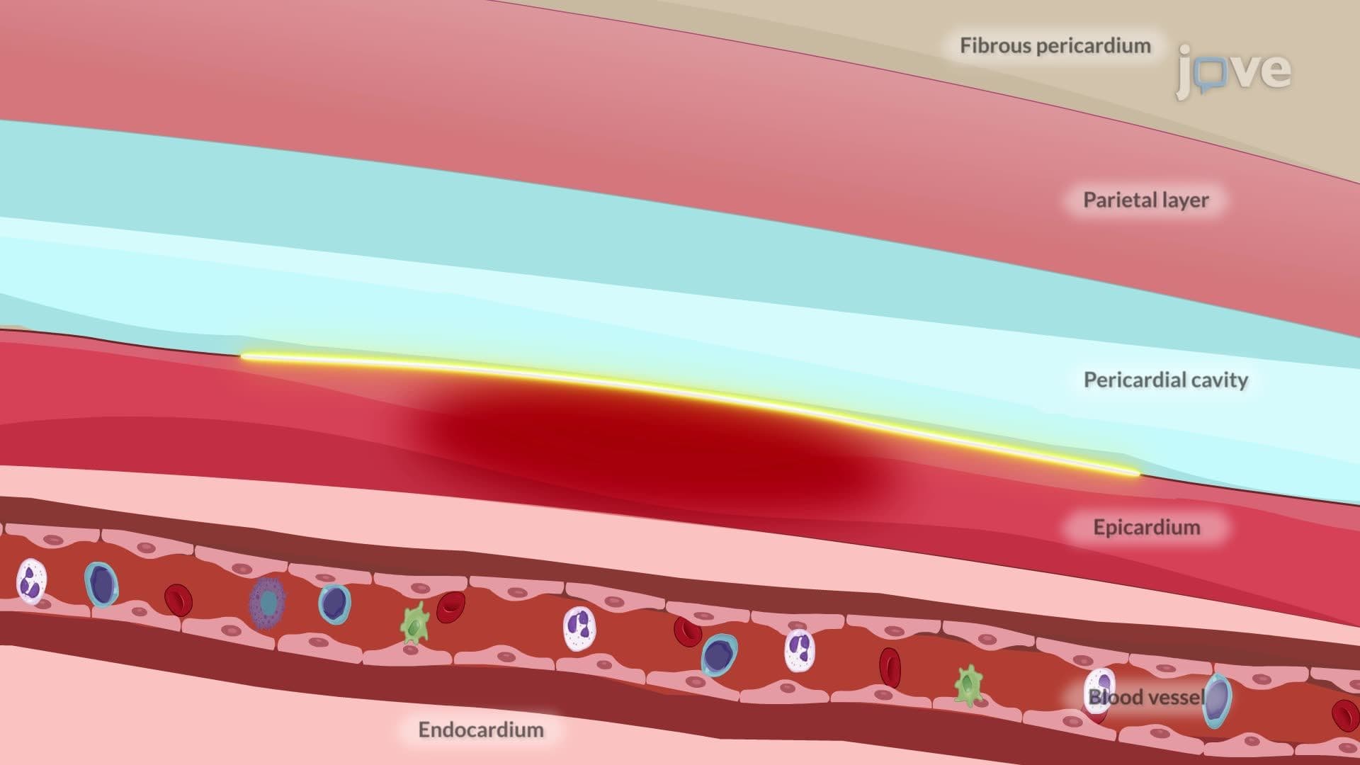
The pericardial friction rub represents the pathognomonic physical finding, occurring in approximately 60-85% of acute pericarditis cases. This high-pitched, scratchy sound results from inflamed pericardial layers rubbing against each other during cardiac motion. Medical students preparing for the USMLE Step 2 CK examination should remember that this rub has three components corresponding to atrial systole, ventricular systole, and ventricular diastole, though all three are not always audible.
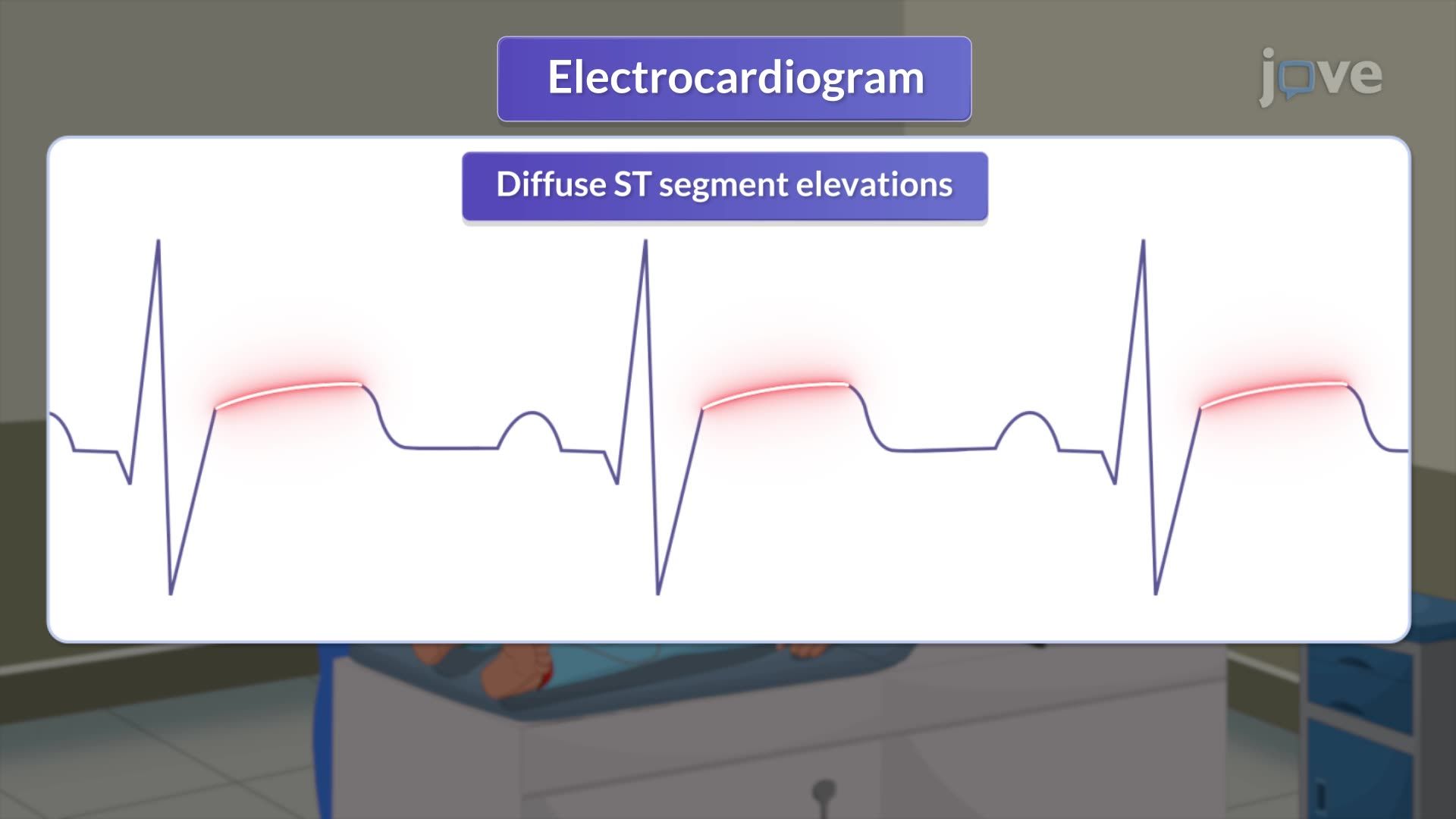
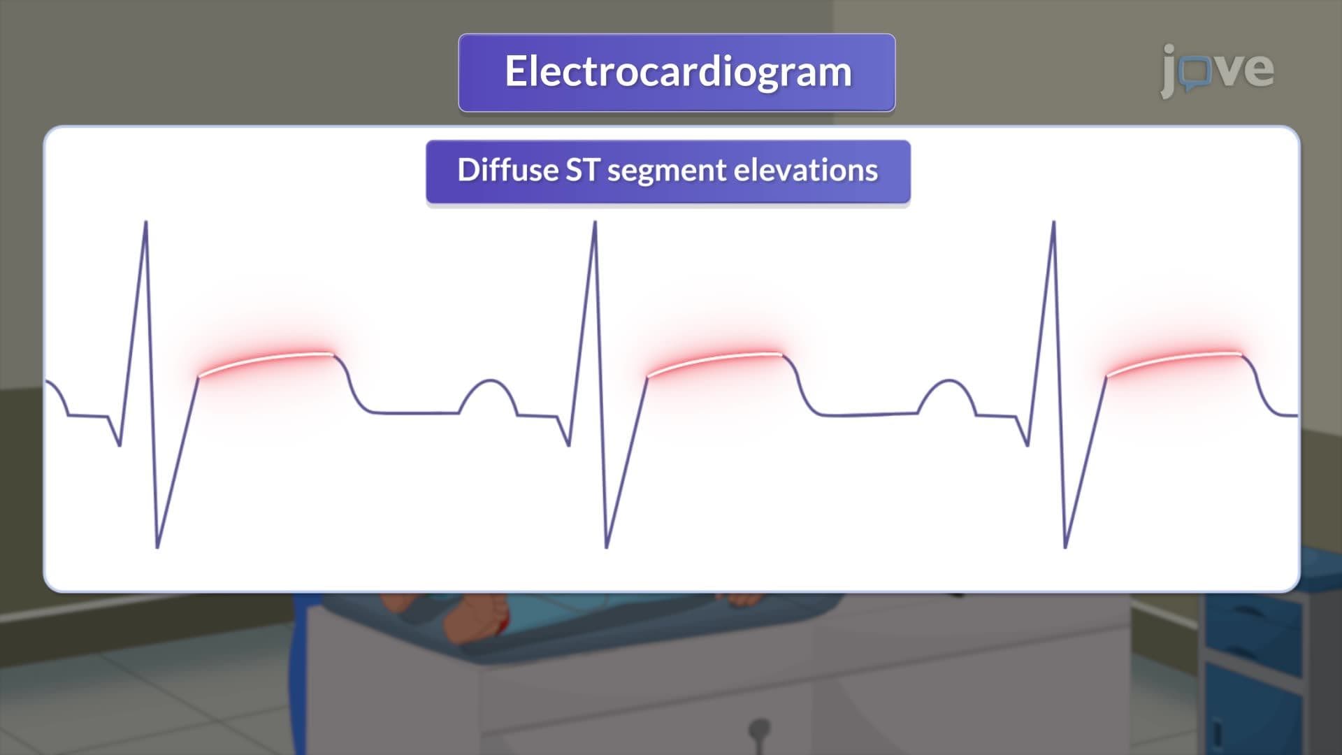
Electrocardiographic findings in pericarditis follow a predictable four-stage evolution, crucial knowledge for MCAT test-takers studying cardiovascular pathophysiology. Stage I shows diffuse ST-elevation with upward concavity, contrasting with the convex ST-elevation seen in myocardial infarction. This distinction has saved countless patients from unnecessary cardiac catheterization procedures in American emergency departments.
Echocardiography serves as the primary imaging modality for detecting pericardial effusion, with sensitivity exceeding 95% when performed by experienced sonographers. The technique can quantify effusion size and assess for hemodynamic compromise-critical information that influences treatment decisions in intensive care units nationwide.
Laboratory evaluation reveals the inflammatory nature of pericarditis through elevated acute-phase reactants. C-reactive protein and erythrocyte sedimentation rate provide objective measures of systemic inflammation, while troponin elevation may indicate concurrent myocardial involvement-a finding that affects prognosis and treatment strategies.
For AP Biology students studying inflammatory responses, pericarditis exemplifies how cytokine-mediated inflammation manifests clinically through measurable biomarkers that correlate with disease severity and treatment response.
Related Micro-courses