Loading subject page…

1,657 views

Video Summary: Rheumatic Heart Disease Ii Clinical Explained
Did you know that rheumatic heart disease II clinical presentations can vary dramatically, from subtle heart murmurs in teenagers to life-threatening heart failure in young adults? Understanding rheumatic heart disease II clinical manifestations is crucial for recognizing this preventable condition that affects approximately 15.6 million people worldwide, including thousands of cases annually in underserved US communities. From acute carditis symptoms like chest pain and new murmurs to chronic complications including atrial fibrillation and valvular damage, mastering Rheumatic Heart Disease II Clinical Explained concepts helps identify critical diagnostic clues and treatment priorities. Watch the full video on JoVE Coach to master this concept with expert-led visuals and step-by-step explanations.
Rheumatic heart disease represents one of the most significant preventable cardiac conditions affecting young people globally, with particular relevance in underserved US populations. The clinical manifestations occur in two distinct phases: acute rheumatic fever and chronic rheumatic heart disease, each presenting unique diagnostic challenges and treatment considerations.
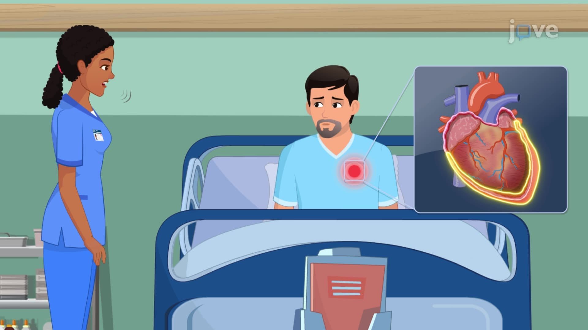
The acute phase centers on carditis, representing active inflammation of heart tissues. Patients typically present with new-onset heart murmurs, often detected during routine physical examinations in schools or primary care settings. Chest pain may be described as sharp or aching, frequently accompanied by palpitations that patients describe as "heart racing" or "skipping beats." Dyspnea, particularly with exertion, reflects early cardiac compromise and should prompt immediate medical evaluation.
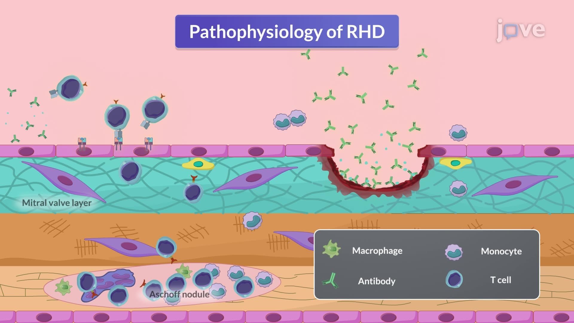
For students preparing for the MCAT or advanced placement biology exams, understanding that acute carditis represents an autoimmune response following group A streptococcal infection is crucial. The molecular mimicry between streptococcal proteins and cardiac tissue antigens triggers cross-reactive antibodies, leading to inflammation and tissue damage.
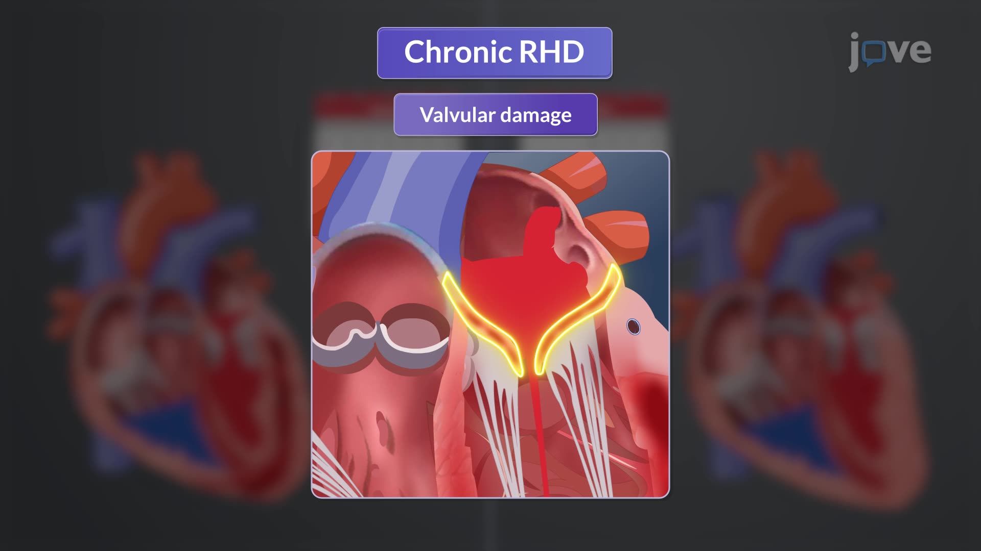
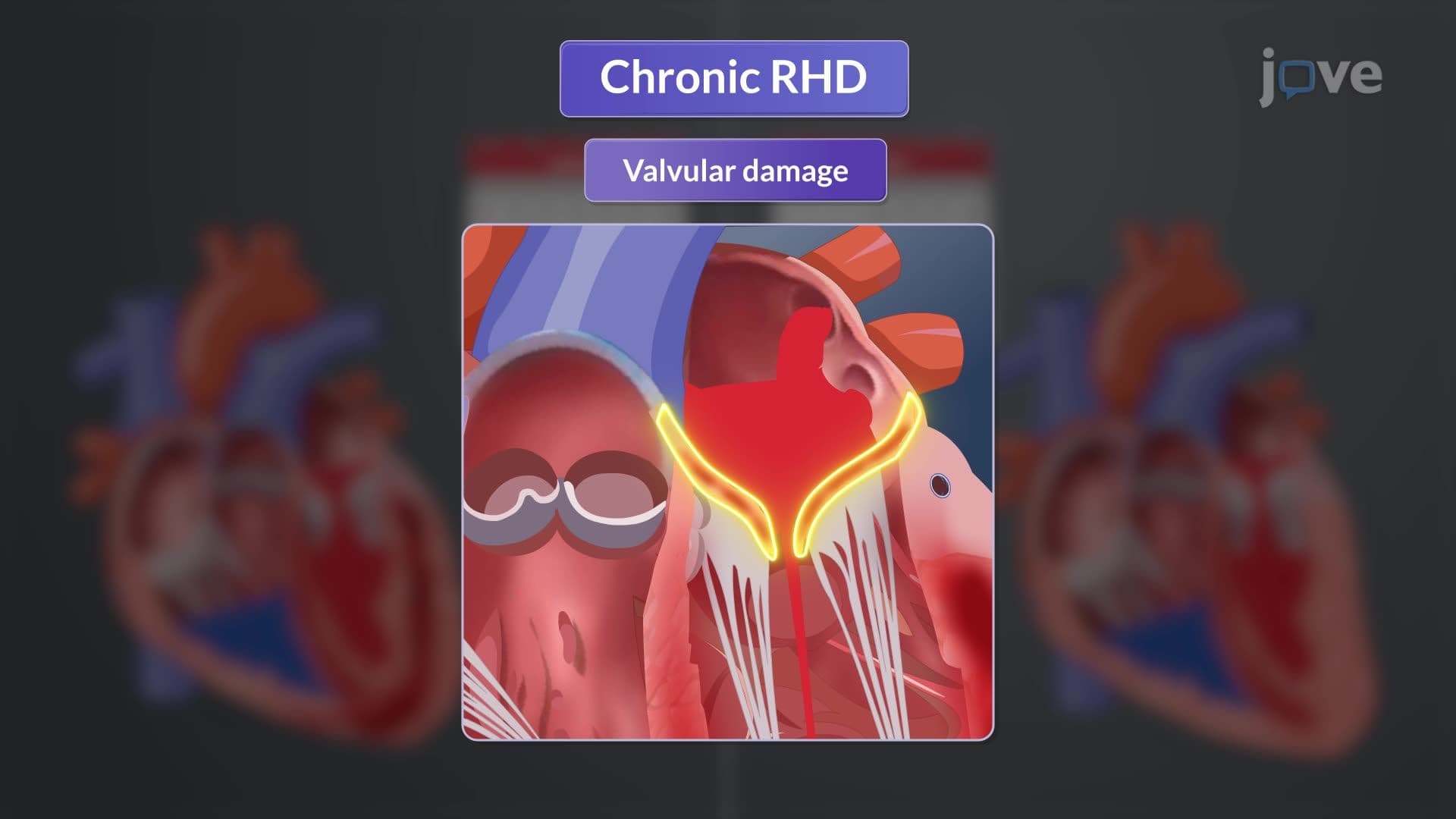
Chronic rheumatic heart disease develops from repeated episodes of acute rheumatic fever, resulting in permanent valvular damage. The mitral valve bears the greatest impact, followed by the aortic valve. Patients experience persistent heart murmurs with characteristic acoustic patterns that trained clinicians can identify through careful auscultation.
Atrial fibrillation represents a serious complication, increasing stroke risk and requiring anticoagulation therapy. Signs of chronic heart failure include peripheral edema, particularly in the ankles and feet, fatigue, and progressive exercise intolerance. These findings are particularly relevant for NCLEX preparation, as nursing students must recognize early heart failure symptoms.
Modern diagnostic evaluation relies heavily on advanced imaging techniques. Echocardiography provides detailed visualization of cardiac structures, allowing assessment of left atrial enlargement, ventricular function, and valvular abnormalities. This technique can identify vegetation (abnormal growths) or thrombi (blood clots) that may complicate the clinical course.
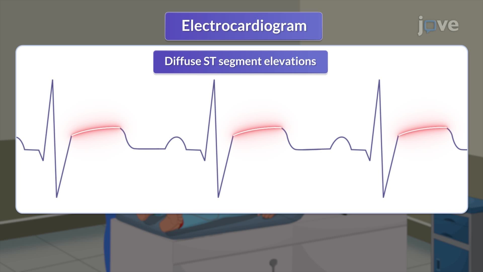
Electrocardiography detects rhythm abnormalities, with atrial fibrillation being the most clinically significant finding. Chest X-rays reveal cardiomegaly (enlarged heart) and pulmonary congestion, indicating advanced disease progression. For USMLE Step 1 preparation, students should understand that Doppler ultrasound provides quantitative assessment of valvular stenosis and regurgitation severity, guiding treatment decisions.
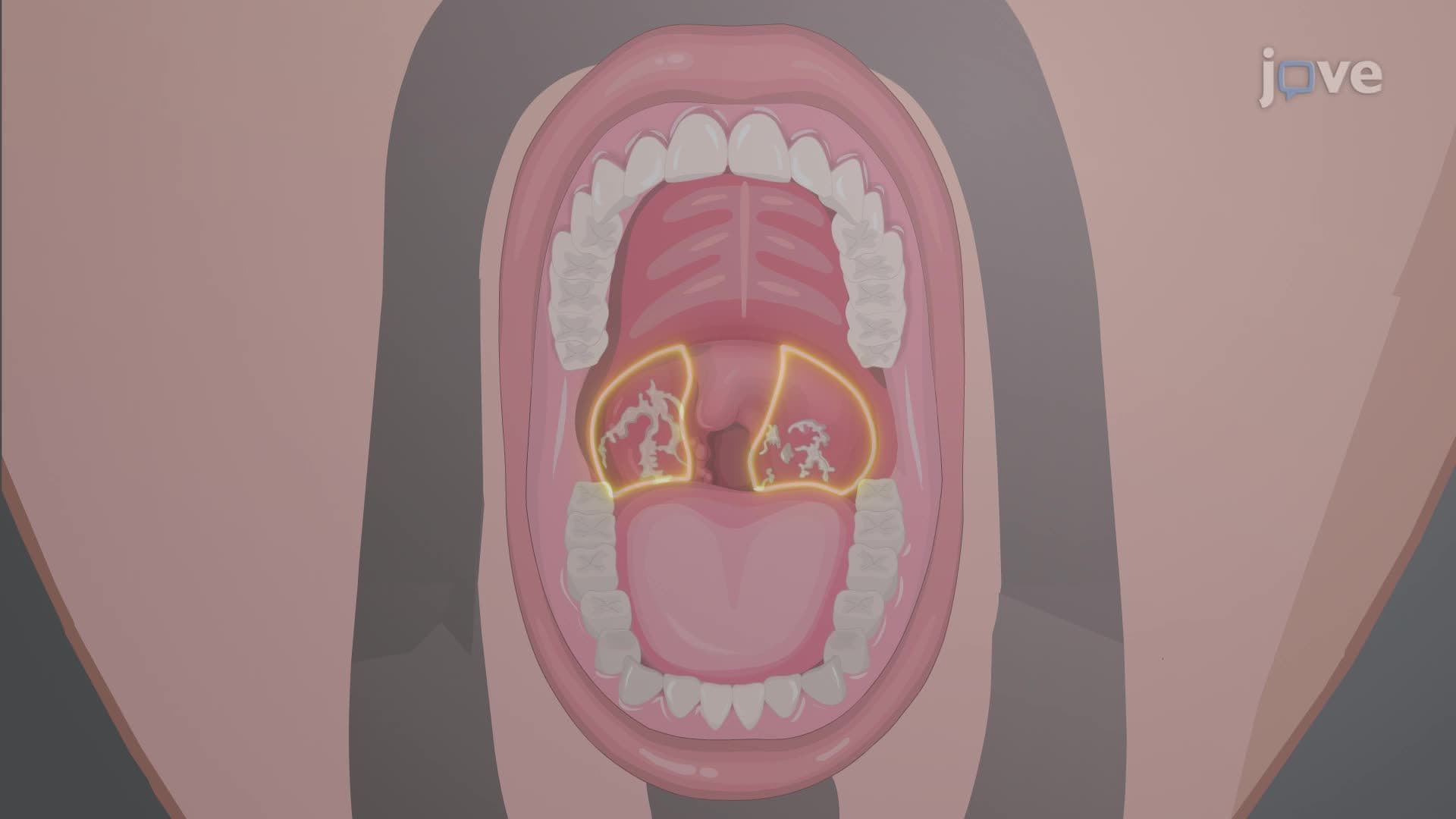
Laboratory testing supports diagnosis through inflammatory markers and evidence of recent streptococcal infection. Elevated antistreptolysin O titers, C-reactive protein, and erythrocyte sedimentation rate (ESR) provide objective evidence supporting the clinical diagnosis.
Related Micro-courses