Loading subject page…


5,820 views


Video Summary: Endoscopic Studies I Bronchoscopy and Thoracoscopy Explained
Did you know that doctors can actually see inside your lungs and chest cavity using tiny cameras? Endoscopic studies i bronchoscopy and thoracoscopy allow medical professionals to diagnose lung diseases, remove blockages, and collect tissue samples without major surgery. At Johns Hopkins Hospital, pulmonologists use these minimally invasive procedures daily to treat everything from lung cancer to pleural effusions. Watch the full video on JoVE Coach to master this concept with expert-led visuals and step-by-step explanations.
Endoscopic respiratory procedures represent revolutionary advances in pulmonary medicine, allowing physicians to visualize, diagnose, and treat conditions within the respiratory system using minimally invasive techniques. These procedures have transformed patient care at major medical centers like Mayo Clinic and Cleveland Clinic, reducing recovery times and improving diagnostic accuracy.
Bronchoscopy utilizes a flexible or rigid endoscope to examine the respiratory tract from the larynx through the bronchial tree. The procedure serves dual purposes: diagnostic evaluation and therapeutic intervention. During diagnostic bronchoscopy, physicians can identify tumors, inflammation, infections, and structural abnormalities while collecting bronchoalveolar lavage samples and performing transbronchial biopsies. This technique proves invaluable for staging lung cancer and diagnosing conditions like sarcoidosis or pneumonia in immunocompromised patients.
Therapeutic applications extend beyond diagnosis. Pulmonologists use bronchoscopy to remove foreign bodies, clear mucus plugs causing atelectasis, control hemorrhage, and deliver targeted treatments like brachytherapy for endobronchial tumors. At institutions like MD Anderson Cancer Center, interventional pulmonologists routinely perform complex therapeutic bronchoscopies to improve patient outcomes.
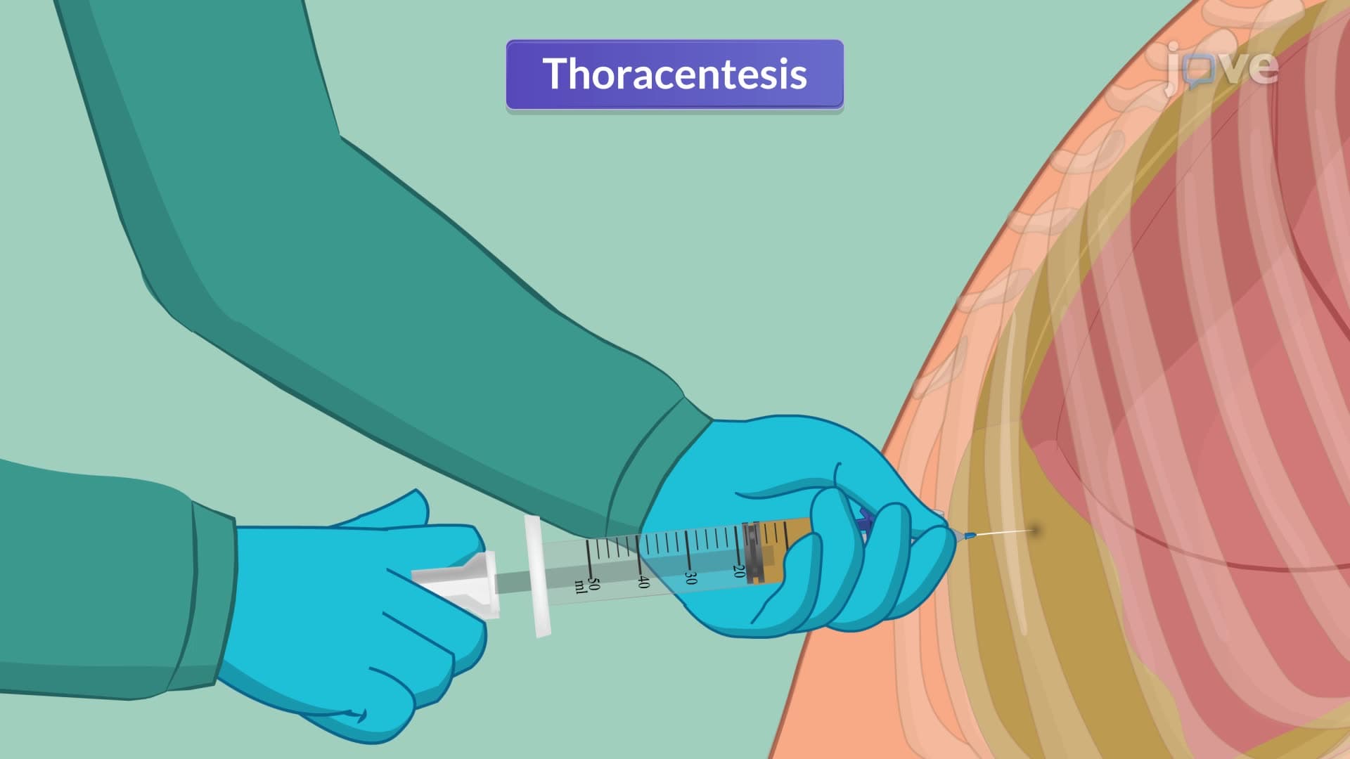
Thoracoscopy, also known as pleuroscopy, provides direct visualization of the pleural cavity through small intercostal incisions. This procedure excels in diagnosing unexplained pleural effusions, particularly when cytological analysis proves inconclusive. The technique allows for comprehensive pleural biopsy collection, adhesiolysis, and pleurodesis procedures.
The diagnostic yield of thoracoscopy significantly exceeds that of blind pleural biopsy, making it the gold standard for evaluating suspected pleural malignancy. Post-procedure chest tube insertion facilitates lung re-expansion and monitors for potential complications like pneumothorax or bleeding.
These procedures frequently appear in NCLEX questions focusing on respiratory nursing care, MCAT passages about diagnostic techniques, and USMLE clinical vignettes. Students should understand the critical importance of pre-procedure NPO status, post-procedure monitoring for complications, and the rationale behind specific nursing interventions. Understanding these concepts proves essential for success in advanced pathophysiology courses and clinical rotations in pulmonary medicine.
Related Micro-courses