Loading subject page…


5,820 views


Video Summary: Radiological Investigation I X Ray Explained
Did you know that chest X-rays can detect life-threatening conditions like pneumonia within seconds, making them the most crucial first-line diagnostic tool in emergency rooms across America? Radiological Investigation I X Ray encompasses the fundamental imaging techniques that healthcare professionals use daily to visualize internal body structures. From detecting collapsed lungs in trauma patients at Johns Hopkins Hospital to identifying early-stage tumors through CT scans, these imaging methods save thousands of lives annually. Understanding both basic X-ray imaging and advanced CT technology is essential for anyone pursuing healthcare careers. Watch the full video on JoVE Coach to master this concept with expert-led visuals and step-by-step explanations.
Radiological Investigation I X Ray represents the cornerstone of modern diagnostic imaging, encompassing both traditional X-ray radiography and advanced computed tomography. These techniques utilize ionizing radiation to create detailed images of internal body structures, enabling healthcare providers to diagnose conditions ranging from simple bone fractures to complex malignancies. In the United States healthcare system, approximately 280 million X-ray examinations are performed annually, making this knowledge essential for students preparing for healthcare careers through programs like pre-med tracks, nursing school, or radiologic technology degrees.
Chest X-rays serve as the primary screening tool for respiratory and cardiac conditions in American hospitals and clinics. These images reveal critical information about lung fields, heart size and position, major blood vessels, and the mediastinum. Healthcare providers at institutions like the Mayo Clinic and Cleveland Clinic rely on chest X-rays to detect pneumonia, pleural effusions, pneumothorax, and cardiac enlargement. For students taking courses like Human Anatomy & Physiology or preparing for the MCAT, understanding how X-rays differentiate between air-filled lungs (appearing dark) and solid structures like the heart (appearing light) is fundamental. This contrast principle explains why radiologists can identify abnormalities such as lung consolidation in pneumonia or fluid accumulation in heart failure.
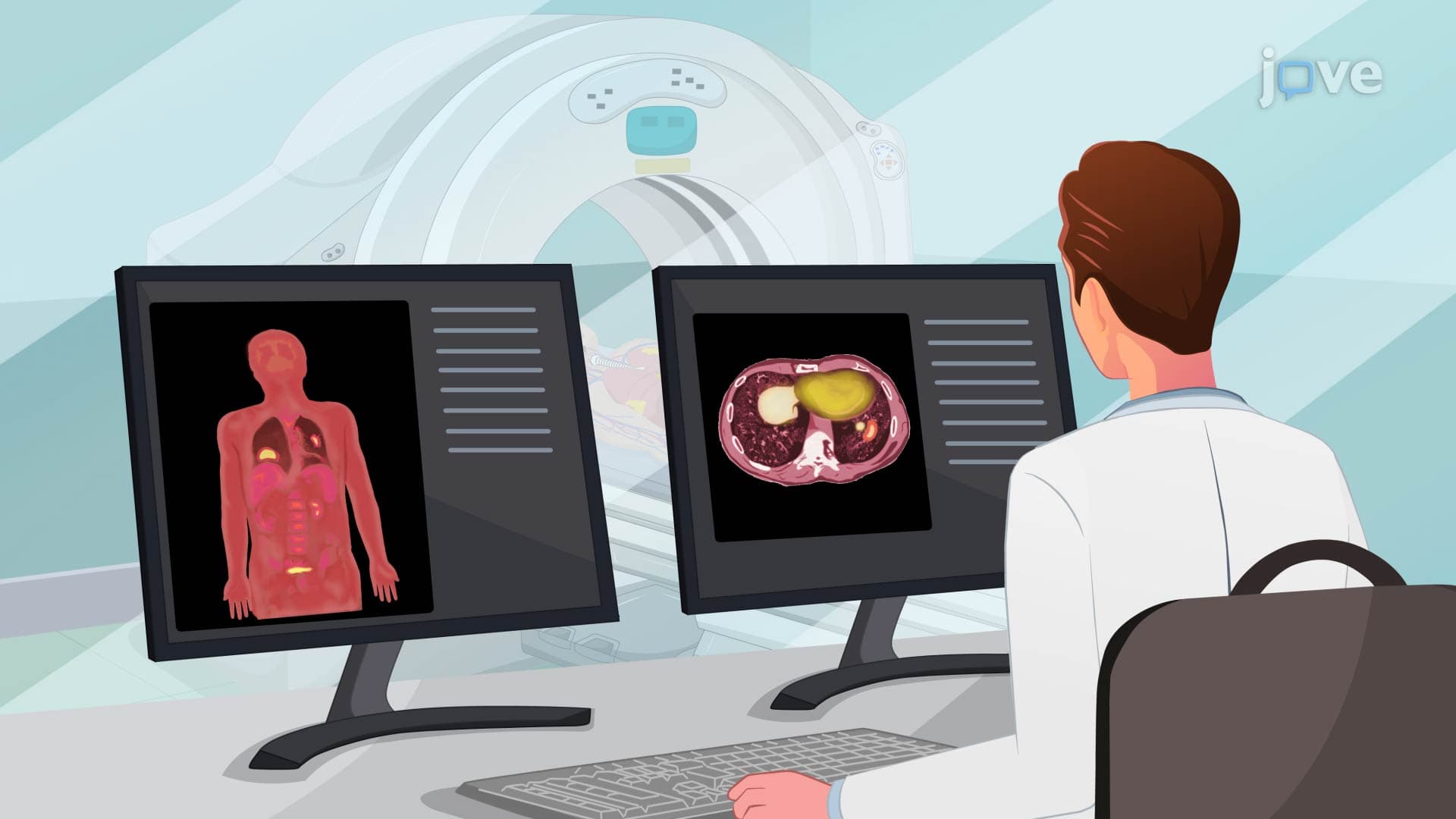
CT scans revolutionize diagnostic capabilities by providing detailed cross-sectional images that overcome the limitations of traditional X-rays. While standard chest X-rays may miss small lesions hidden behind the heart or ribs, CT technology creates multiple "slices" through the body, revealing structures in unprecedented detail. This advancement proves particularly valuable for detecting pulmonary nodules smaller than 1 centimeter, evaluating hilar lymph nodes, and assessing mediastinal masses. Students studying for the NCLEX or HESI A2 exams must understand that CT scans expose patients to higher radiation doses than standard X-rays, making clinical judgment crucial in determining appropriate imaging protocols.
Professional nursing practice requires comprehensive understanding of radiological procedure protocols and patient safety measures. Before any imaging study, nurses must ensure proper patient preparation, including removal of metallic objects that could interfere with image quality or pose safety risks. For contrast-enhanced CT studies, pre-procedural assessment becomes critical, particularly evaluating renal function through BUN and creatinine levels. Since contrast agents are iodine-based, nurses must screen for shellfish allergies and previous contrast reactions. The four-hour fasting requirement before contrast studies prevents aspiration risks if adverse reactions occur. These protocols, emphasized in nursing programs and tested on the NCLEX examination, directly impact patient outcomes and procedural success rates in clinical settings across American healthcare institutions.
Related Micro-courses