Loading video…


 10 Concepts
 5.8K views

Video Summary: Radiological Investigation Ii Mri and Ventilation Perfusion Scans
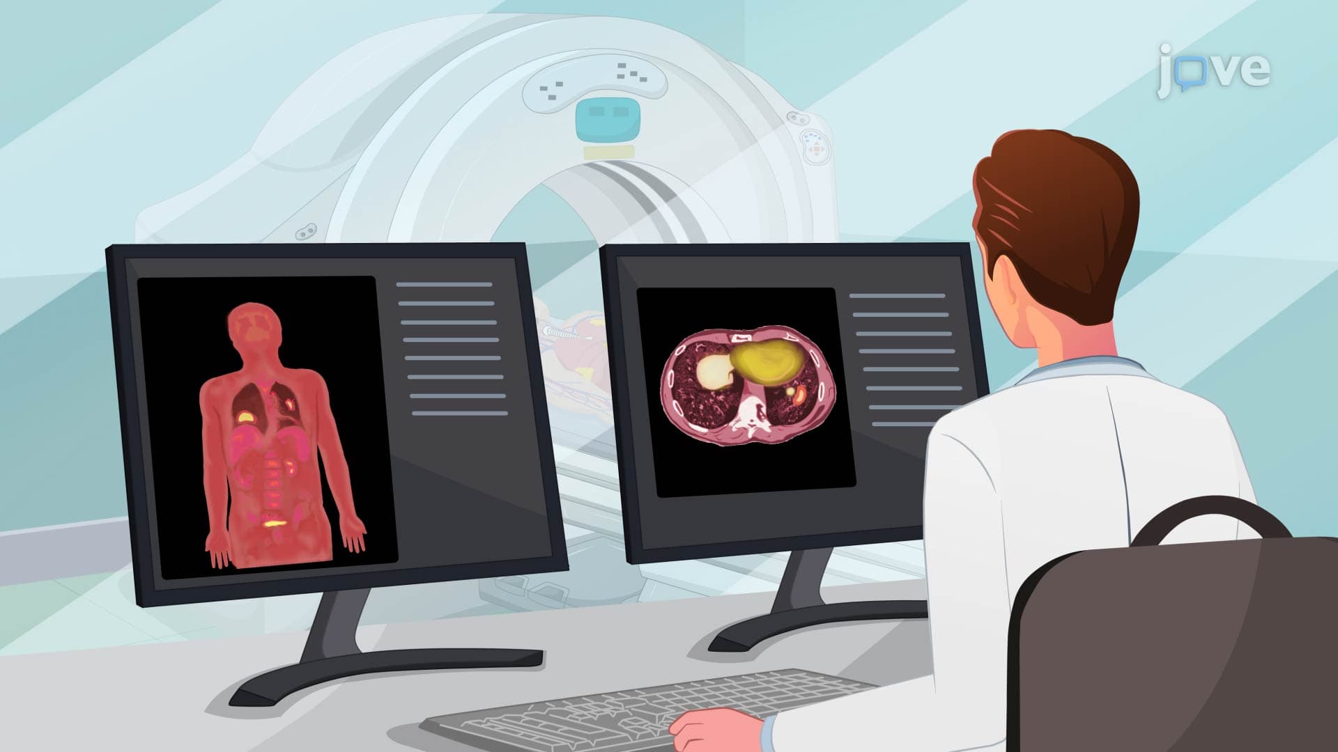
Did you know that radiological investigation II: MRI and ventilation-perfusion scans can detect lung cancer months before symptoms appear? These advanced imaging techniques revolutionize respiratory diagnostics by combining magnetic field technology with radioactive tracers to create detailed lung maps. At Cleveland Clinic, radiologists use MRI scans to identify pulmonary nodules smaller than a penny, while V/Q scans reveal blood flow patterns that indicate conditions like pulmonary embolism. Watch the full video on JoVE Coach to master this concept with expert-led visuals and step-by-step explanations.
Advanced radiological investigations represent a cornerstone of modern respiratory medicine, with MRI and ventilation-perfusion (V/Q) scans leading the diagnostic revolution. These sophisticated imaging modalities provide healthcare professionals with unprecedented insight into lung structure and function, enabling early detection and precise treatment planning for complex respiratory conditions.
Magnetic Resonance Imaging utilizes powerful magnetic fields ranging from 1.5 to 3.0 Tesla-roughly 30,000 times stronger than Earth's magnetic field-combined with radiofrequency pulses to generate detailed cross-sectional images. Unlike conventional X-rays or CT scans, MRI provides superior soft tissue contrast without ionizing radiation exposure, making it particularly valuable for evaluating pulmonary masses, pleural diseases, and mediastinal structures.
The technology excels in detecting bronchogenic carcinoma, with sensitivity rates exceeding 95% for tumors larger than 1 centimeter. Major medical centers like Mayo Clinic and Johns Hopkins routinely employ cardiac-gated MRI sequences to minimize respiratory motion artifacts, producing images that reveal tumor invasion into chest wall structures, vascular involvement, and lymph node metastases. For students preparing for the MCAT or NCLEX examinations, understanding MRI's role in staging lung cancer according to TNM classification systems proves essential.
V/Q scans represent nuclear medicine's contribution to respiratory diagnostics, employing dual-phase imaging to assess both air distribution (ventilation) and blood flow (perfusion) within the lungs. The ventilation phase utilizes inhaled xenon-133 or technetium-99m aerosols, while perfusion imaging involves intravenous injection of technetium-99m macroaggregated albumin particles.
This dual approach proves particularly powerful in diagnosing pulmonary embolism, with sensitivity rates approaching 90% when interpreted using established criteria. At institutions like UCLA Medical Center, radiologists analyze V/Q mismatch patterns-areas showing normal ventilation but reduced perfusion-to identify blood clots with remarkable precision. The technique also demonstrates exceptional utility in evaluating chronic obstructive pulmonary disease progression and assessing regional lung function before surgical interventions.
Both imaging modalities require specialized nursing protocols to ensure patient safety and diagnostic accuracy. MRI screening protocols must identify ferromagnetic implants, pacemakers, and metallic foreign bodies that pose significant safety risks in magnetic fields. The FDA mandates comprehensive screening questionnaires, while institutions like Cleveland Clinic employ handheld metal detectors as additional safety measures.
V/Q scan preparation involves establishing reliable intravenous access and confirming recent chest radiographs to establish baseline lung appearance. Post-procedure protocols emphasize increased fluid intake to accelerate radioisotope elimination through renal excretion, typically achieving background radiation levels within 24-48 hours. These concepts frequently appear in HESI A2 and TEAS nursing entrance examinations, emphasizing the critical nature of proper preparation and monitoring.
Related Micro-courses