Loading subject page…


5,820 views

Video Summary: Radiological Investigation Iii Pulmonary Angiogram Explained
Every year, over 600,000 Americans are diagnosed with pulmonary embolism, making radiological investigation iii pulmonary angiogram one of the most critical diagnostic procedures in emergency medicine. This invasive imaging technique uses a catheter threaded through the heart to inject contrast dye directly into lung arteries, creating detailed X-ray images that can save lives by detecting dangerous blood clots. Watch the full video on JoVE Coach to master this concept with expert-led visuals and step-by-step explanations.
Radiological investigation iii pulmonary angiogram represents a gold-standard diagnostic approach for evaluating pulmonary vascular disease, particularly when rapid, definitive answers are needed. This procedure combines interventional cardiology techniques with advanced imaging to create a roadmap of lung blood vessels. At major medical centers like Johns Hopkins and Mayo Clinic, pulmonary angiograms are performed daily to diagnose conditions ranging from acute pulmonary embolism to chronic thromboembolic pulmonary hypertension.
The procedure begins with femoral vein access, typically in the right groin area. A specialized catheter follows the venous return pathway: femoral vein → inferior vena cava → right atrium → right ventricle → pulmonary artery. This anatomical journey requires precise navigation skills and real-time fluoroscopic guidance. The contrast medium, typically iodine-based, temporarily highlights blood vessels by absorbing X-rays differently than surrounding tissue. Students preparing for the MCAT should understand that this contrast creates the "negative image" effect where blood vessels appear bright white against darker lung tissue.
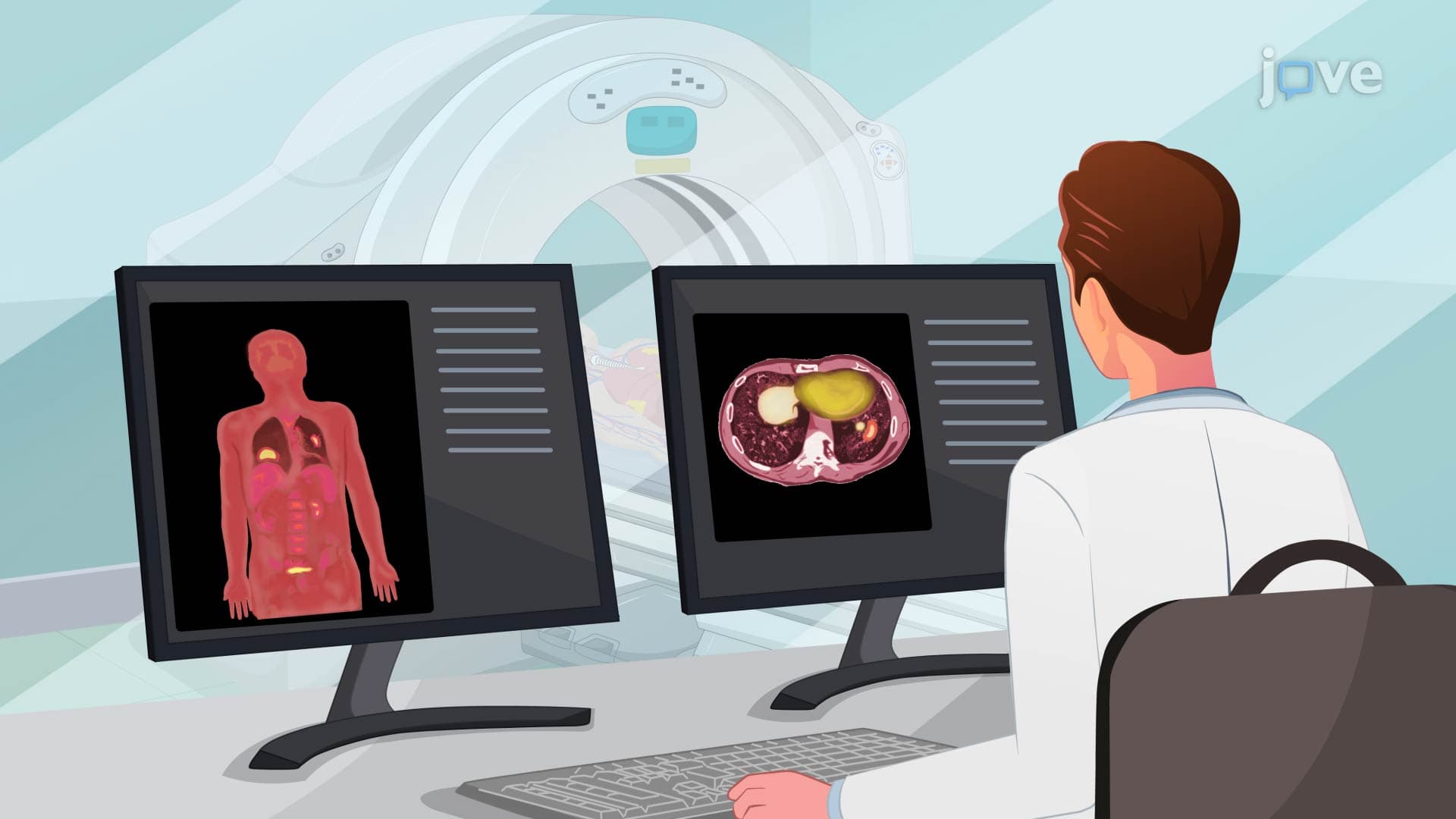
While pulmonary angiography shows vessel structure, PET scans reveal functional information through metabolic activity. The radioactive glucose tracer (fluorodeoxyglucose or FDG) accumulates in cells with high glucose consumption-cancer cells consume up to 20 times more glucose than normal tissue. This principle makes PET scans invaluable for staging lung cancer and monitoring treatment response. The 30-60 minute waiting period allows tracer distribution throughout the body, while the gamma ray detectors create three-dimensional metabolic maps.
Modern pulmonary angiography has evolved from purely diagnostic to therapeutic applications. During the same procedure, physicians can perform clot removal (thrombectomy) or balloon angioplasty. Nursing responsibilities reflect the procedure's invasive nature: anticoagulation screening prevents excessive bleeding, allergy assessment avoids contrast reactions, and NPO status reduces aspiration risk during sedation. Post-procedure monitoring focuses on access site complications, contrast-induced kidney injury prevention through hydration, and early detection of vessel injury through vital sign changes.
Related Micro-courses