Loading subject page…

1,417 views

Video Summary: Acute Pyelonephritis Ii Diagnostic Studies Explained
Did you know that over 250,000 Americans develop pyelonephritis annually, yet many cases go undiagnosed due to subtle symptoms? Acute pyelonephritis ii diagnostic studies involve a systematic approach using urinalysis, blood work, and imaging to identify kidney infections before they become life-threatening. Consider a college student at UCLA presenting with fever and back pain-proper diagnostic testing can distinguish between a simple UTI and serious kidney involvement. Watch the full video on JoVE Coach to master this concept with expert-led visuals and step-by-step explanations.
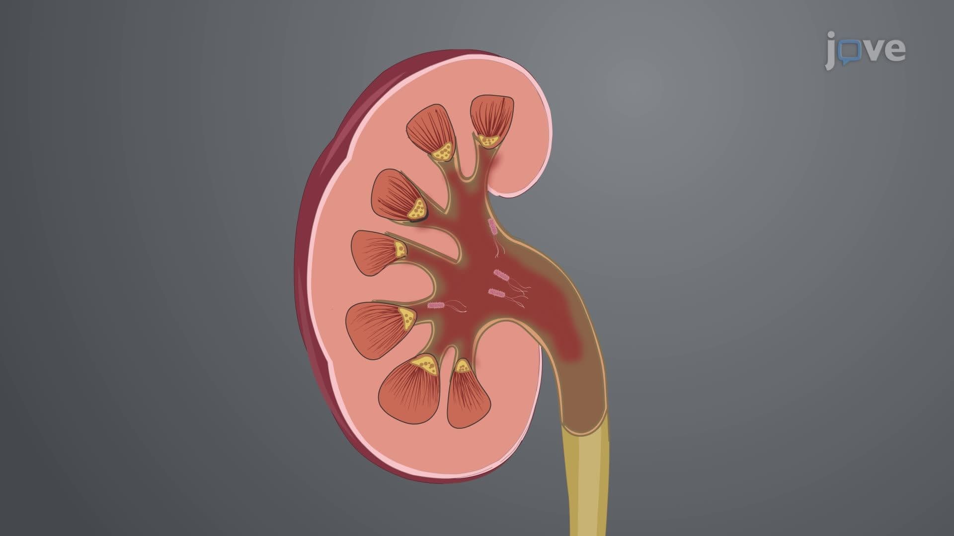
Acute pyelonephritis represents a serious kidney infection that affects approximately 15-17 cases per 10,000 women and 3-4 cases per 10,000 men annually in the United States. The diagnostic approach requires a methodical evaluation combining clinical assessment with laboratory and imaging studies. Unlike simple cystitis, pyelonephritis involves the kidney parenchyma and collecting system, making accurate diagnosis crucial for preventing complications like sepsis or chronic kidney disease.
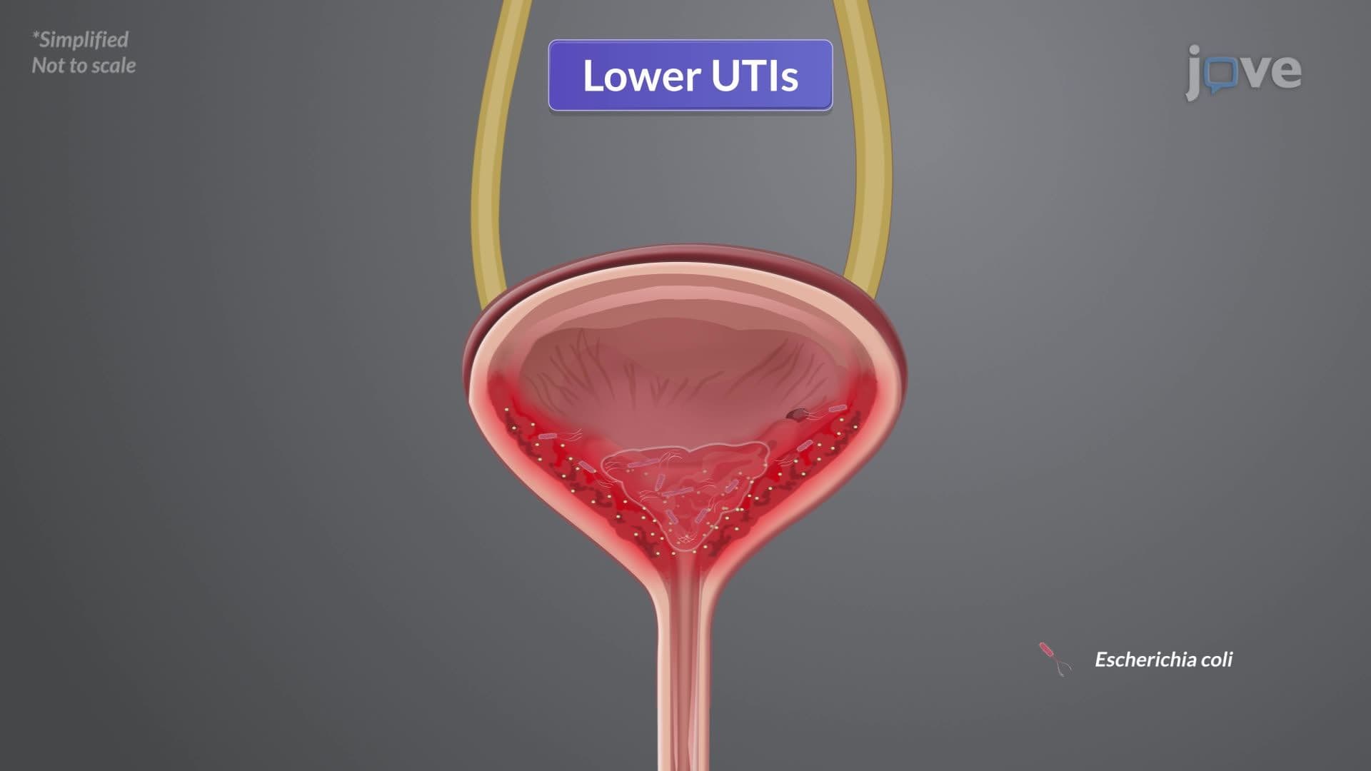
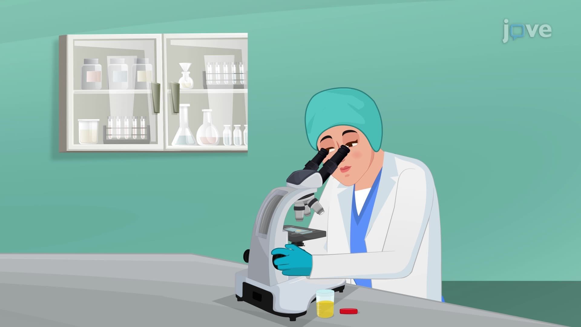
The cornerstone of pyelonephritis diagnosis begins with comprehensive urinalysis and urine culture. Urinalysis typically reveals pyuria (>10 white blood cells per high-power field), bacteriuria, and positive nitrites-particularly important since nitrite-positive bacteria like E. coli cause 80-85% of uncomplicated cases. The presence of white blood cell casts specifically indicates upper urinary tract involvement, distinguishing pyelonephritis from lower UTIs.
Blood work plays an equally critical role, with complete blood count revealing leukocytosis (typically >12,000 cells/μL) and left shift indicating systemic inflammation. Blood cultures prove positive in approximately 20-30% of pyelonephritis cases, with positive results suggesting more severe disease requiring hospitalization. At institutions like Johns Hopkins Hospital, emergency departments routinely obtain both urine and blood cultures before initiating antibiotic therapy.
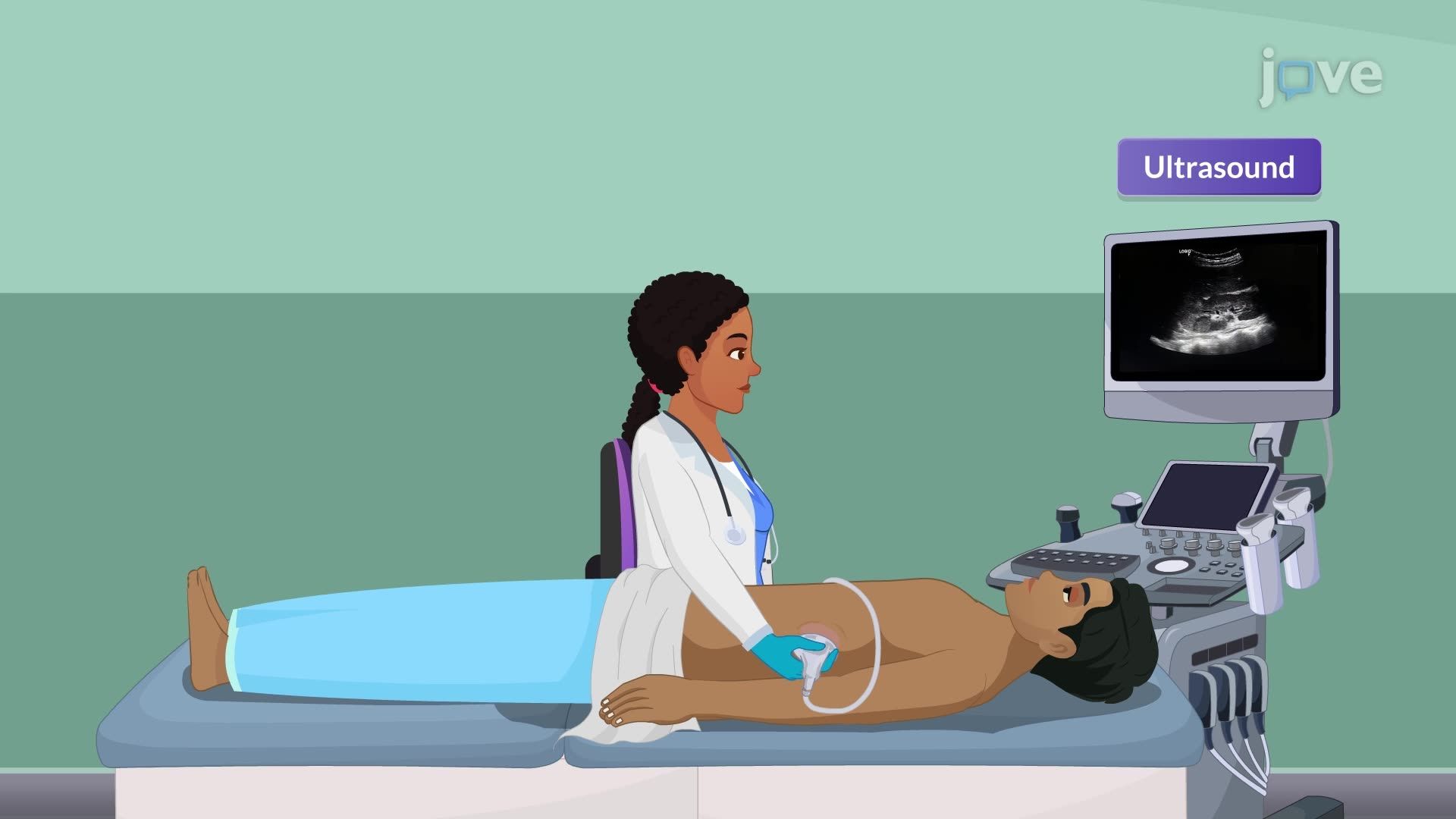
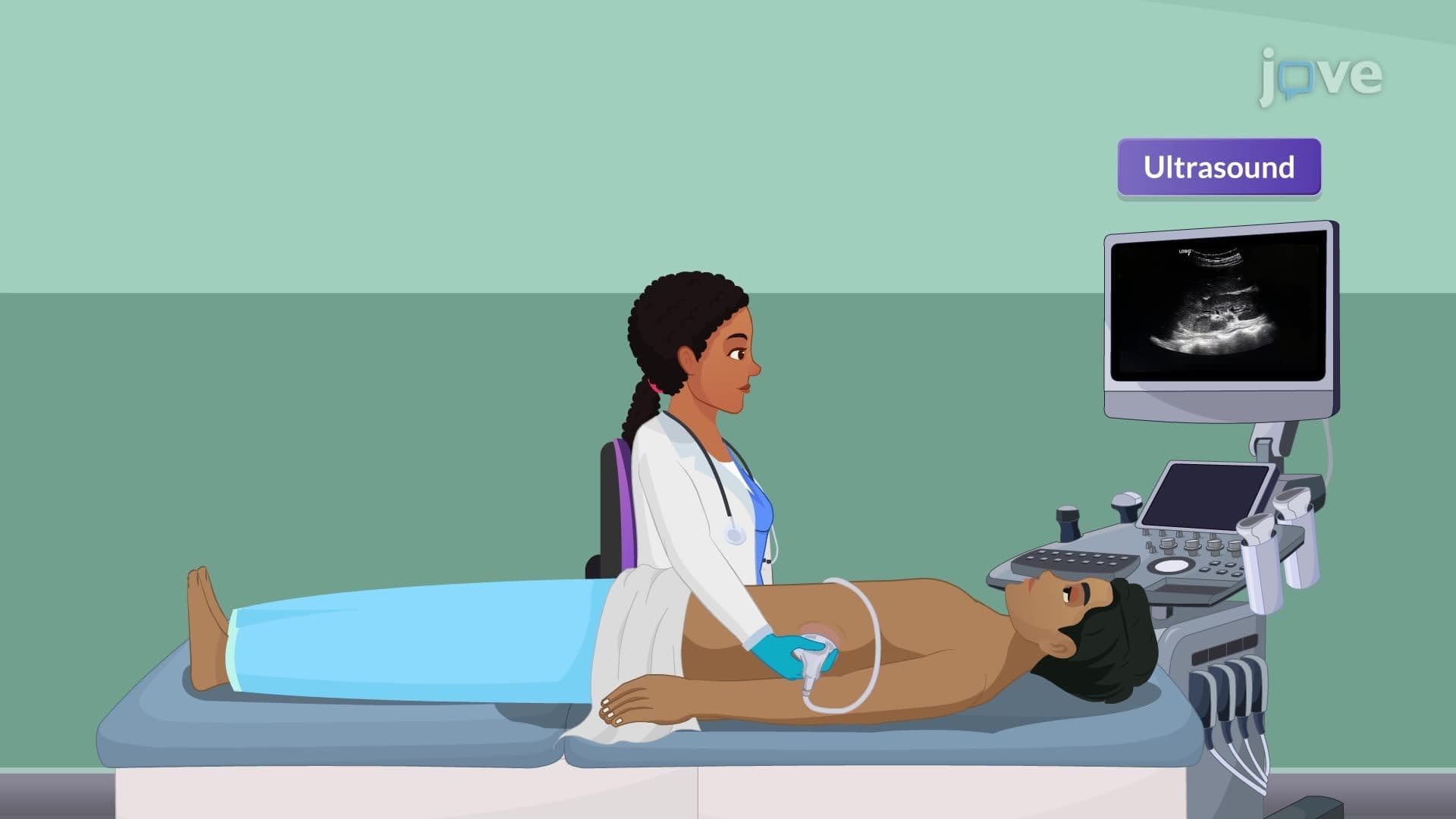
Imaging studies provide valuable anatomical information, particularly when complications are suspected. Renal ultrasound serves as the first-line imaging modality, effectively detecting hydronephrosis, abscesses, or structural abnormalities without radiation exposure. For students preparing for the MCAT or USMLE Step 1, understanding that CT scans are reserved for complex cases-such as suspected emphysematous pyelonephritis or perinephric abscesses-demonstrates advanced clinical reasoning.
Cystoscopy remains a specialized procedure typically performed by urologists when recurrent infections suggest anatomical abnormalities or obstruction. This invasive procedure allows direct visualization of the bladder and can identify foreign bodies, stones, or structural defects contributing to infection risk.
Diagnostic findings directly influence treatment decisions and prognosis. Mild cases with normal vital signs, minimal systemic symptoms, and ability to tolerate oral medications can often be managed outpatient with fluoroquinolones like ciprofloxacin or oral cephalosporins. However, patients presenting with high fever (>101.3°F), vomiting, or signs of sepsis require hospitalization and IV antibiotics such as ceftriaxone or piperacillin-tazobactam.
For AP Biology students studying homeostasis and disease, pyelonephritis demonstrates how bacterial invasion disrupts normal kidney function, triggering inflammatory responses that appear in diagnostic tests. College pre-med students should understand that antibiotic selection initially relies on local resistance patterns and empirical guidelines, then adjusts based on culture sensitivity results-a principle tested on standardized exams like the MCAT.
Related Micro-courses