Loading subject page…

1,417 views

Video Summary: Urinary Tract Infection Iii Diagnostic Explained
Did you know that over 8 million Americans visit healthcare providers annually for UTIs, yet many cases go undiagnosed due to incomplete testing? Understanding urinary tract infection iii diagnostic methods is crucial for accurate identification and treatment of these common infections. From basic urinalysis detecting bacteria and leukocytes to advanced imaging like CT scans, the Urinary Tract Infection III Diagnostic process involves multiple sophisticated testing approaches that healthcare providers use daily in clinics across the United States. Watch the full video on JoVE Coach to master this concept with expert-led visuals and step-by-step explanations.
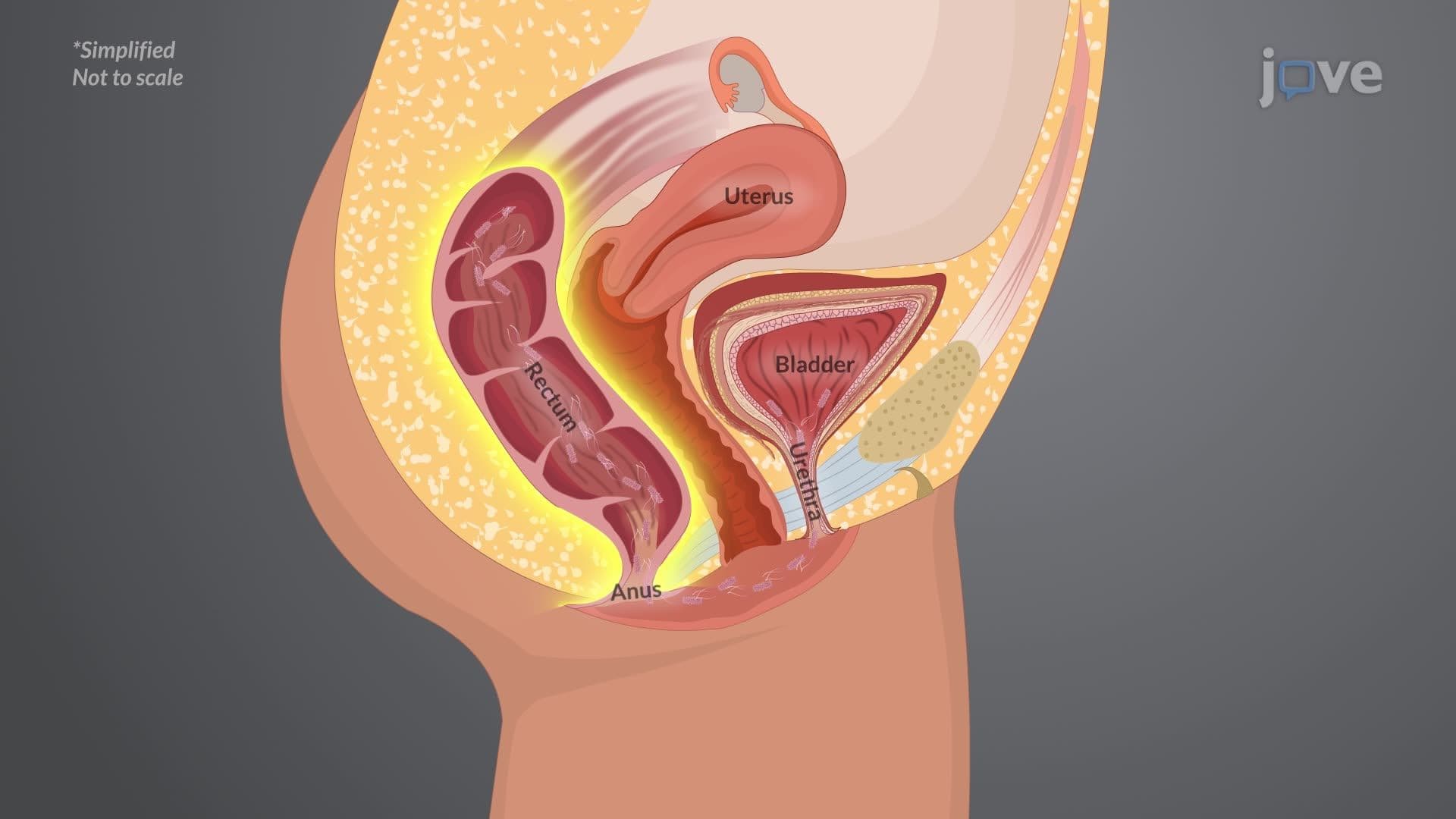
The Urinary Tract Infection III Diagnostic process represents a systematic approach to identifying and characterizing urinary tract infections through multiple complementary testing methods. This comprehensive diagnostic framework goes beyond simple symptom recognition to provide definitive bacterial identification and treatment guidance.
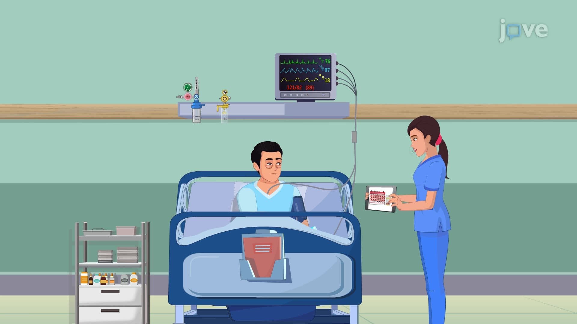
Healthcare providers begin UTI diagnosis by evaluating characteristic symptoms including dysuria (burning during urination), urinary frequency, urgency, and suprapubic pain. However, symptoms alone cannot definitively diagnose UTIs, particularly in populations like elderly patients who may present atypically. The Centers for Disease Control and Prevention emphasizes that proper diagnostic testing prevents unnecessary antibiotic use, which contributes to antibiotic resistance-a growing concern in US healthcare systems.
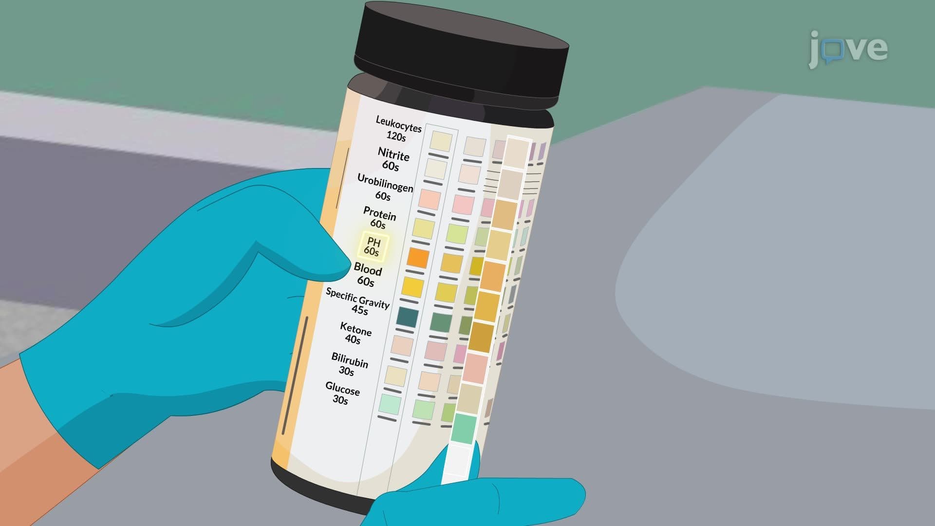
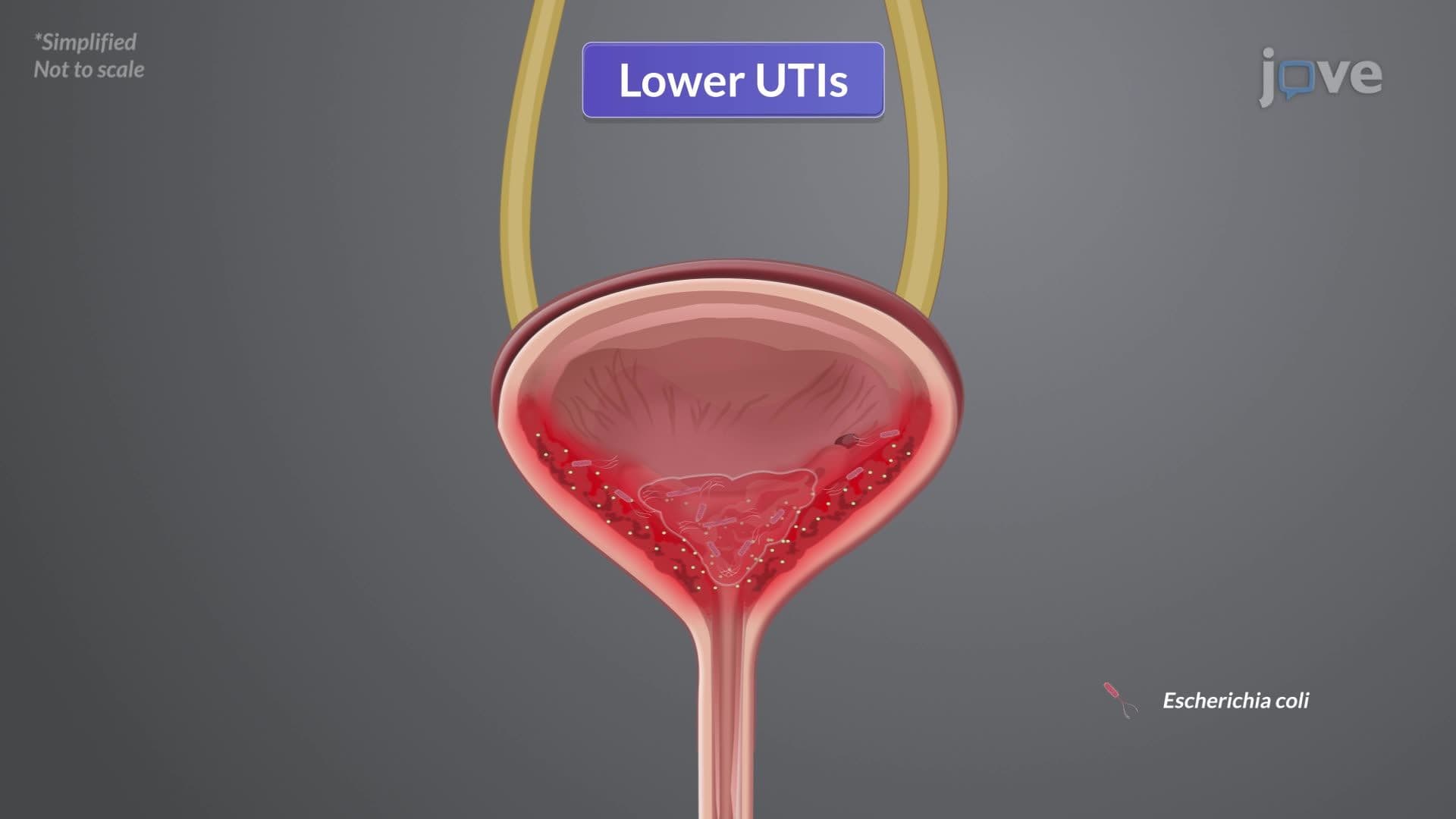
Urinalysis serves as the primary screening tool, detecting leukocyte esterase (indicating white blood cell presence) and nitrites (produced by certain bacteria like E. coli). A positive nitrite test strongly suggests bacterial infection, while leukocyte esterase indicates inflammatory response. However, some bacteria don't produce nitrites, making urine culture essential for definitive diagnosis.
Urine culture remains the gold standard for UTI diagnosis, requiring 24-48 hours for bacterial growth and identification. The critical threshold of 100,000 colony-forming units (CFU) per milliliter distinguishes true infection from contamination, though lower counts may be significant in symptomatic patients. Modern US clinical laboratories use automated systems for rapid bacterial identification and antibiotic susceptibility testing, crucial for selecting appropriate treatment.
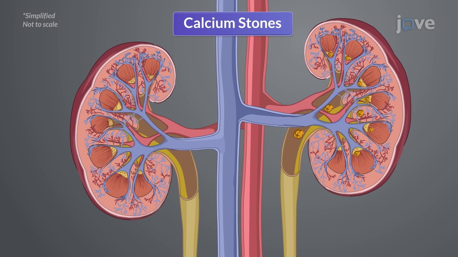
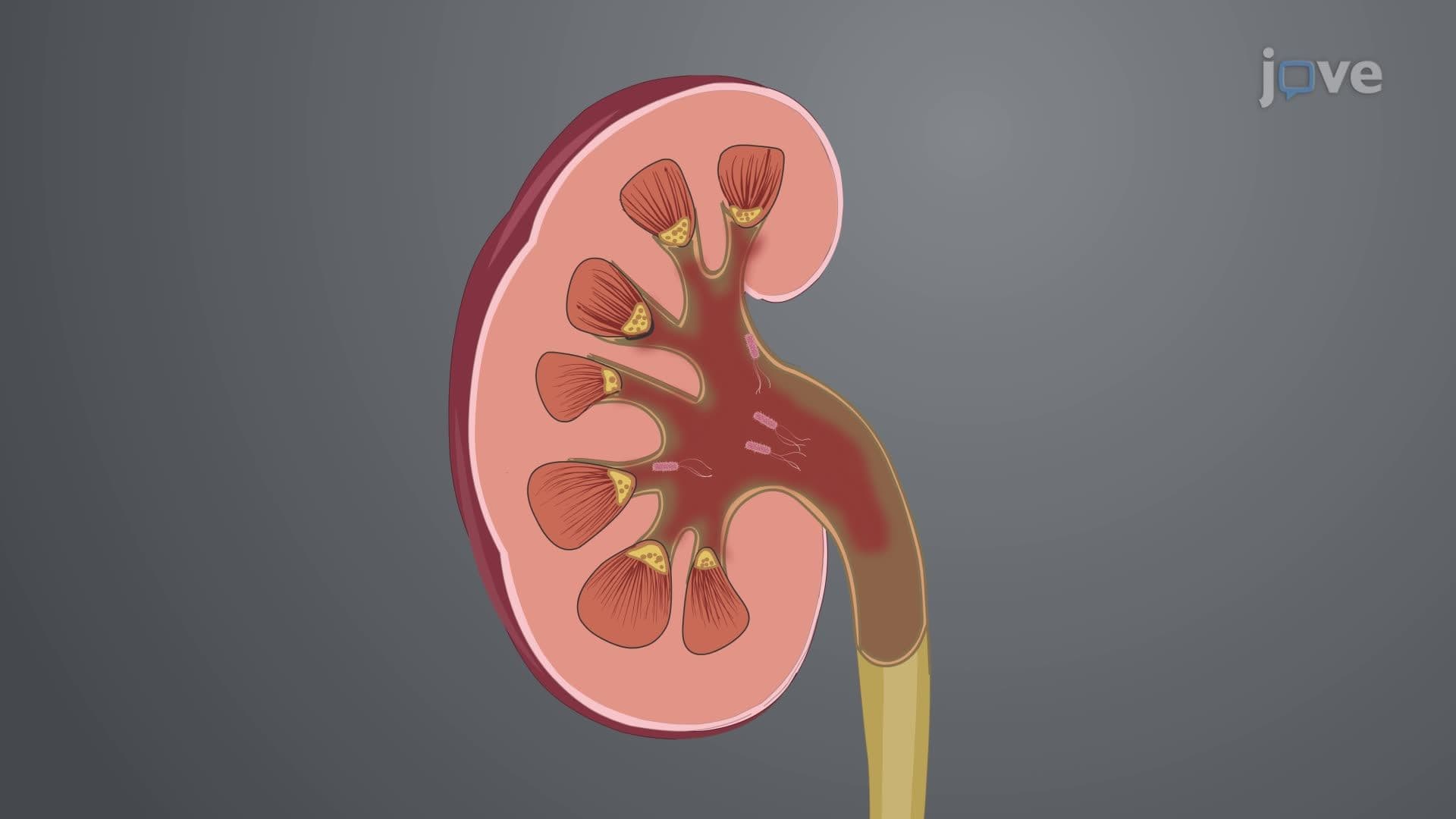
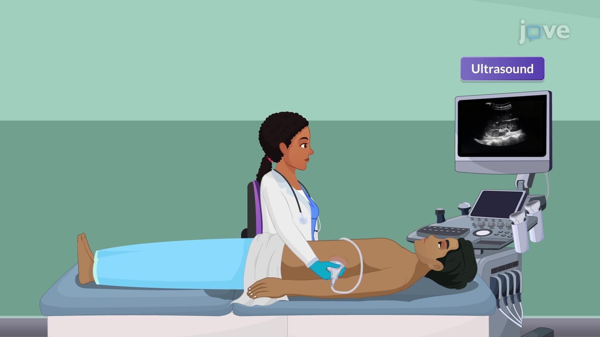
For complicated UTIs or recurrent infections, advanced imaging becomes necessary. CT urography provides detailed visualization of the entire urinary tract, identifying structural abnormalities, kidney stones, or abscesses that may predispose patients to infection. The American College of Radiology recommends CT imaging for patients with treatment-resistant UTIs or concerning symptoms suggesting upper tract involvement.
Cystoscopy allows direct bladder visualization, particularly valuable for identifying bladder tumors, stones, or anatomical abnormalities in patients with recurrent UTIs. This procedure is commonly performed in urology practices across the United States and provides real-time assessment of bladder and urethral conditions that may contribute to infection susceptibility.
Related Micro-courses