Loading subject page…

1,733 views

Video Summary: Aortic Regurgitation Ii Clinical Features Explained
Did you know that some heart valve conditions can remain completely silent for years before suddenly causing life-threatening symptoms? Aortic regurgitation II clinical features represent the progressive stage where patients begin experiencing noticeable symptoms as their heart struggles with backward blood flow. At leading US cardiac centers like the Mayo Clinic, cardiologists rely on recognizing these specific clinical patterns to diagnose and treat patients before irreversible heart damage occurs. Understanding Aortic Regurgitation II Clinical Features Explained helps future healthcare professionals identify critical warning signs through physical examination techniques and diagnostic approaches. Watch the full video on JoVE Coach to master this concept with expert-led visuals and step-by-step explanations.
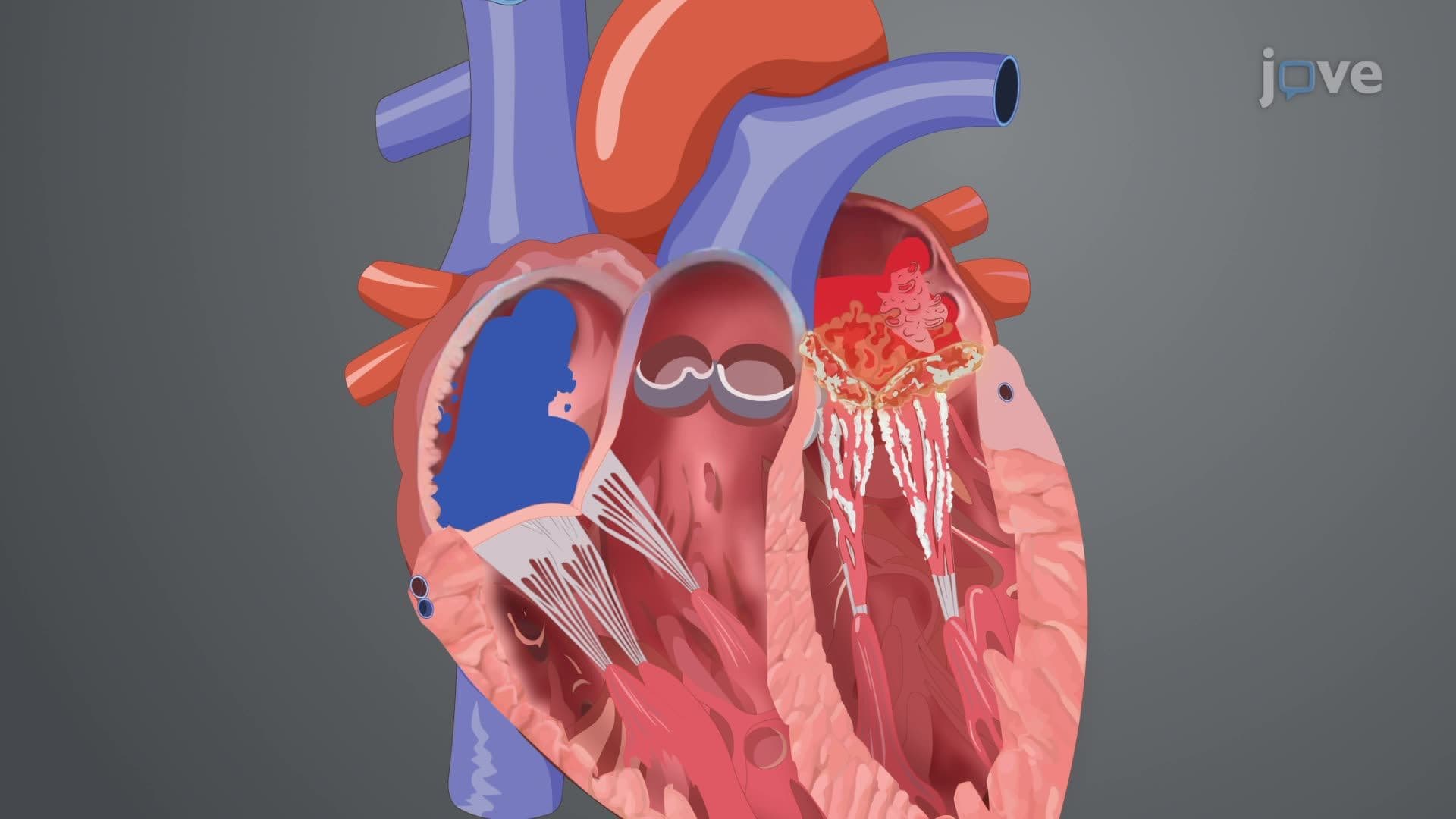
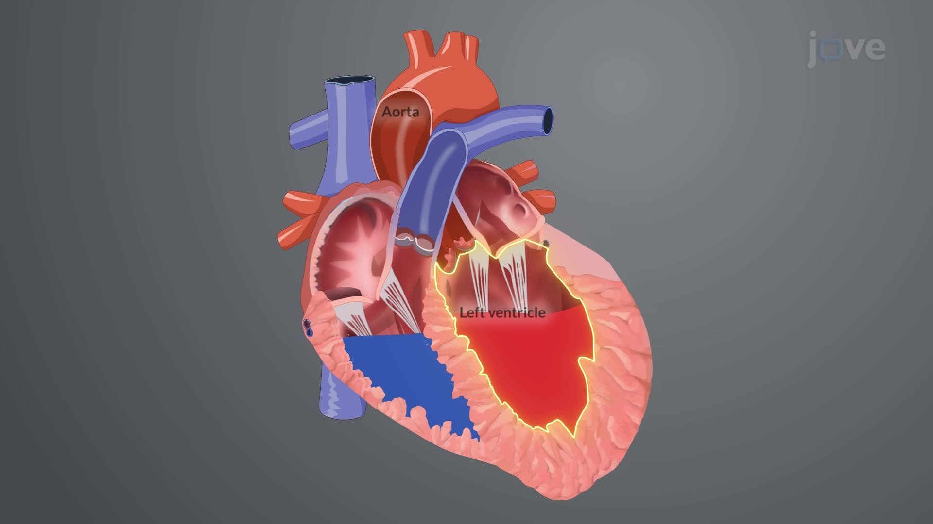
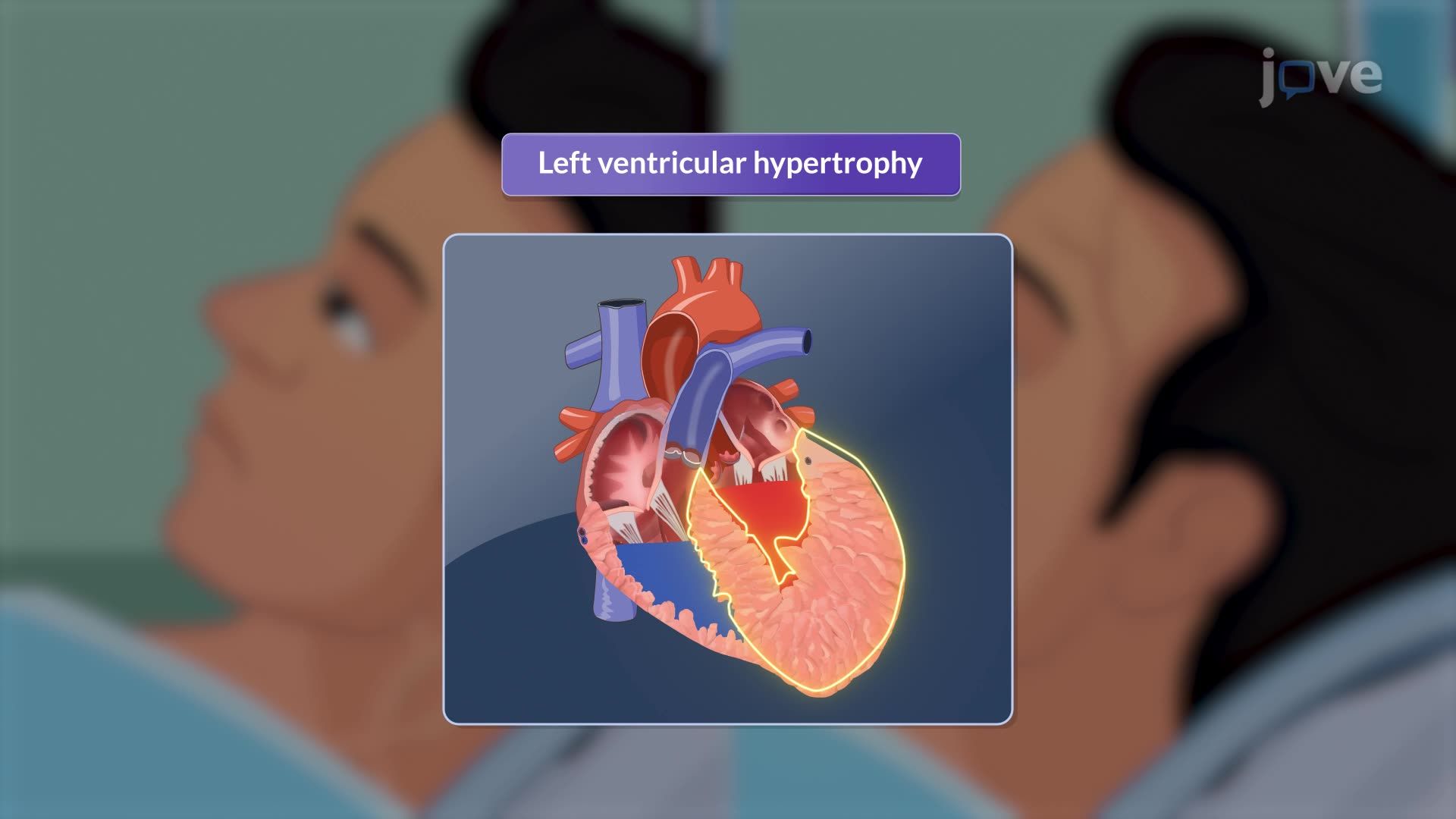
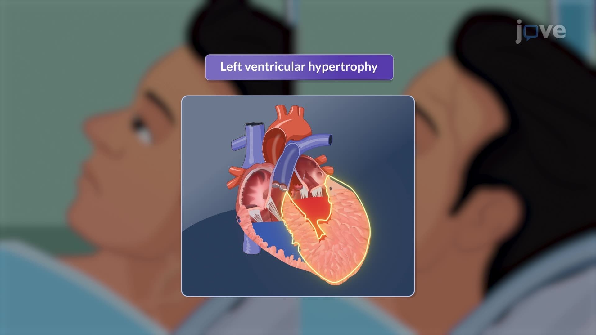
Aortic regurgitation represents a critical cardiovascular condition where the aortic valve fails to close properly, allowing blood to flow backward from the aorta into the left ventricle during diastole. This backward flow creates a volume overload that initially triggers compensatory mechanisms, but eventually leads to progressive left ventricular dysfunction and clinical symptoms that define the advanced stages of this condition.
The clinical presentation of aortic regurgitation follows a predictable pattern as the heart's compensatory mechanisms become overwhelmed. Patients typically remain asymptomatic during the early chronic phase, which can last for decades. However, as left ventricular function deteriorates, classic heart failure symptoms emerge. Dyspnea on exertion represents the earliest symptom, followed by orthopnea and paroxysmal nocturnal dyspnea as pulmonary congestion develops.
At major US medical centers like Johns Hopkins and Cleveland Clinic, cardiologists note that patients often describe a distinctive "pounding" sensation in their chest, neck, or head. This occurs because the large stroke volume creates visible and palpable arterial pulsations, particularly noticeable at the carotid and temporal arteries. For students preparing for the MCAT or USMLE Step 1, understanding this pathophysiology connects the mechanical valve dysfunction to the clinical presentation.
The physical examination reveals several pathognomonic findings that distinguish aortic regurgitation from other cardiac conditions. The pulse examination demonstrates Corrigan's pulse (also called water-hammer pulse), characterized by a rapid upstroke followed by quick collapse. This finding reflects the large stroke volume and rapid diastolic runoff back into the left ventricle.
Auscultation reveals the classic high-pitched, blowing diastolic murmur best heard at the third or fourth intercostal space along the left sternal border. Students should note that this murmur begins immediately after the second heart sound and may extend throughout diastole in severe cases. The intensity and duration of the murmur correlate with regurgitation severity, making this a crucial diagnostic skill for future physicians.
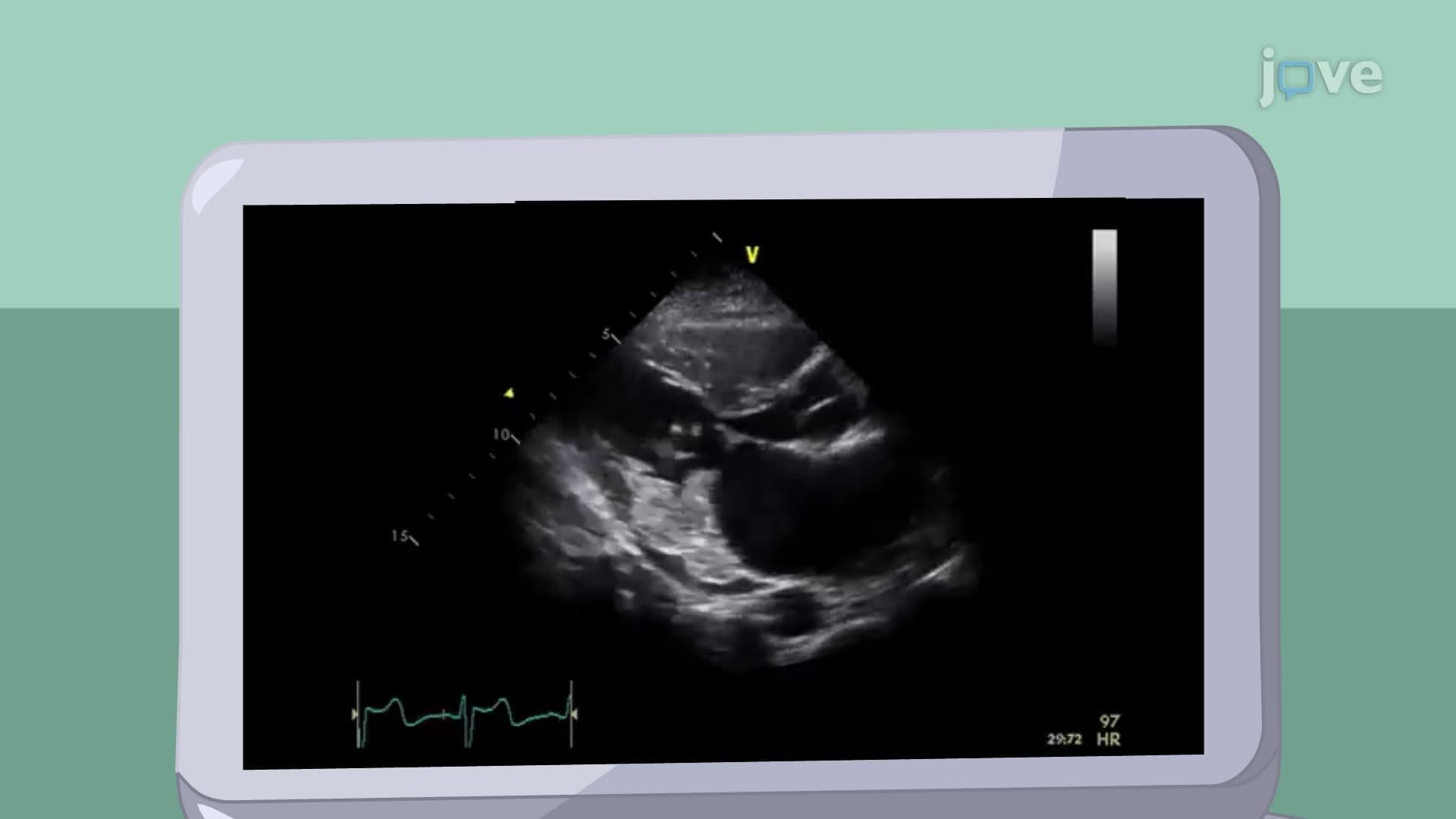
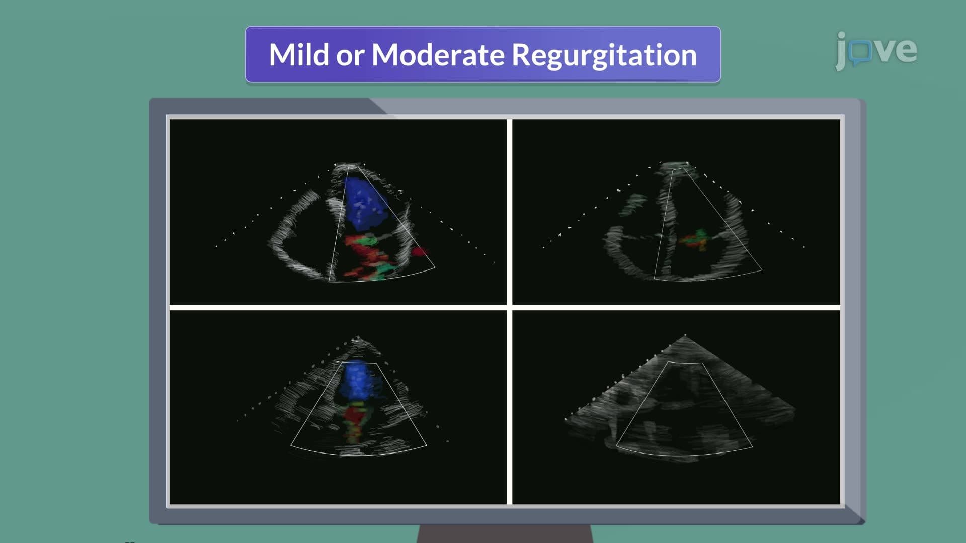
Contemporary diagnostic evaluation relies heavily on echocardiographic assessment. Transthoracic echocardiography serves as the initial imaging modality, providing essential information about regurgitation severity, left ventricular dimensions and function, and potential underlying causes. Advanced techniques allow cardiologists to quantify regurgitant volume and fraction, guiding treatment decisions.
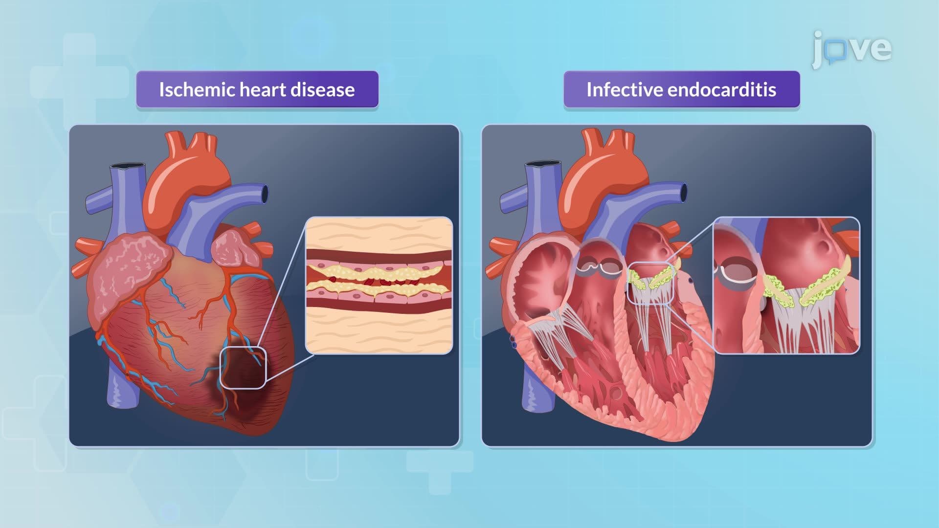
Transesophageal echocardiography offers superior visualization of the aortic valve structure and aortic root anatomy. This technique proves particularly valuable when evaluating for infective endocarditis or assessing bicuspid valve morphology. For students studying for AP Biology or college physiology courses, understanding these diagnostic modalities illustrates how technology enhances clinical assessment of cardiovascular pathology.
Related Micro-courses