Loading subject page…


1,733 views


Video Summary: Mitral Stenosis Ii Clinical Features Explained
Did you know that the distinctive "rumbling" heart murmur of mitral stenosis II clinical features can be so subtle that cardiologists at Mayo Clinic position patients on their left side just to detect it? This progressive valve condition creates a cascade of symptoms from simple exercise fatigue to life-threatening breathing difficulties at rest. Understanding Mitral Stenosis II Clinical Features Explained is crucial for recognizing how this heart valve disorder manifests in real patients and affects their daily lives. Watch the full video on JoVE Coach to master this concept with expert-led visuals and step-by-step explanations.
Mitral Stenosis II Clinical Features Explained encompasses the comprehensive clinical presentation of this progressive valvular heart disease. Unlike acute conditions, mitral stenosis develops gradually, often over decades, allowing patients to unconsciously adapt their activity levels. This adaptation means that by the time symptoms become apparent, significant valve narrowing has already occurred.
The hallmark symptom progression begins with exertional dyspnea-patients initially notice shortness of breath during activities like climbing stairs at their workplace or walking uphill. As stenosis worsens, this dyspnea occurs with less exertion and eventually at rest. Johns Hopkins cardiologists emphasize that patients often don't recognize early symptoms because the progression is so gradual.
Fatigue becomes prominent as cardiac output fails to meet metabolic demands during physical activity. Palpitations may indicate the development of atrial fibrillation, a common complication affecting up to 40% of patients with severe mitral stenosis. Orthopnea-difficulty breathing when lying flat-forces patients to sleep propped up on multiple pillows or in recliners.
More concerning symptoms include hemoptysis (coughing up blood) and recurrent respiratory infections. The hemoptysis results from elevated pulmonary venous pressure causing small vessel rupture, while recurrent pneumonia occurs due to pulmonary congestion creating an environment conducive to bacterial growth.
The physical examination requires specific positioning and techniques taught in medical schools across the United States. The left lateral decubitus position-patient lying on their left side-brings the cardiac apex closer to the chest wall, optimizing detection of the characteristic murmur.
The mitral stenosis murmur is distinctively low-pitched and rumbling, heard during diastole when the mitral valve should be open. This timing differentiates it from systolic murmurs associated with other valve disorders. The murmur's intensity doesn't always correlate with severity-some patients with severe stenosis have barely audible murmurs due to reduced cardiac output.
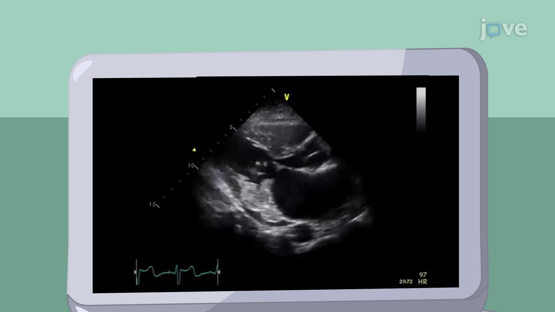
Modern cardiology relies heavily on echocardiography for quantitative assessment. The mitral valve area measurement is crucial-normal area is 4-6 cm², mild stenosis is 1.5-2.5 cm², moderate is 1.0-1.5 cm², and severe stenosis is less than 1.0 cm². These measurements directly impact treatment decisions at institutions like Cleveland Clinic and Massachusetts General Hospital.
Electrocardiographic findings provide valuable insights into disease progression. P mitrale-broad, notched P waves in lead II exceeding 0.12 seconds-indicates left atrial enlargement. This finding often precedes the development of atrial fibrillation, a complication that significantly increases stroke risk and requires anticoagulation therapy.
Students preparing for the MCAT or USMLE Step 1 should understand that chest X-ray findings complement other diagnostic modalities. Left atrial enlargement appears as a "double density" behind the right heart border, while pulmonary artery enlargement indicates elevated pulmonary pressures-both consequences of mitral stenosis progression.
Related Micro-courses