Loading subject page…

1,733 views

Video Summary: Mitral Valve Prolapse I Introduction Explained
Did you know that mitral valve prolapse affects up to 3% of Americans, making it one of the most common heart valve disorders? Understanding mitral valve prolapse I introduction reveals how structural changes in heart valve leaflets cause them to bulge backward during each heartbeat. Consider a patient at Johns Hopkins Hospital presenting with chest pain and a distinctive heart murmur-this could indicate MVP complications. Watch the full video on JoVE Coach to master this concept with expert-led visuals and step-by-step explanations.
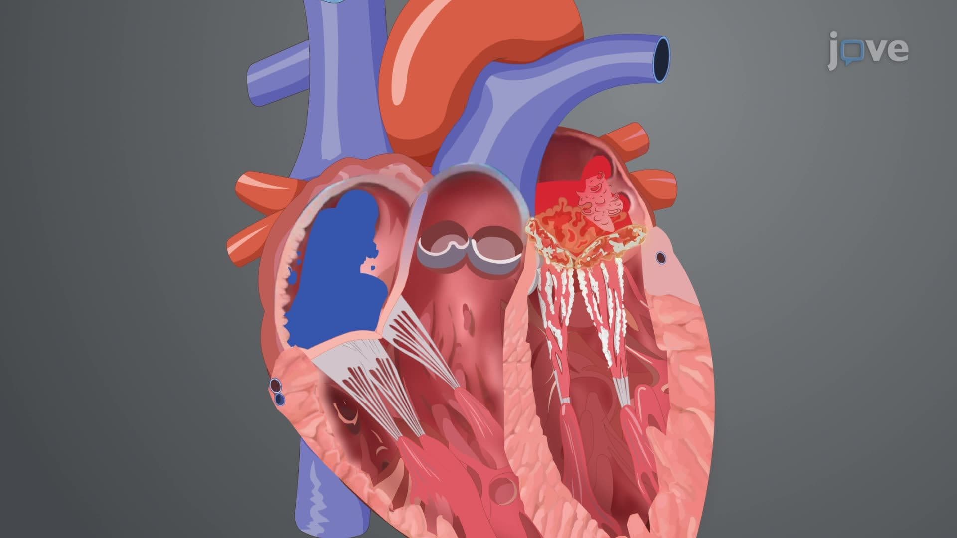
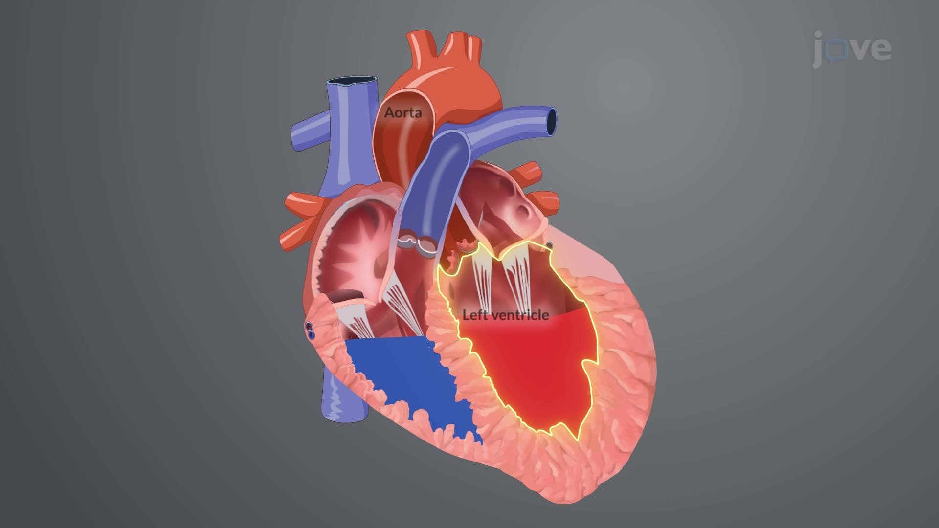
Mitral valve prolapse represents a fascinating intersection of cardiac anatomy, genetics, and pathophysiology that every pre-med and health science student must master. This condition occurs when the mitral valve's bicuspid leaflets fail to close properly during systole, instead bulging backward into the left atrium like a parachute caught in reverse wind.
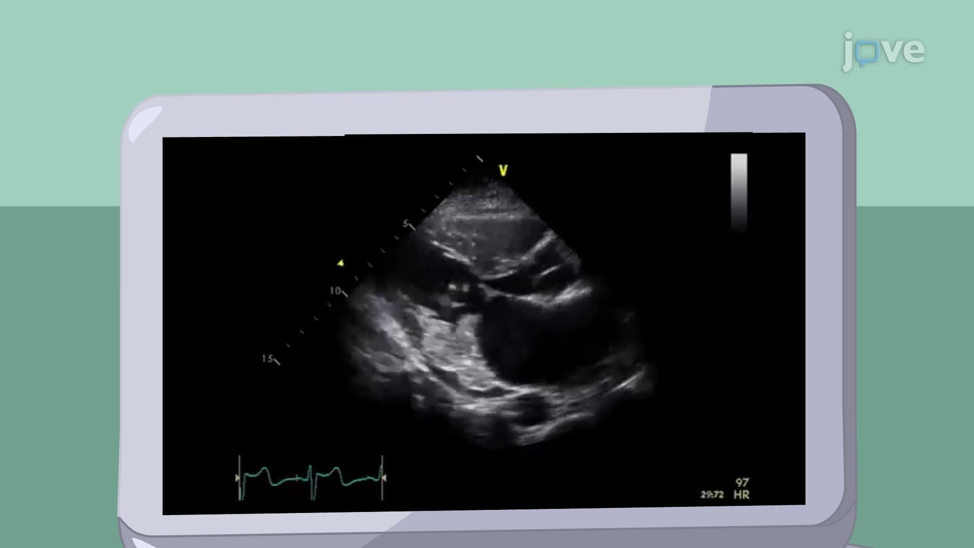
The pathological hallmark involves myxomatous degeneration-a process where excess collagen and proteoglycans accumulate within valve leaflets. Picture the valve tissue becoming increasingly "floppy," similar to how overcooked pasta loses its structural integrity. This degenerative process particularly affects the spongiosa layer of the valve, creating the characteristic thickened, redundant tissue seen in echocardiographic studies at major US medical centers like Mayo Clinic and Cleveland Clinic.
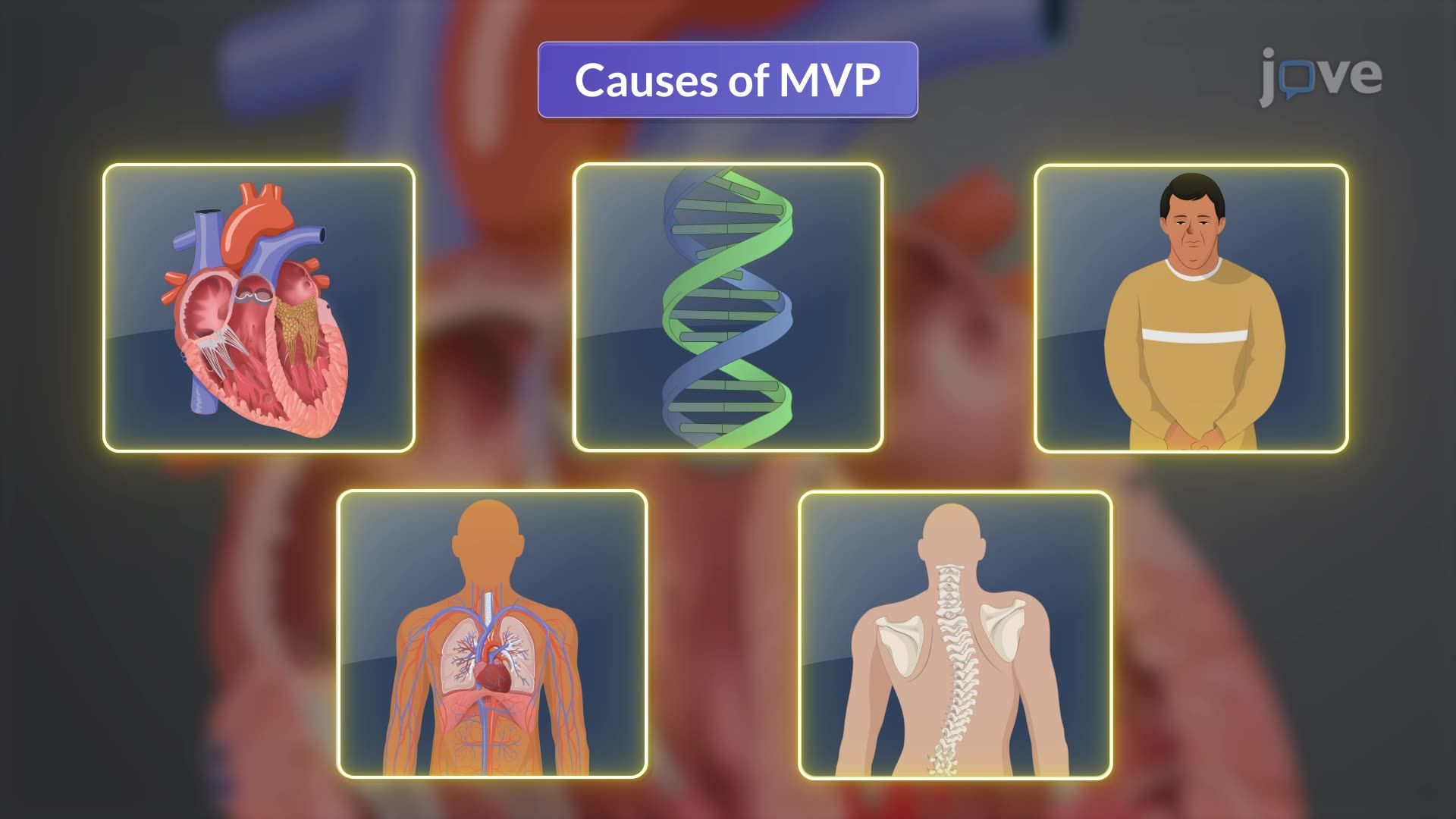
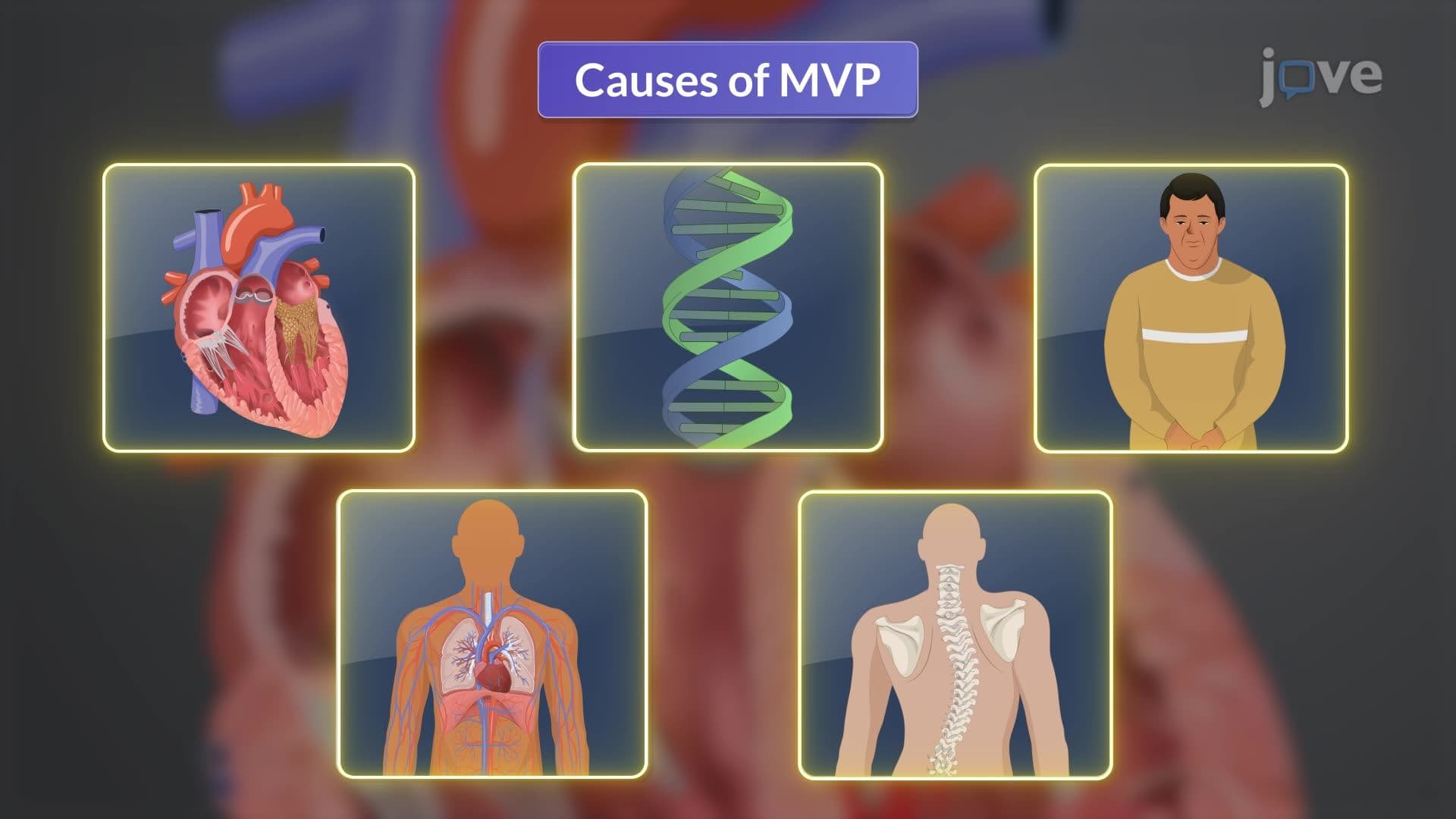
MVP demonstrates significant genetic heterogeneity, with familial clustering patterns documented in American populations. Connective tissue disorders create particularly high-risk scenarios-Marfan syndrome patients show MVP prevalence rates exceeding 80%. The condition also associates with skeletal abnormalities including scoliosis, pectus deformities, and muscular dystrophy. For MCAT preparation, students should understand how these systemic connective tissue abnormalities manifest cardiac complications.
The progression from simple prolapse to mitral regurgitation represents a critical concept for AP Biology and college-level physiology courses. When chordae tendineae weaken or rupture, the support system fails catastrophically, creating severe regurgitation. Blood volume overload in the left atrium triggers compensatory mechanisms including atrial enlargement and eventual atrial fibrillation-complications frequently tested on USMLE Step 1 examinations.
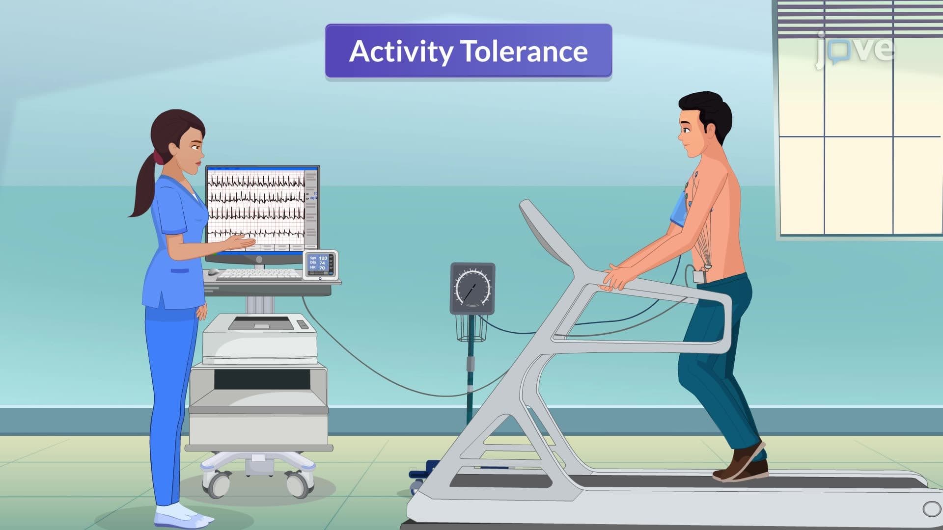
Understanding these pathophysiological cascades prepares students for clinical scenarios where MVP patients present with palpitations, chest pain, or exercise intolerance at US emergency departments nationwide.
Related Micro-courses