Loading subject page…

1,733 views

Video Summary: Mitral Valve Prolapse Ii Assessment Explained
Did you know that mitral valve prolapse affects up to 3% of the U.S. population, making it one of the most common heart valve conditions? Mitral valve prolapse ii assessment involves systematic evaluation techniques that help clinicians identify this condition where heart valve leaflets bulge backward into the left atrium. For instance, at Johns Hopkins Hospital, cardiologists routinely use the characteristic "mid-systolic click" sound during physical exams as a key diagnostic clue. This Mitral Valve Prolapse II Assessment Explained demonstrates the step-by-step diagnostic approach from patient history to advanced imaging. Watch the full video on JoVE Coach to master this concept with expert-led visuals and step-by-step explanations.
Mitral valve prolapse ii assessment represents a cornerstone skill in cardiovascular diagnostics that medical students encounter extensively in their clinical rotations. This systematic evaluation process begins with recognizing that MVP occurs when one or both mitral valve leaflets bulge backward into the left atrium during ventricular contraction. At prestigious medical centers like the Cleveland Clinic, this condition affects approximately 2-3% of the general population, with women being affected twice as frequently as men.
The mitral valve prolapse ii assessment tutorial emphasizes that while many patients remain asymptomatic, others present with a constellation of symptoms that can significantly impact quality of life. Palpitations represent the most common complaint, often described by patients as a "fluttering" or "racing" heart sensation. Chest pain in MVP patients differs from typical angina, presenting as sharp, stabbing sensations rather than the crushing pressure associated with coronary artery disease. Healthcare providers at institutions like Mayo Clinic have documented that anxiety and panic attacks frequently accompany MVP, creating a complex clinical picture that requires careful assessment.
Understanding how mitral valve prolapse ii assessment works requires mastery of cardiac auscultation techniques. The pathognomonic mid-systolic click occurs due to sudden tensing of the mitral valve apparatus as the leaflets prolapse into the left atrium. This click may be followed by a late systolic murmur if mitral regurgitation is present. Medical students preparing for the USMLE Step 2 CK must understand that certain maneuvers can alter these findings: standing or Valsalva maneuver moves the click earlier in systole, while squatting delays it.
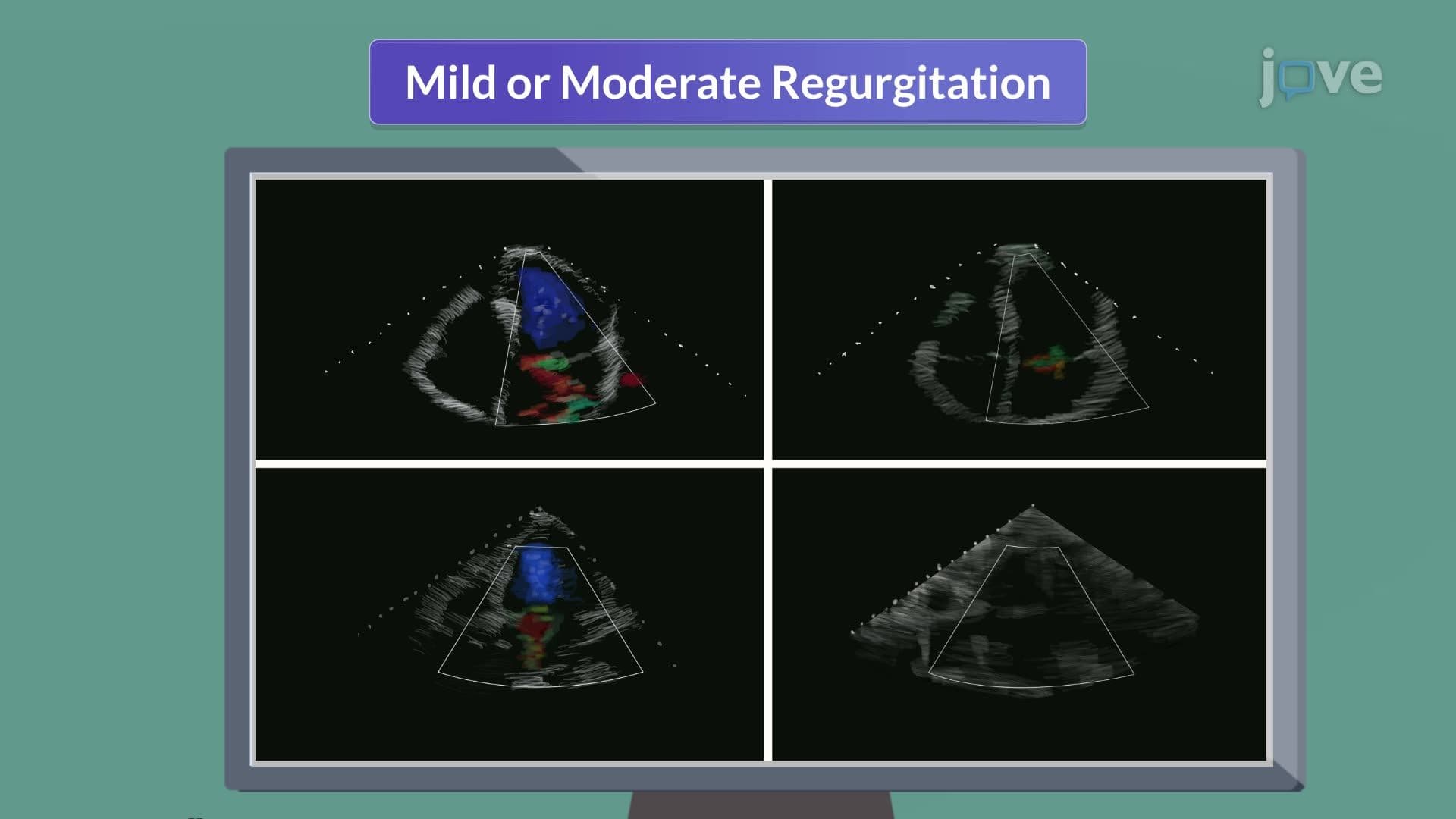
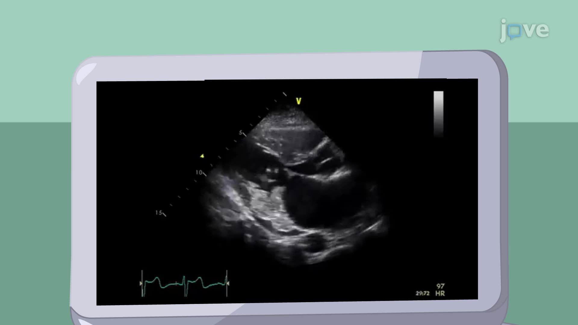
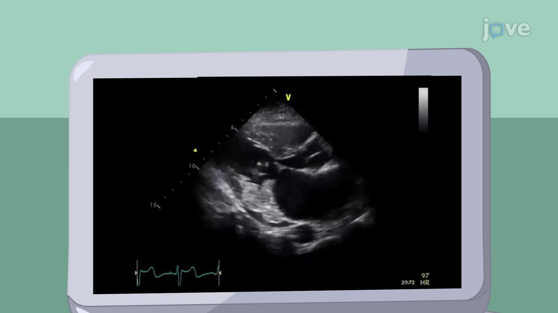
The mitral valve prolapse ii assessment concept relies heavily on transthoracic echocardiography, which provides definitive visualization of valve structure and function. This imaging modality allows clinicians to measure the extent of leaflet prolapse, assess mitral regurgitation severity, and evaluate left ventricular function. Students studying for the MCAT should understand that echocardiography uses ultrasound principles to create real-time images of cardiac structures. Complementary studies include 12-lead electrocardiograms to detect arrhythmias and chest radiographs to assess cardiac silhouette and pulmonary vasculature.
Related Micro-courses