Loading subject page…

1,733 views

Video Summary: What Is Aortic Regurgitation I Introduction
Did you know that nearly 5% of Americans over 65 have moderate to severe aortic regurgitation, yet many remain undiagnosed until serious complications arise? Aortic regurgitation I introduction explains how blood flows backward from the aorta into the left ventricle when the aortic valve fails to close properly during diastole. This condition affects thousands of patients annually at major US medical centers like Cleveland Clinic and Mayo Clinic. Watch the full video on JoVE Coach to master this concept with expert-led visuals and step-by-step explanations.
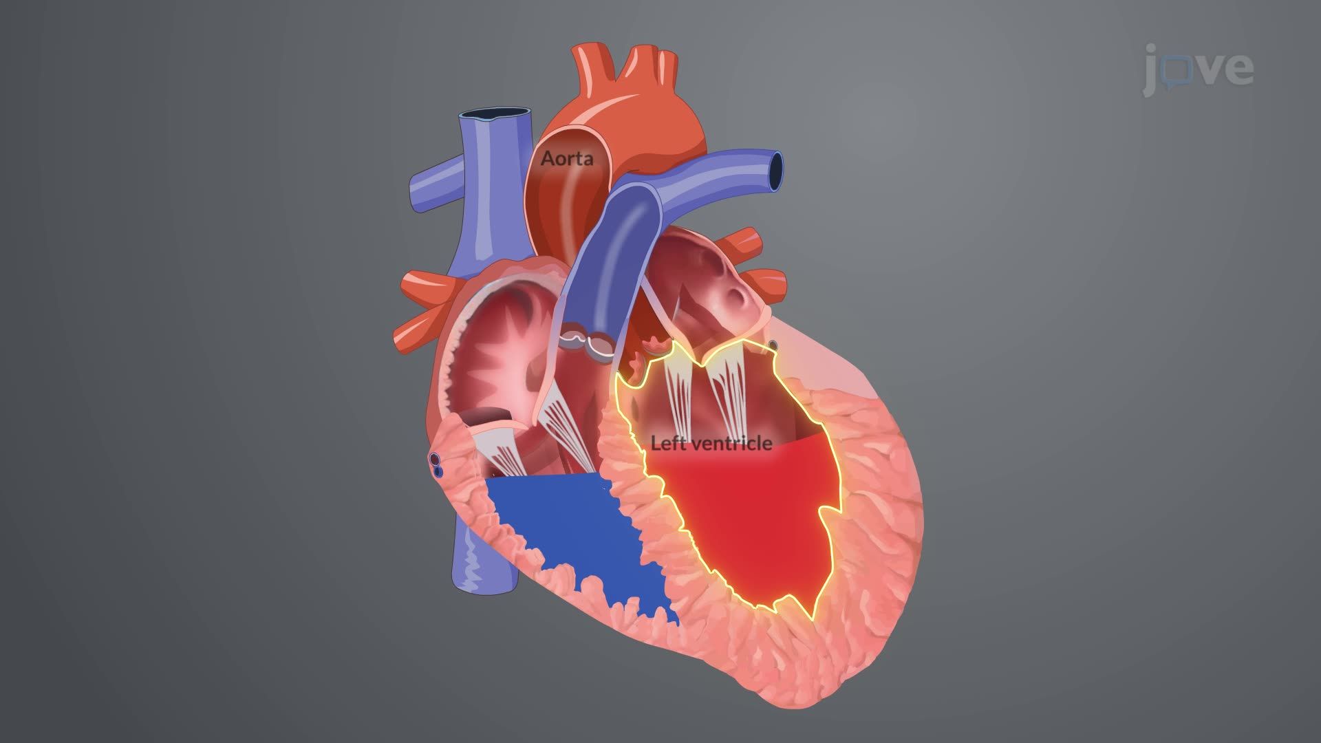
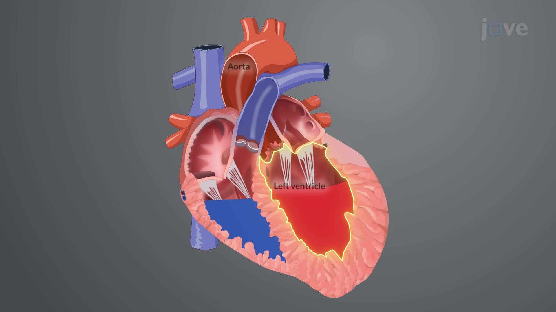
Aortic regurgitation I introduction represents a critical cardiac pathology where the aortic valve fails to create a competent seal during diastole, allowing oxygenated blood to flow retrograde from the aorta back into the left ventricle. This seemingly simple mechanical failure triggers a cascade of compensatory mechanisms that initially maintain cardiac output but ultimately lead to heart failure if left untreated.
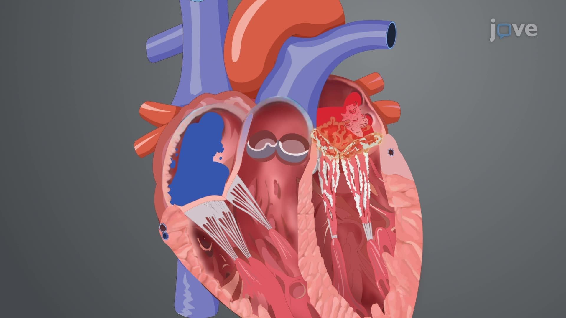
The normal aortic valve consists of three cusps that close tightly when ventricular pressure falls below aortic pressure during diastole. In aortic regurgitation, structural abnormalities or damage prevent complete closure, creating a regurgitant orifice. This defect forces the left ventricle to handle both its normal preload from the left atrium plus the additional regurgitant volume from the aorta.
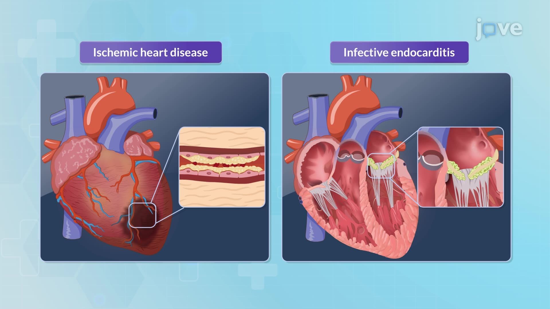
Acute aortic regurgitation often results from sudden valve destruction due to infective endocarditis (particularly common with Staphylococcus aureus infections in IV drug users), blunt chest trauma from motor vehicle accidents, or aortic dissection. The left ventricle has no time to adapt, leading to rapid onset of pulmonary edema and cardiogenic shock.
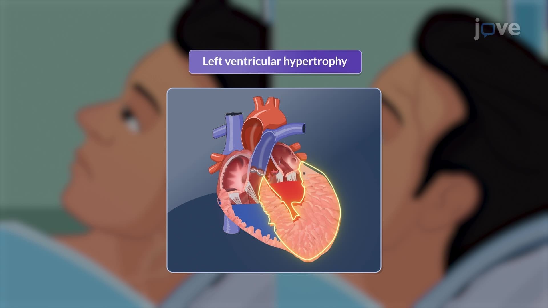
Chronic aortic regurgitation develops gradually from conditions like bicuspid aortic valve (present in 1-2% of the US population), rheumatic heart disease, or connective tissue disorders such as Marfan syndrome. The left ventricle gradually dilates and develops eccentric hypertrophy to accommodate the increased volume load while maintaining forward stroke volume.
Initially, the left ventricle compensates through the Frank-Starling mechanism, where increased preload enhances contractility. The chamber dilates to accommodate extra volume while wall thickness increases to normalize wall stress. This compensated phase can last years to decades, during which patients remain asymptomatic despite significant regurgitation.
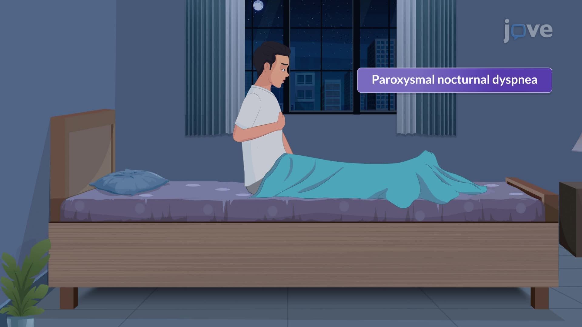
However, chronic volume overload eventually overwhelms compensatory mechanisms. Left ventricular end-diastolic pressure rises, transmitting backward pressure to the left atrium, pulmonary veins, and pulmonary capillaries. This progression explains why patients with severe chronic aortic regurgitation eventually develop exertional dyspnea, orthopnea, and paroxysmal nocturnal dyspnea.
Understanding aortic regurgitation proves essential for MCAT preparation, particularly in cardiovascular physiology sections. AP Biology students encounter this concept when studying circulatory systems and homeostatic mechanisms. College anatomy and physiology courses extensively cover valvular heart disease as a prime example of structure-function relationships.
Pre-med students should recognize that aortic regurgitation represents a classic example of how mechanical cardiac problems evolve into complex pathophysiological states. This knowledge foundation proves crucial for USMLE Step 1 questions involving cardiac cycle analysis, pressure-volume relationships, and heart failure mechanisms.
Related Micro-courses