Loading subject page…

1,733 views

Video Summary: What Is Mitral Regurgitation I Introduction
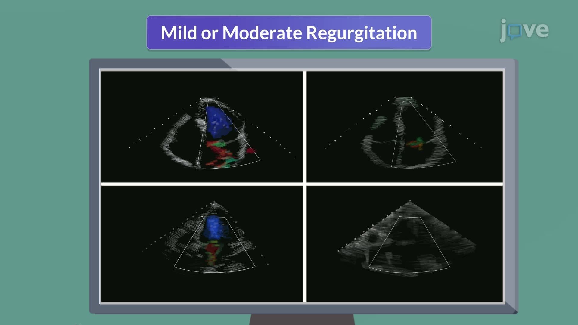
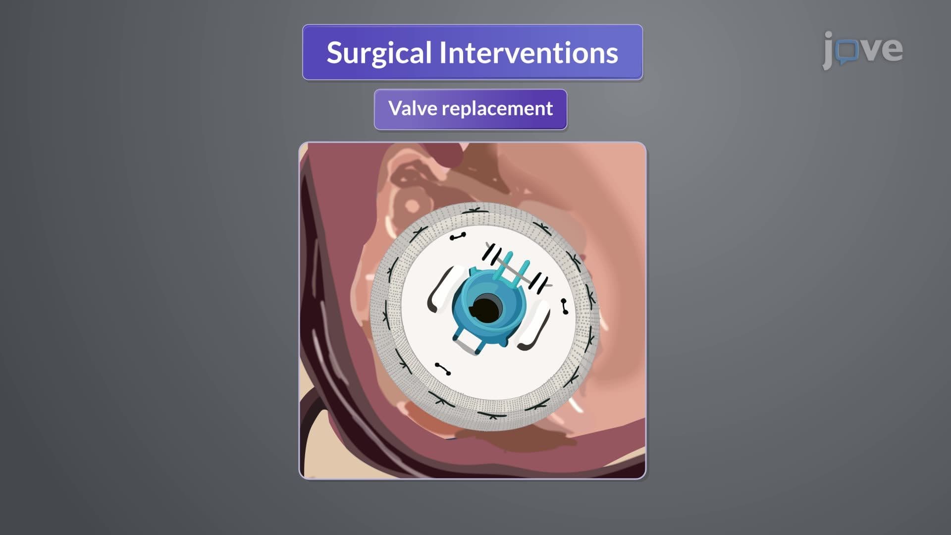
Did you know that mitral regurgitation affects approximately 10% of Americans over age 75, making it one of the most common heart valve disorders? Mitral regurgitation I introduction reveals how this condition occurs when the heart's mitral valve fails to seal properly, allowing blood to flow backward into the left atrium during each heartbeat. Patients at major US cardiac centers like Cleveland Clinic frequently present with this condition, which can range from mild leakage requiring only monitoring to severe cases demanding surgical intervention. Watch the full video on JoVE Coach to master this concept with expert-led visuals and step-by-step explanations.
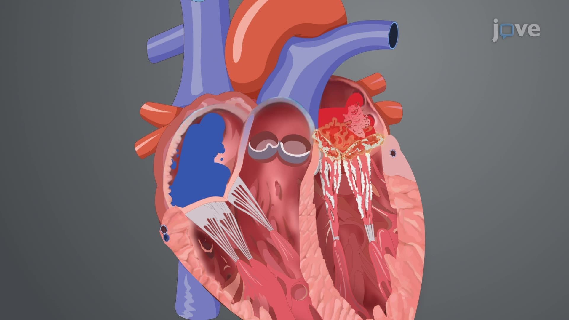
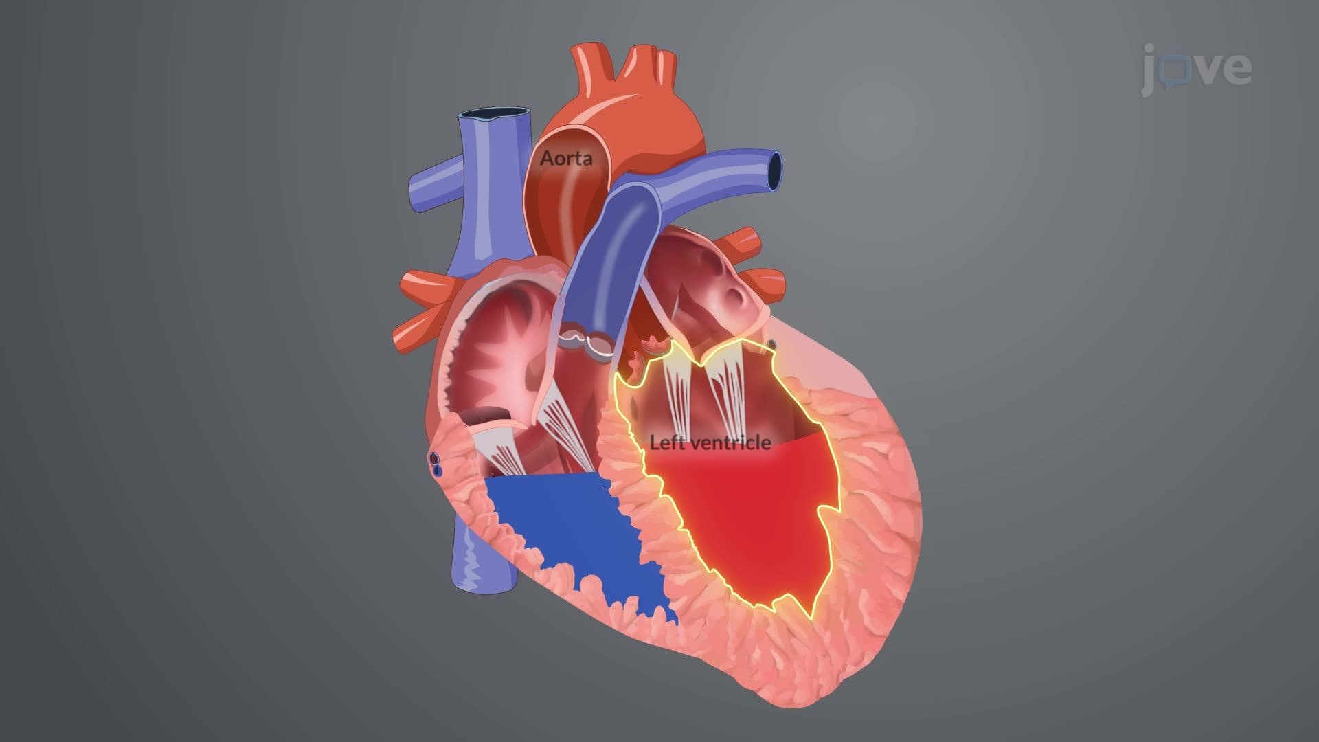
Mitral regurgitation I introduction encompasses the foundational understanding of one of cardiology's most prevalent valve disorders. The mitral valve, positioned between the left atrium and left ventricle, normally creates a one-way seal during systole (heart contraction). When this valve becomes incompetent, blood regurgitates backward, creating a volume overload state that progressively damages the heart's structure and function.
This concept appears frequently on the MCAT's biology section and forms essential knowledge for pre-med students studying cardiovascular physiology. Understanding mitral regurgitation requires grasping both normal cardiac anatomy and the pathological processes that disrupt valve function.
The mitral regurgitation I introduction definition distinguishes between two fundamental etiologic categories. Primary mitral regurgitation stems from intrinsic valve pathology-the valve itself is diseased. Common causes include mitral valve prolapse (affecting 2-3% of Americans), degenerative changes from aging, and rheumatic heart disease (though rare in the US due to antibiotic availability).
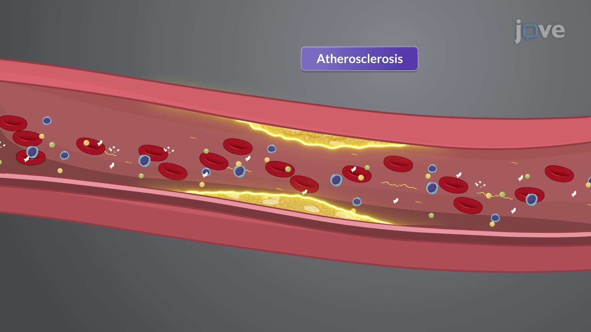
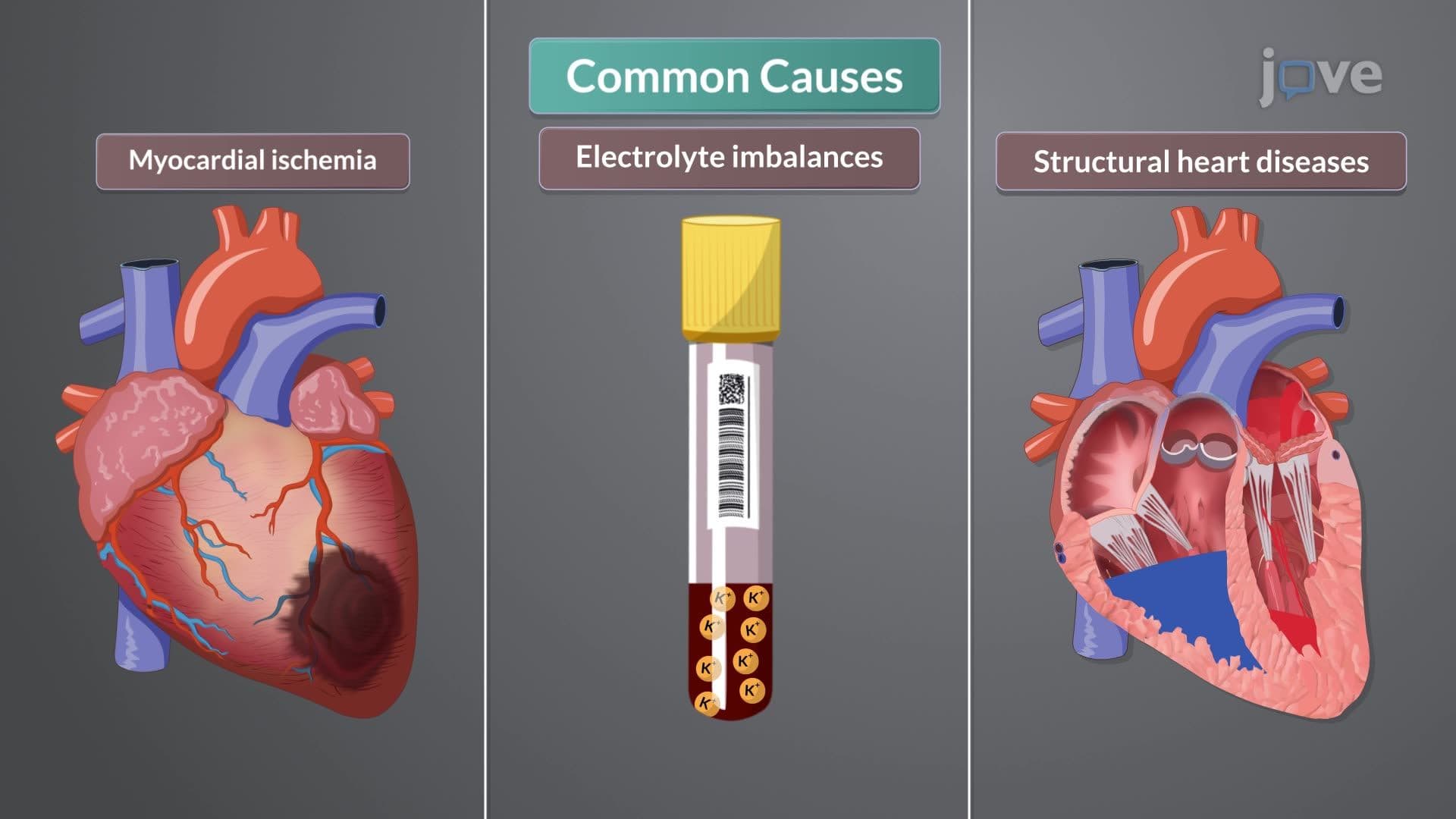
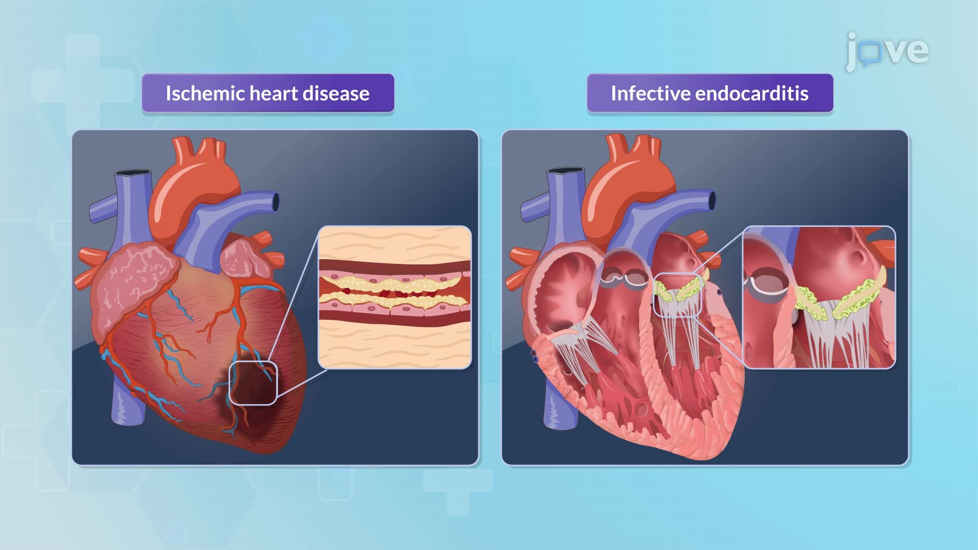
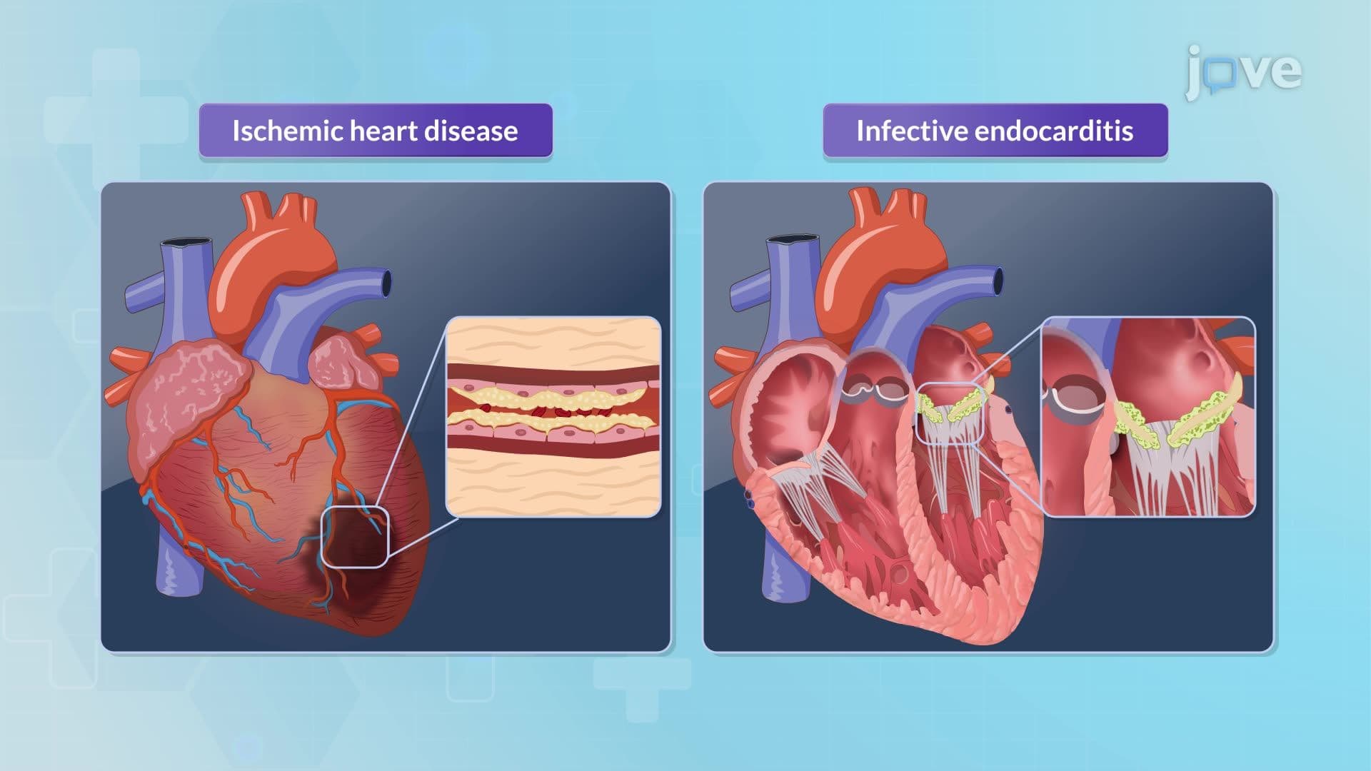
Secondary mitral regurgitation results from external factors affecting valve function. Ischemic heart disease, prevalent in American adults with coronary artery disease, can damage papillary muscles or alter ventricular geometry, preventing proper valve closure. This distinction proves crucial for treatment decisions at major US cardiac surgery centers.
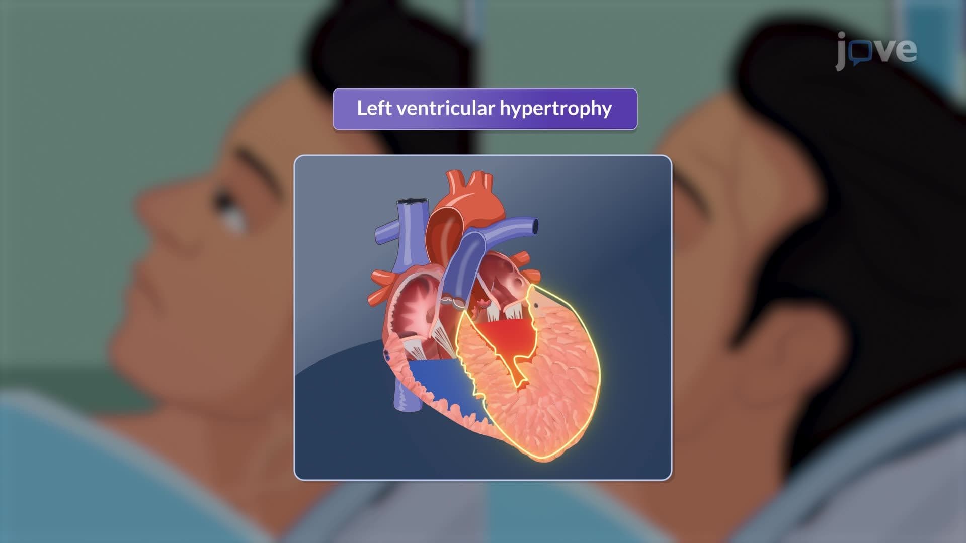
What is mitral regurgitation I introduction in detail involves understanding the complex hemodynamic alterations. Initially, the left ventricle compensates for volume overload through eccentric hypertrophy-enlarging to accommodate increased blood volume. Simultaneously, the left atrium dilates to handle regurgitant blood flow.
These compensatory mechanisms eventually fail, leading to systolic dysfunction and reduced forward cardiac output. Elevated left atrial pressures transmit backward through pulmonary veins, causing pulmonary congestion and potentially pulmonary hypertension. This progression explains why patients with severe mitral regurgitation develop shortness of breath, fatigue, and exercise intolerance-symptoms frequently tested on USMLE Step 1 examinations.
For students preparing for healthcare careers, mitral regurgitation represents a cornerstone cardiovascular concept. AP Biology students encounter this topic when studying circulatory systems, while college anatomy and physiology courses dedicate substantial time to valve disorders. Understanding this condition's pathophysiology provides essential foundation knowledge for future clinical courses and standardized examinations like the MCAT, where cardiovascular physiology questions commonly appear.
Related Micro-courses