Loading video…

 15 Concepts
 1.7K views

Video Summary: What Is Mitral Stenosis I Introduction
Did you know that mitral stenosis I introduction can cause a heart valve to narrow to just 25% of its normal size? This mitral stenosis I introduction definition explains how the mitral valve restricts blood flow between heart chambers, most commonly due to rheumatic fever complications seen in patients at centers like Cleveland Clinic. Understanding mitral stenosis I introduction helps explain why some patients experience shortness of breath and fatigue during everyday activities. Watch the full video on JoVE Coach to master this concept with expert-led visuals and step-by-step explanations.
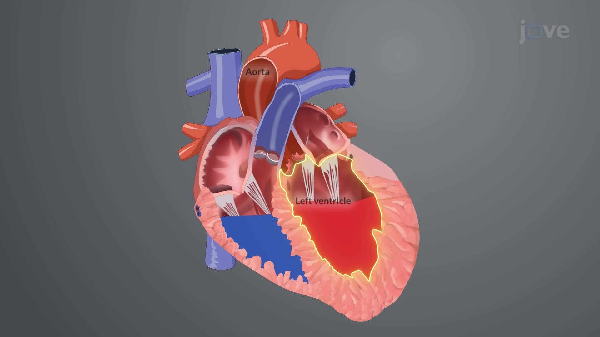
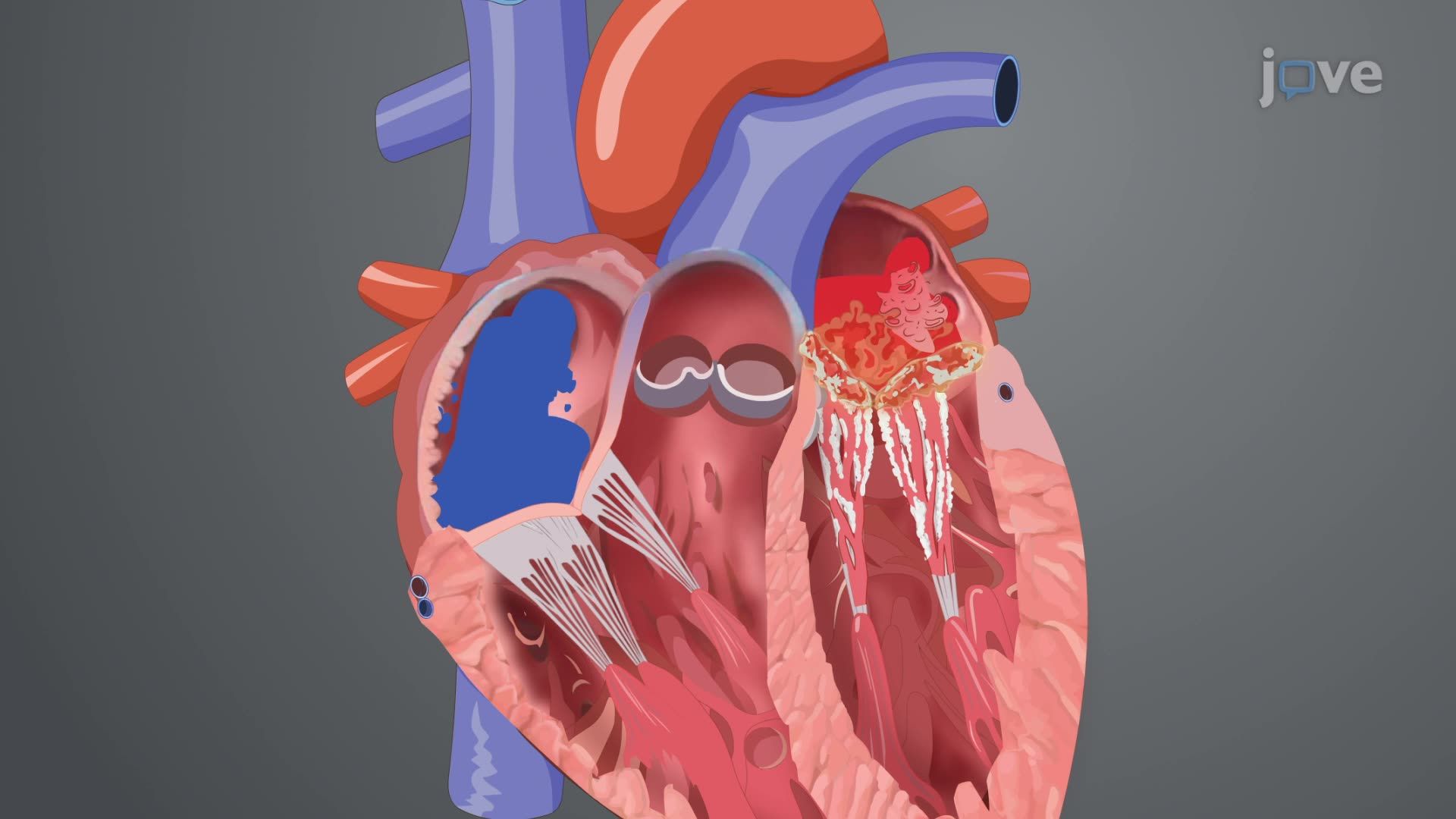
Mitral stenosis I introduction centers on understanding how the mitral valve-one of four heart valves-becomes pathologically narrowed, creating a bottleneck in blood circulation. Located between the left atrium and left ventricle, this bicuspid valve normally opens widely during diastole (heart relaxation) to allow oxygen-rich blood to flow efficiently into the heart's main pumping chamber.
While rheumatic fever remains the leading cause globally, mitral stenosis I introduction in US patients increasingly involves other etiologies. Rheumatic endocarditis, following untreated strep throat infections, causes inflammatory scarring that fuses valve leaflets together. However, American cardiologists at institutions like Mayo Clinic also encounter cases from congenital malformations, autoimmune conditions like systemic lupus erythematosus, radiation therapy complications in cancer survivors, and severe mitral annular calcification in elderly patients.
The mitral stenosis I introduction concept emphasizes understanding sequential cardiovascular changes. As the valve orifice narrows from a normal 4-6 cm² to less than 1.5 cm² in severe cases, blood backs up into the left atrium during diastole. This creates elevated pressures that the left atrium initially compensates for through hypertrophy and dilation.
However, sustained pressure elevation triggers a domino effect: pulmonary veins experience increased pressure, leading to pulmonary congestion and potentially life-threatening pulmonary edema. The enlarged, pressure-overloaded left atrium becomes electrically unstable, predisposing patients to atrial fibrillation-an irregular heart rhythm that further compromises cardiac efficiency.
Students preparing for medical school entrance exams should understand that mitral stenosis I introduction connects cardiovascular anatomy with pathophysiology principles. MCAT questions often test understanding of pressure-volume relationships and compensatory mechanisms. The concept appears frequently in cardiology rotations and USMLE Step 1 examinations, where students must integrate knowledge of valve anatomy, hemodynamics, and clinical presentations to answer complex scenario-based questions about patient management and prognosis.
Related Micro-courses