Loading video…

 27 Concepts
 3.6K views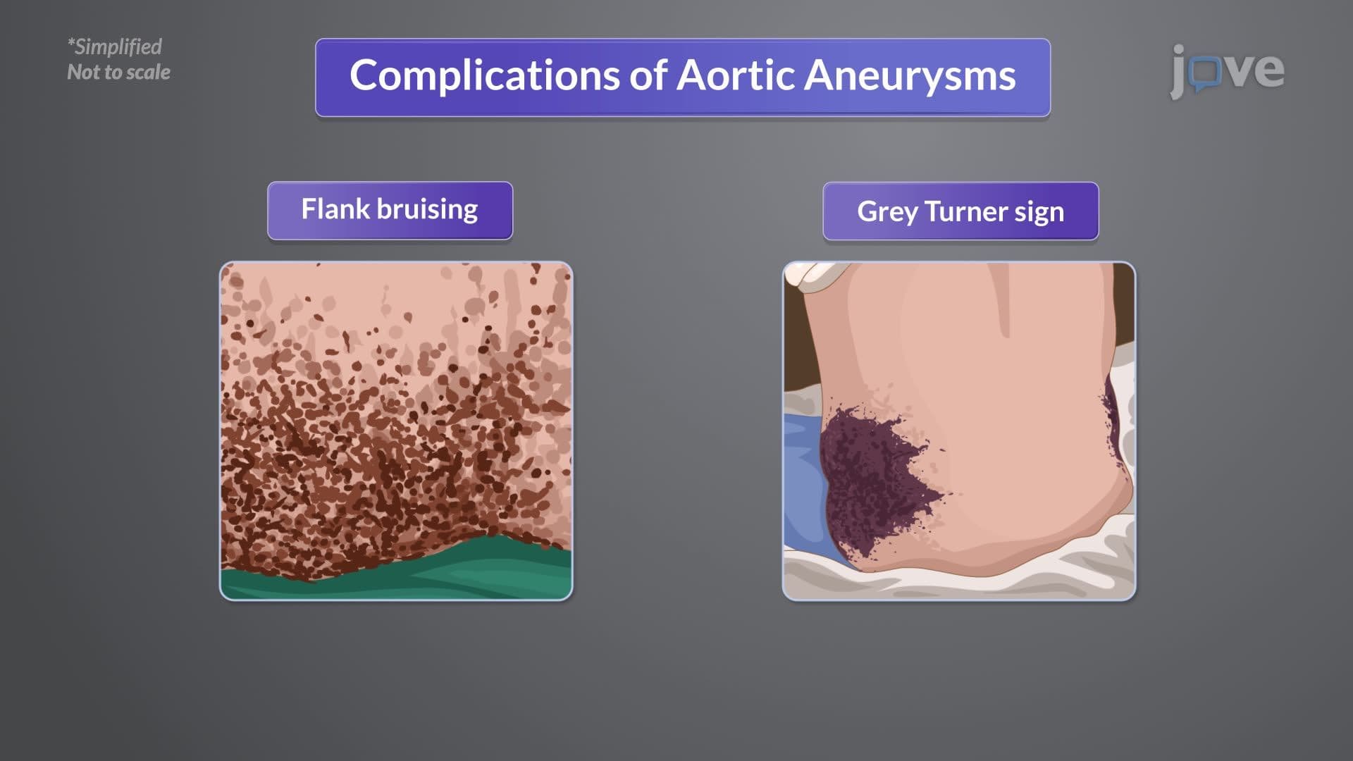

Video Summary: Aneurysm Ii Clinical Manifestations and Diagnostic Evaluation
Did you know that some aortic aneurysms can grow silently for years before causing a distinctive "brassy cough" that alerts doctors to a life-threatening condition? Understanding aneurysm II: clinical manifestations and diagnostic evaluation reveals how these vascular bulges create recognizable symptoms and diagnostic patterns. For instance, patients at Cleveland Clinic often present with chest pain radiating to their back-a classic sign of thoracic aortic involvement. This comprehensive examination of Aneurysm II: Clinical Manifestations and Diagnostic Evaluation covers symptom recognition and modern imaging approaches. Watch the full video on JoVE Coach to master this concept with expert-led visuals and step-by-step explanations.
The clinical presentation of aortic aneurysms depends critically on their anatomical location, size, and rate of expansion. This systematic approach to aneurysm II: clinical manifestations and diagnostic evaluation forms the foundation for early detection and appropriate management in clinical practice.
Thoracic aortic aneurysms create symptoms through mass effect and vascular compromise. Patients typically develop dyspnea when aneurysmal expansion compresses bronchi or lung parenchyma. The characteristic deep, diffuse chest pain radiating to the interscapular region results from aortic wall stretching and possible mediastinal structure involvement. A distinctive brassy, paroxysmal cough develops from recurrent laryngeal nerve compression-a finding that emergency physicians at major trauma centers like Harborview Medical Center recognize as highly suggestive of aortic pathology.
Ascending aorta and aortic arch aneurysms pose particular risks by compromising coronary artery perfusion, manifesting as angina pectoris. Superior vena cava compression produces the classic triad of jugular venous distention, facial edema, and upper extremity swelling-collectively known as superior vena cava syndrome.
Abdominal aortic aneurysms present more subtly but with pathognomonic physical findings. The pulsatile mass located in the periumbilical area, characteristically left of midline, represents the most reliable clinical sign. Experienced clinicians can often detect audible bruits over these aneurysms, indicating turbulent blood flow within the dilated vessel.
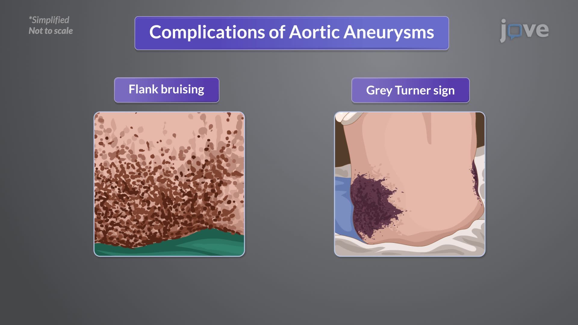
Aneurysm rupture represents a surgical emergency with location-specific manifestations. Retroperitoneal rupture creates the Grey Turner sign-flank ecchymosis resulting from blood tracking along tissue planes. This finding, named after British surgeon George Grey Turner, indicates significant hemorrhage and impending hemodynamic collapse. Rupture into thoracic or abdominal cavities produces rapid hypovolemic shock, requiring immediate surgical intervention.
These concepts frequently appear on MCAT passages testing cardiovascular pathophysiology, while AP Biology courses emphasize the relationship between vessel wall integrity and systemic circulation. Medical students preparing for USMLE Step 1 should focus on correlating anatomical locations with specific symptom clusters and diagnostic approaches.
Related Micro-courses