Loading subject page…

3,563 views

Video Summary: Pulmonary Embolism Ii Diagnostic Studies Explained
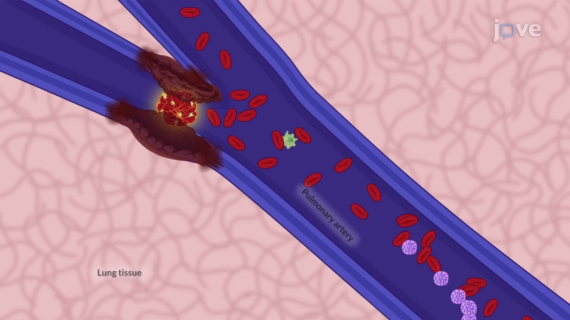
Every year in the US, pulmonary embolism affects over 600,000 Americans, yet diagnosis remains challenging because symptoms often mimic other conditions. Pulmonary embolism II diagnostic studies involve sophisticated imaging techniques like CT angiography and ventilation-perfusion scans, combined with blood tests such as D-dimer analysis. At Massachusetts General Hospital, emergency physicians rely on these diagnostic tools to quickly identify blood clots in lung arteries and save lives. Watch the full video on JoVE Coach to master this concept with expert-led visuals and step-by-step explanations.
Diagnosing pulmonary embolism requires a multi-faceted approach that combines clinical assessment with advanced diagnostic technologies. The condition affects approximately 1-2 per 1,000 people annually in the United States, making accurate diagnosis crucial for patient survival. Healthcare providers must rapidly differentiate PE from other conditions like pneumonia, heart attack, or anxiety disorders that present with similar symptoms.
CT Pulmonary Angiography (CTPA) serves as the gold standard diagnostic tool in most US emergency departments. This imaging technique uses contrast dye to highlight blood vessels in the lungs, allowing physicians to directly visualize blood clots. Major medical centers like Johns Hopkins and Cleveland Clinic report diagnostic accuracy rates exceeding 95% with modern CT scanners.
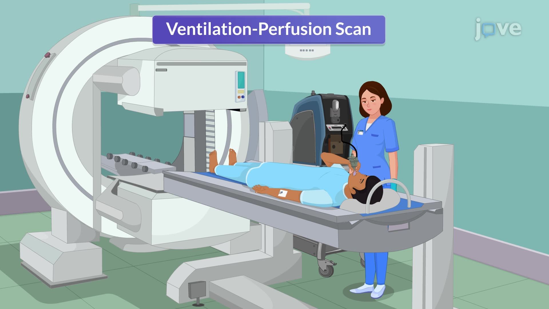
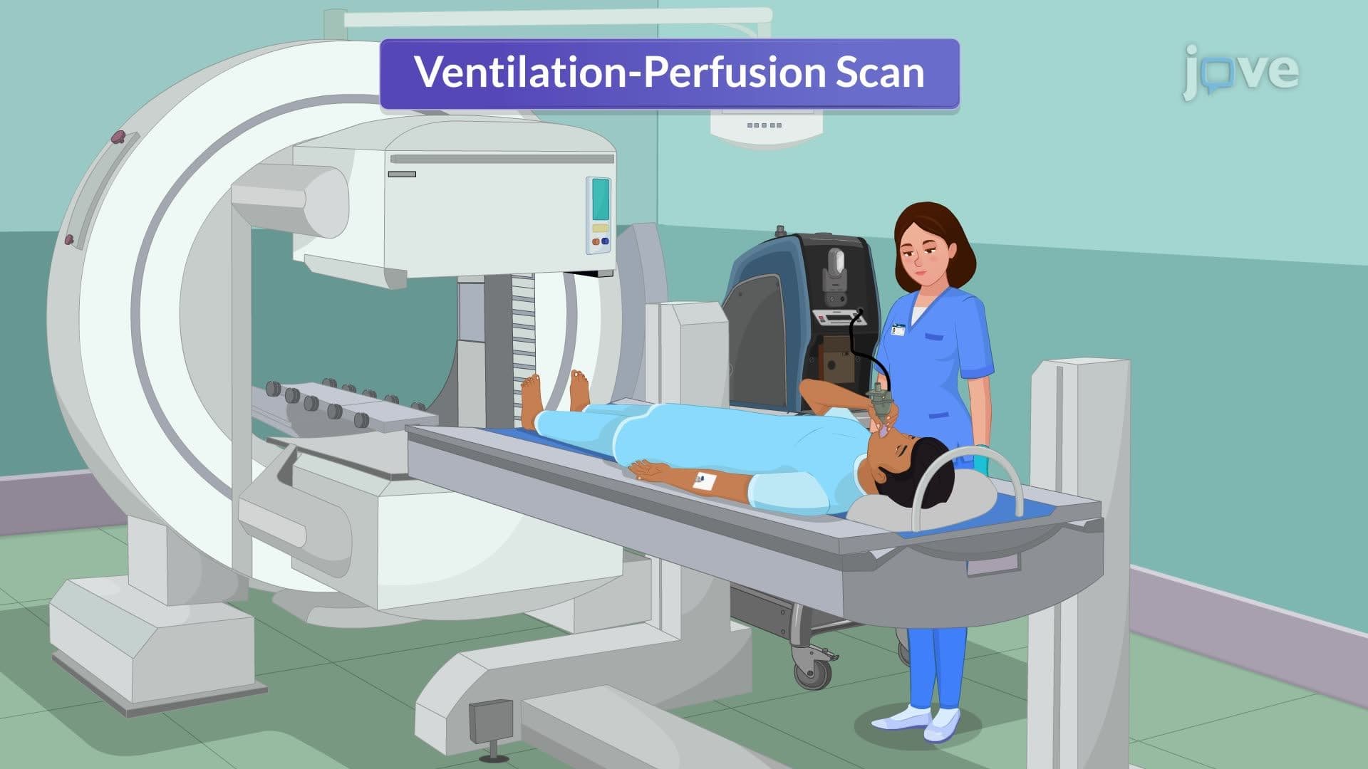
Ventilation-Perfusion (V/Q) scans provide an alternative diagnostic approach, particularly valuable for patients who cannot receive contrast dye due to kidney problems or allergies. This nuclear medicine test compares air distribution (ventilation) with blood flow (perfusion) throughout the lungs. Mismatched areas where air reaches but blood flow is blocked indicate potential embolism sites.
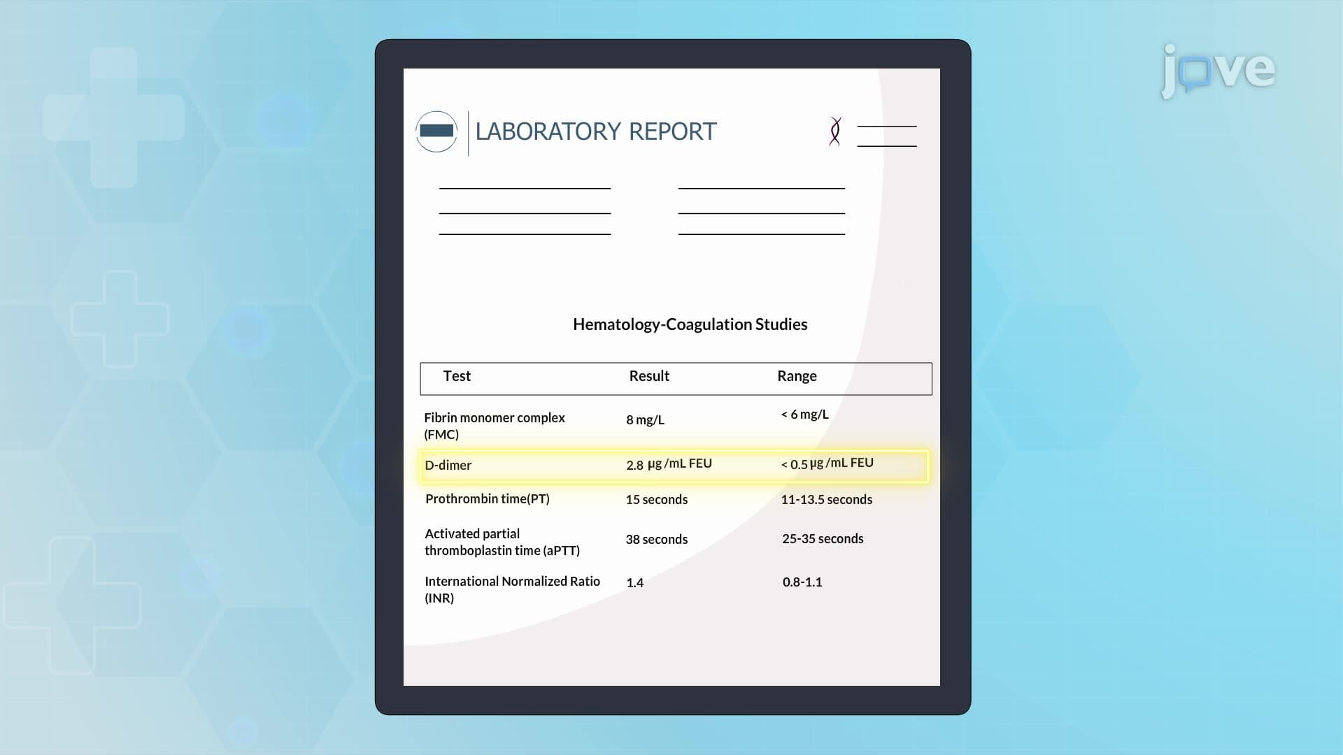
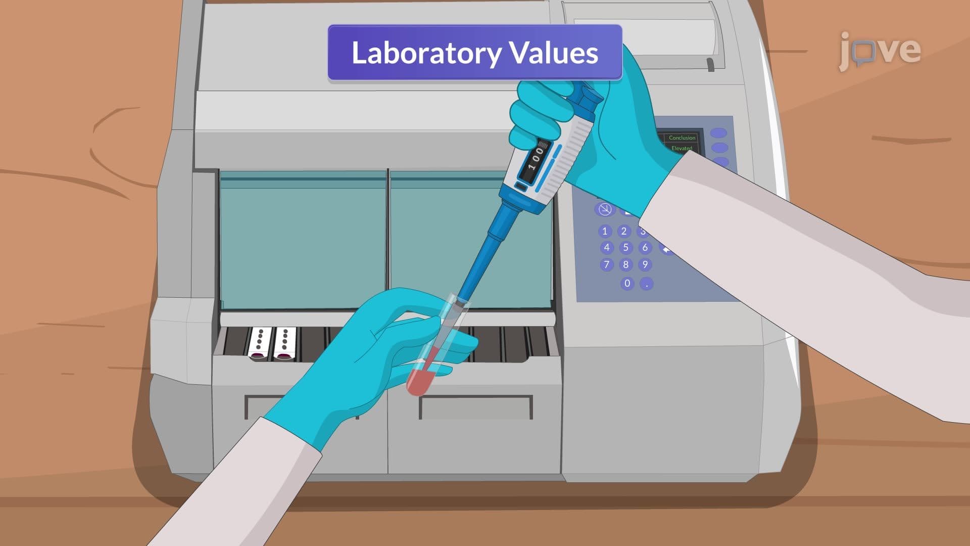
The D-dimer test measures fibrin degradation products in the blood, indicating recent clot formation and breakdown. While elevated D-dimer levels suggest possible PE, this test lacks specificity since many conditions can cause elevation. Emergency physicians at institutions like Mayo Clinic use D-dimer primarily to rule out PE when levels are normal, particularly in low-risk patients.
Arterial Blood Gas (ABG) analysis reveals important physiological changes associated with PE. Patients typically show decreased oxygen levels (hypoxemia) and sometimes decreased carbon dioxide levels due to hyperventilation. These findings, while not specific to PE, help assess disease severity and guide oxygen therapy decisions.
Once PE is confirmed, treatment selection depends on patient stability and embolism severity. Stable patients without cardiovascular compromise typically receive anticoagulation therapy with medications like enoxaparin (low-molecular-weight heparin) or newer oral anticoagulants such as rivaroxaban. These medications prevent clot growth and reduce recurrence risk.
Unstable patients with massive PE and hemodynamic compromise require more aggressive intervention. Thrombolytic therapy using tissue plasminogen activator (tPA) can dissolve existing clots but carries bleeding risks. When thrombolytics are contraindicated, surgical options include catheter-directed thrombolysis or surgical embolectomy procedures available at specialized centers nationwide.
This diagnostic and treatment approach appears frequently on medical licensing exams including the USMLE and NCLEX, where students must demonstrate understanding of appropriate test selection and interpretation in clinical scenarios.
Related Micro-courses