Loading subject page…

3,563 views

Video Summary: Varicose Veins Ii Diagnostic Studies Explained
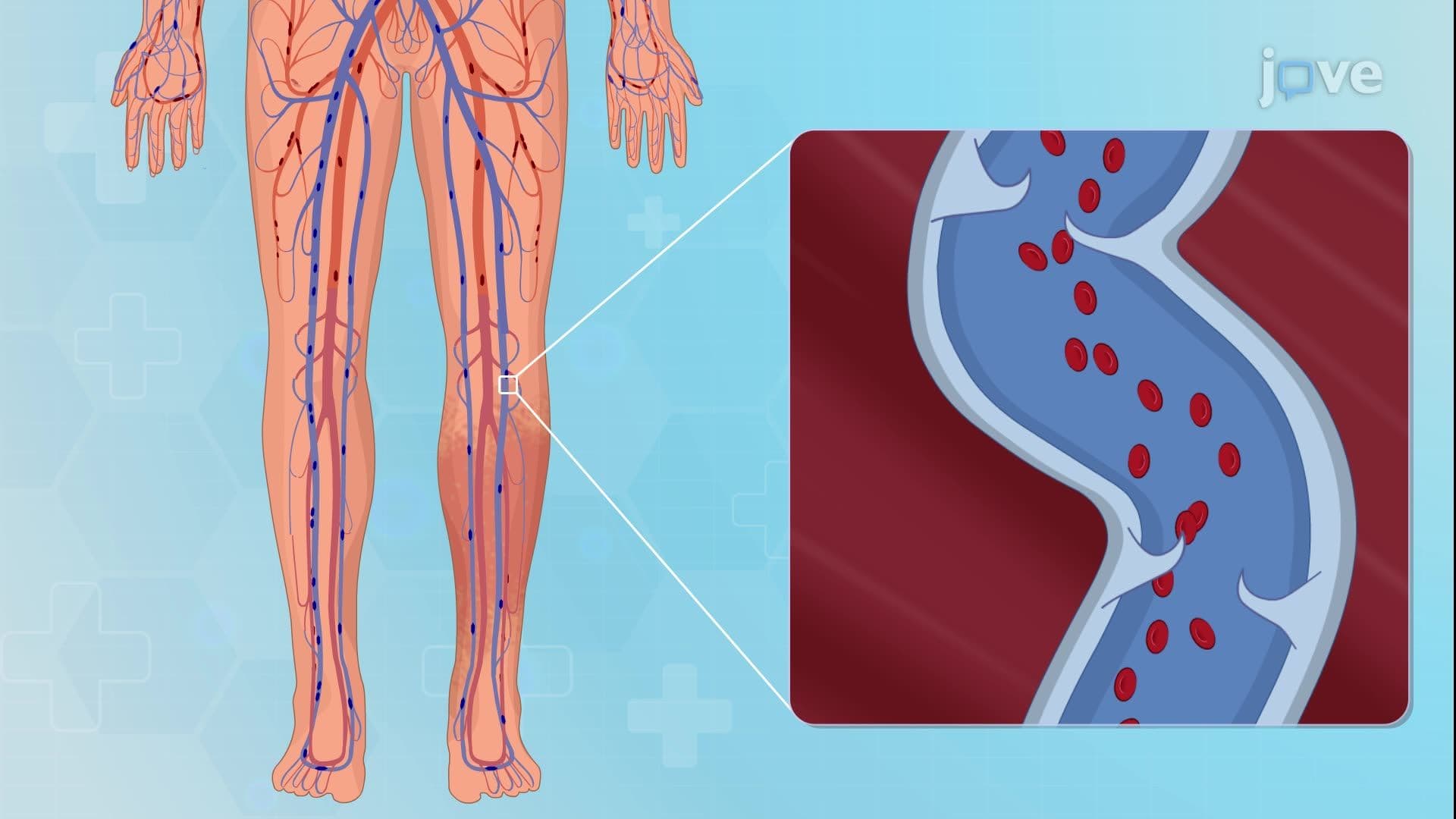
Did you know that over 23% of American adults develop varicose veins, making proper diagnostic techniques essential for healthcare providers? Varicose veins II diagnostic studies involve comprehensive evaluation methods including duplex ultrasound scanning to assess venous anatomy and valve function. At major medical centers like Mayo Clinic, these diagnostic approaches help determine the most effective treatment strategies for millions of patients experiencing leg pain and swelling. Watch the full video on JoVE Coach to master this concept with expert-led visuals and step-by-step explanations.
Varicose veins II diagnostic studies represent a systematic approach to evaluating venous insufficiency that affects millions of Americans annually. These diagnostic protocols extend beyond basic visual inspection to include sophisticated imaging techniques and functional assessments. Healthcare providers at institutions like Cleveland Clinic and Johns Hopkins utilize these comprehensive evaluation methods to develop targeted treatment plans for patients experiencing venous disease complications.
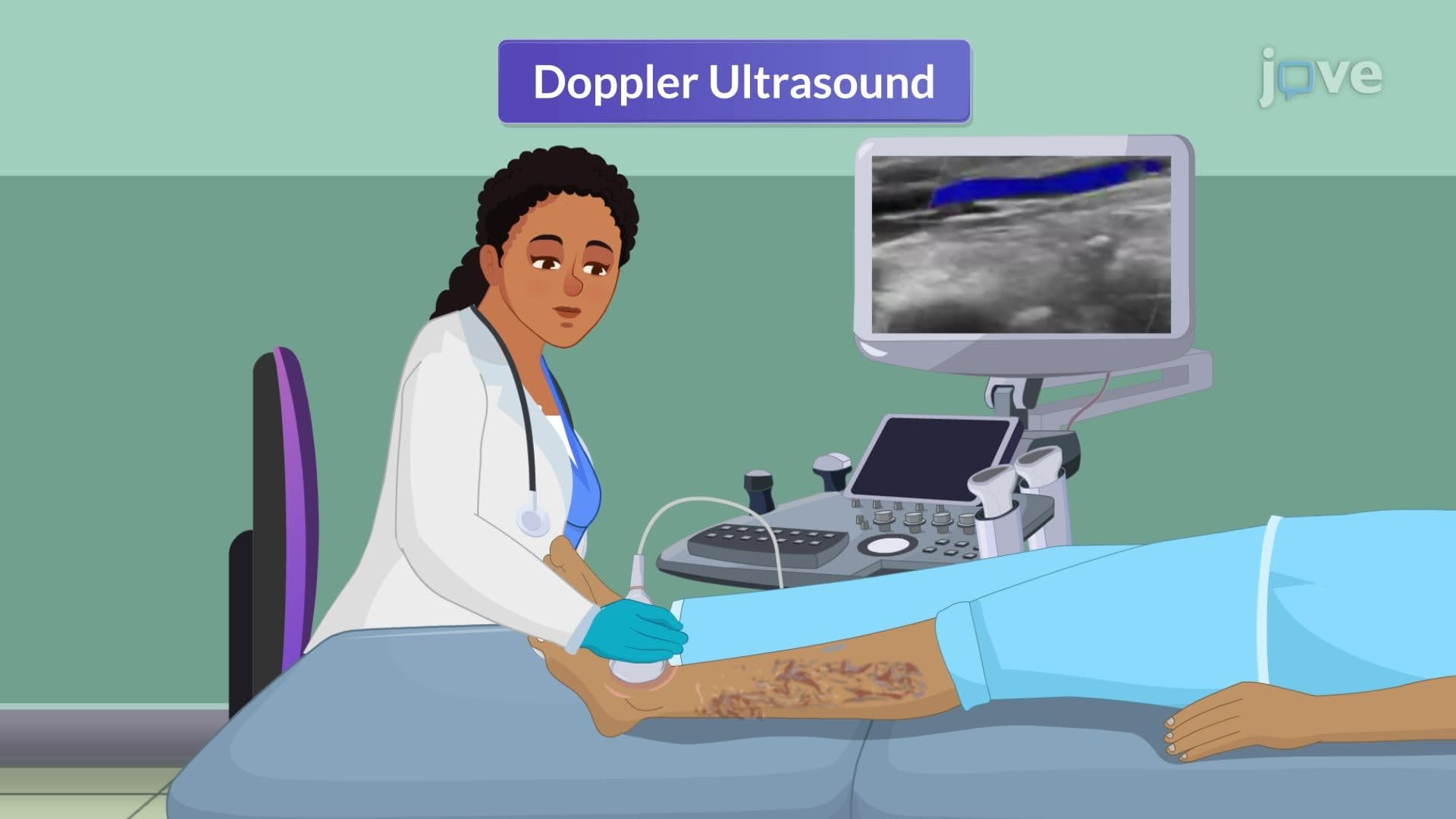
Duplex ultrasound scanning serves as the cornerstone of varicose veins diagnostic evaluation, combining real-time imaging with Doppler flow assessment. This non-invasive technique allows clinicians to visualize venous anatomy while simultaneously measuring blood flow patterns and valve function. The technology identifies incompetent valves, measures reflux duration, and maps the extent of venous disease throughout the lower extremity. Students preparing for the MCAT or nursing entrance exams like NCLEX should understand that duplex scanning provides crucial information including vein diameter, wall thickness, and the presence of blood clots or obstructions.
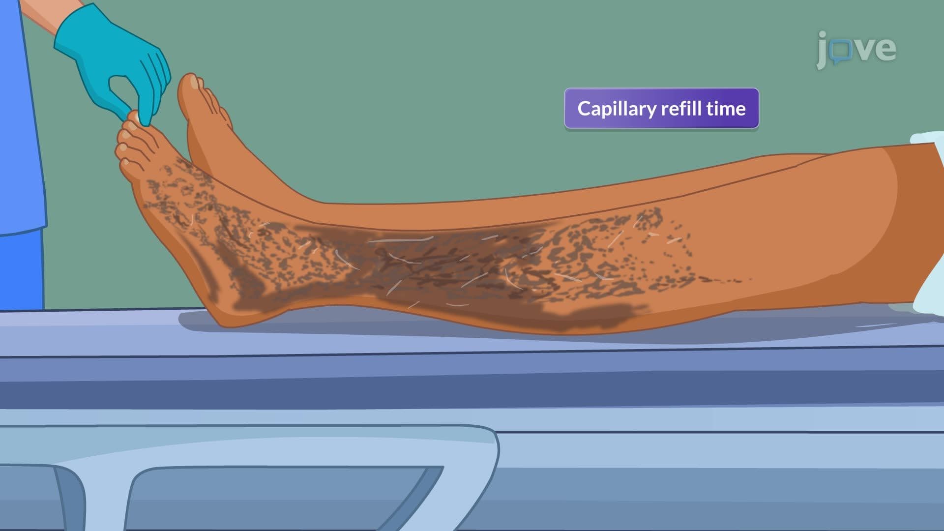
Proper diagnostic evaluation begins with comprehensive symptom assessment, as varicose veins present with characteristic patterns that guide treatment decisions. Patients typically report heaviness, aching sensations after prolonged standing, and exercise intolerance that significantly impacts daily activities. Healthcare providers document the presence of nocturnal leg cramps, localized itching or burning sensations, and progressive edema formation. These symptoms often correlate with specific anatomical locations and severity grades, information crucial for AP Biology students studying circulatory system pathophysiology.
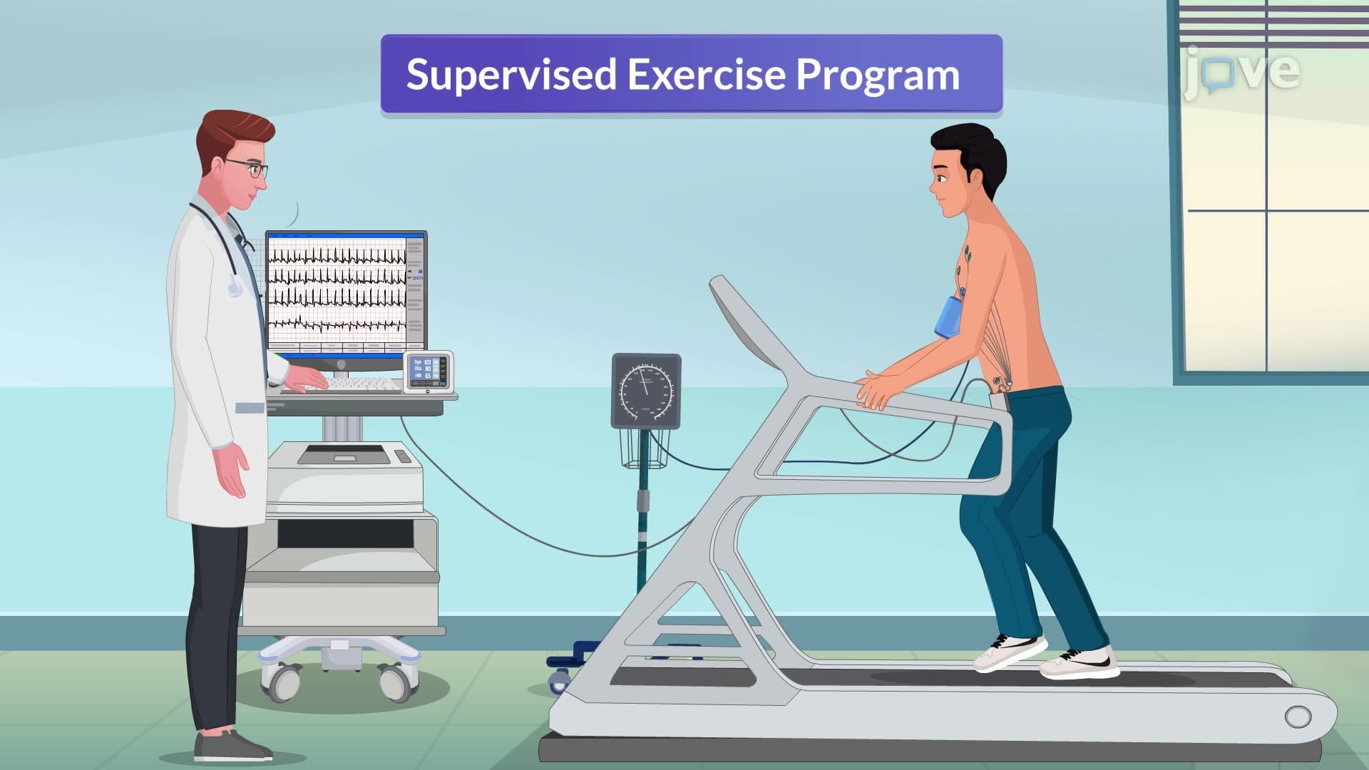
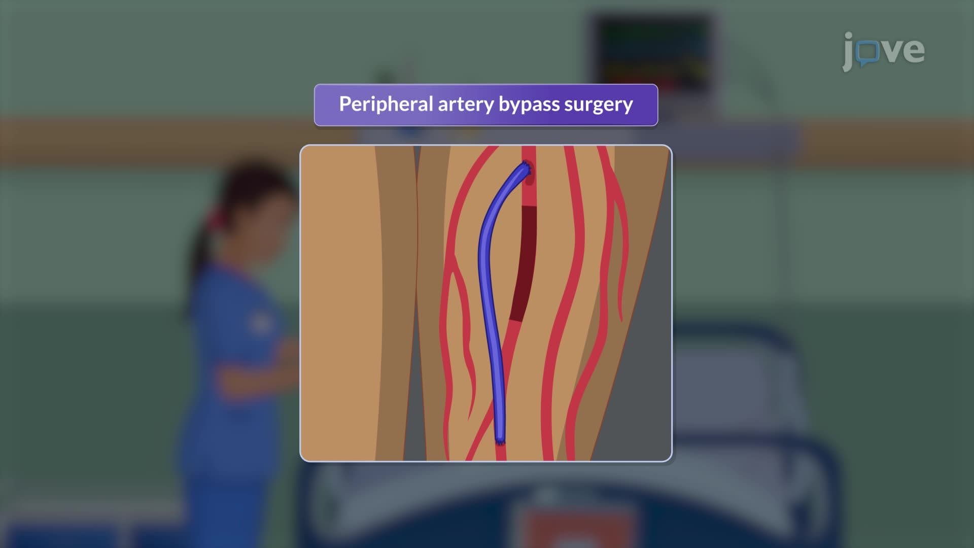
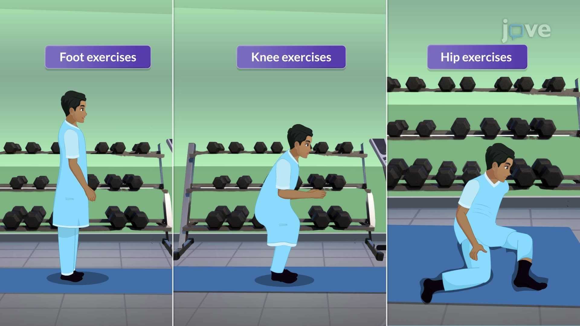
The diagnostic process directly influences treatment selection, ranging from conservative management to advanced surgical interventions. Conservative approaches include graduated compression therapy with pressures between 20-30 mmHg, structured exercise programs emphasizing calf muscle strengthening, and lifestyle modifications. Nonsurgical options progress to sclerotherapy using FDA-approved agents like polidocanol, followed by thermal ablation techniques including radiofrequency and laser treatments. Surgical interventions represent the final treatment tier, incorporating vein ligation procedures and ambulatory phlebectomy for severe cases. Understanding this treatment hierarchy proves essential for pre-med students taking college-level anatomy and physiology courses.
Related Micro-courses