Loading subject page…

3,563 views

Video Summary: Venous Thrombosis Ii Clinical Manifestations Explained
Did you know that over 900,000 Americans develop venous thrombosis annually, yet many cases go undiagnosed because symptoms can be subtle or absent? Venous Thrombosis II Clinical Manifestations Explained reveals how to recognize the critical warning signs of both superficial vein thrombosis (SVT) and deep vein thrombosis (DVT). From the characteristic cord-like feeling of SVT to the unilateral leg swelling that signals DVT, understanding these clinical presentations can be life-saving in emergency departments across the US. Watch the full video on JoVE Coach to master this concept with expert-led visuals and step-by-step explanations.
The clinical presentation of venous thrombosis varies dramatically depending on the location and extent of clot formation. While superficial vein thrombosis typically announces itself with obvious local symptoms, deep vein thrombosis can masquerade silently until life-threatening complications arise. This diagnostic challenge makes understanding clinical manifestations crucial for healthcare providers and students preparing for medical careers.
Superficial vein thrombosis (SVT) presents with a classic constellation of findings that make it relatively straightforward to diagnose. Patients experience localized pain and tenderness that follows the exact pathway of the affected vein, creating a linear pattern of discomfort. The overlying skin becomes erythematous (red) and warm to touch, while palpation reveals the hallmark finding: a firm, cord-like structure beneath the skin that represents the thrombosed vein itself.
In US emergency departments, SVT commonly affects the greater saphenous vein system in the legs, particularly in patients with varicose veins or recent IV catheter placement. The condition typically remains localized and rarely causes systemic complications, though it can extend into the deep venous system if left untreated.
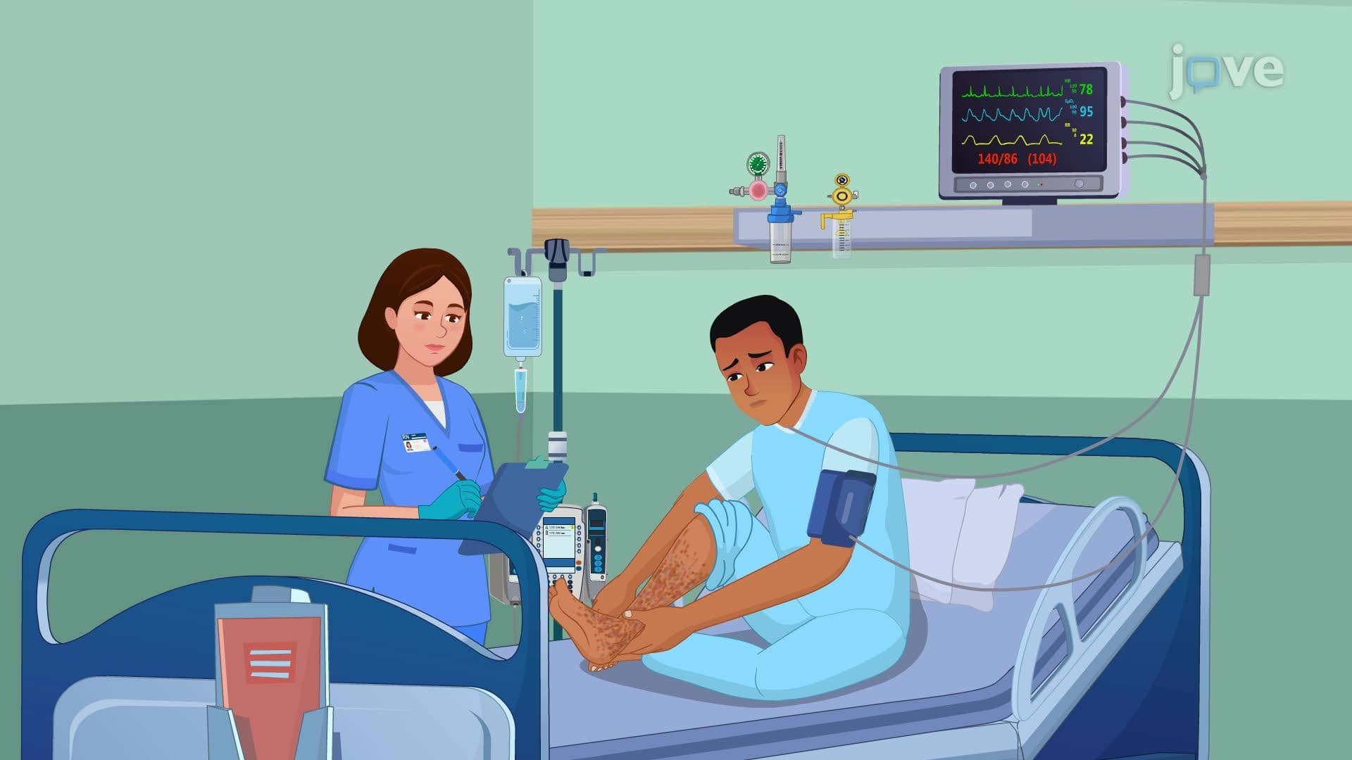
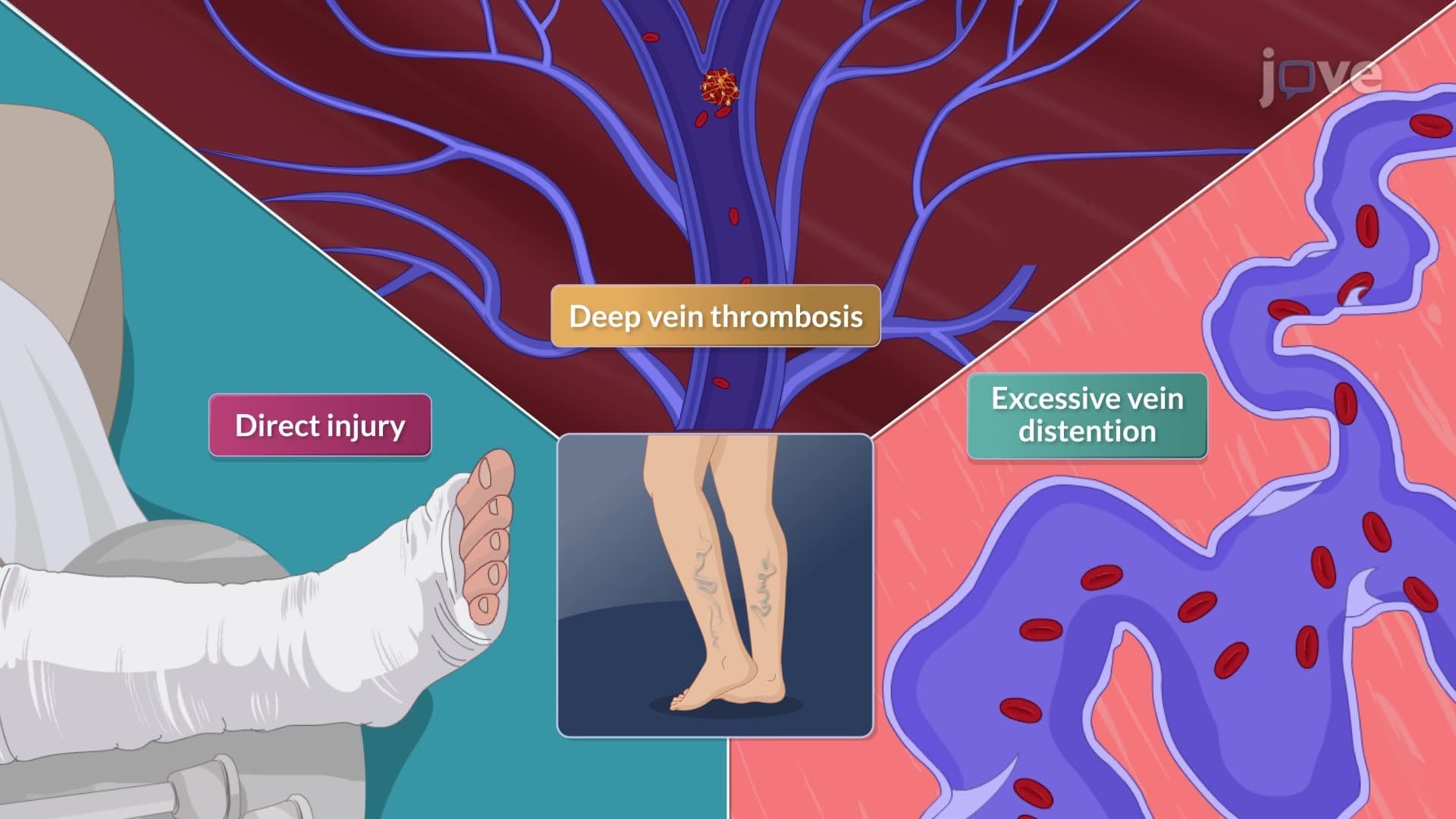
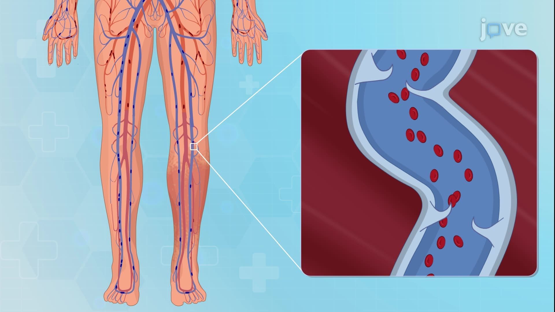
Deep vein thrombosis (DVT) presents a more complex diagnostic challenge because approximately 50% of cases are completely asymptomatic. When symptoms do occur, they typically manifest unilaterally, affecting only one leg. The classic presentation includes progressive leg swelling (edema), aching pain that may worsen with walking, and a sensation of heaviness or fullness in the affected calf or thigh.
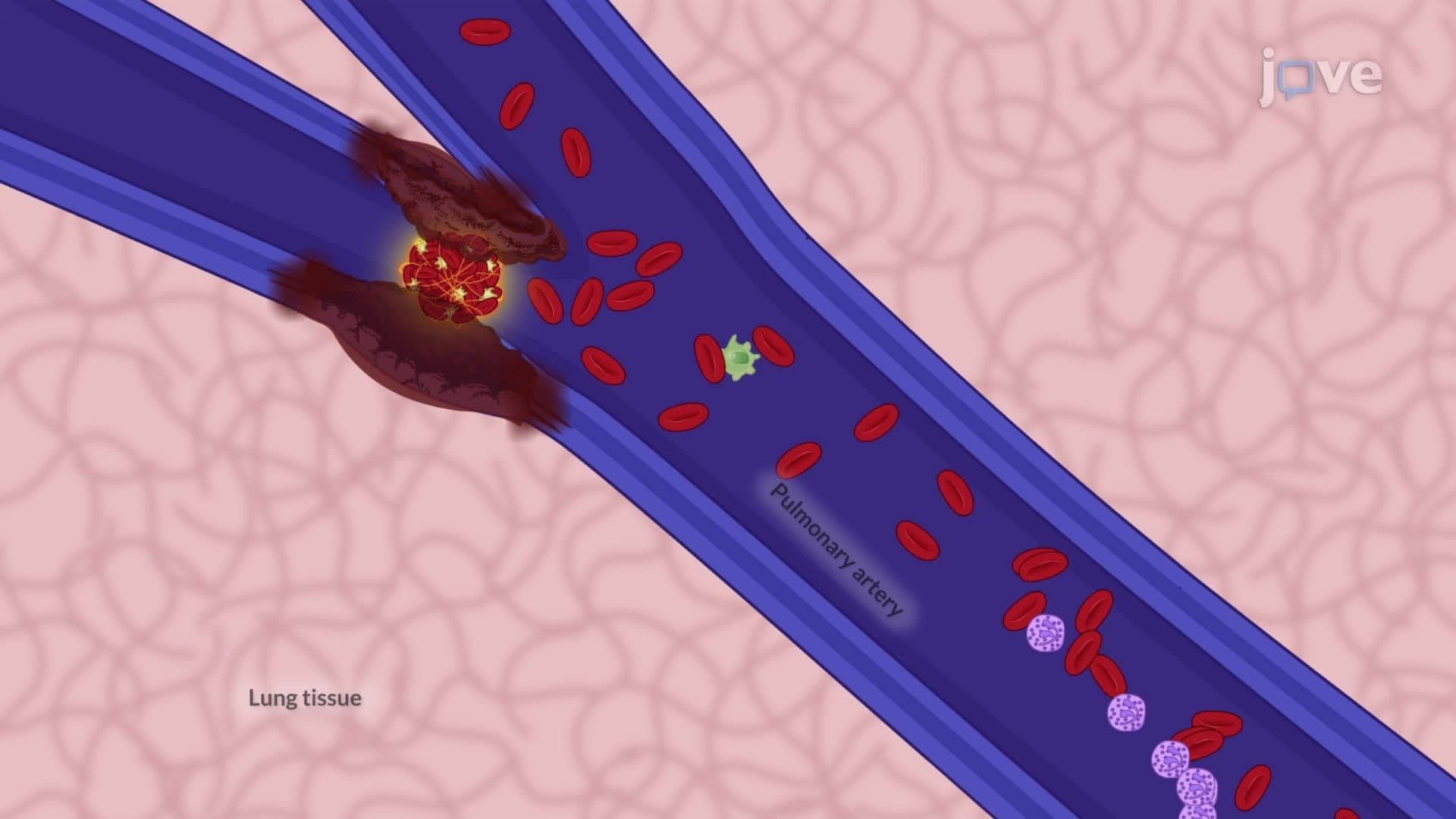
Physical examination reveals several key findings: unilateral edema, warmth over the affected area, dilated superficial veins that serve as collateral circulation, and tenderness with deep palpation. Some patients report paresthesias (tingling sensations) or cramping pain that mimics muscle strain. The absence of symptoms doesn't rule out DVT, making it particularly dangerous for conditions like pulmonary embolism.
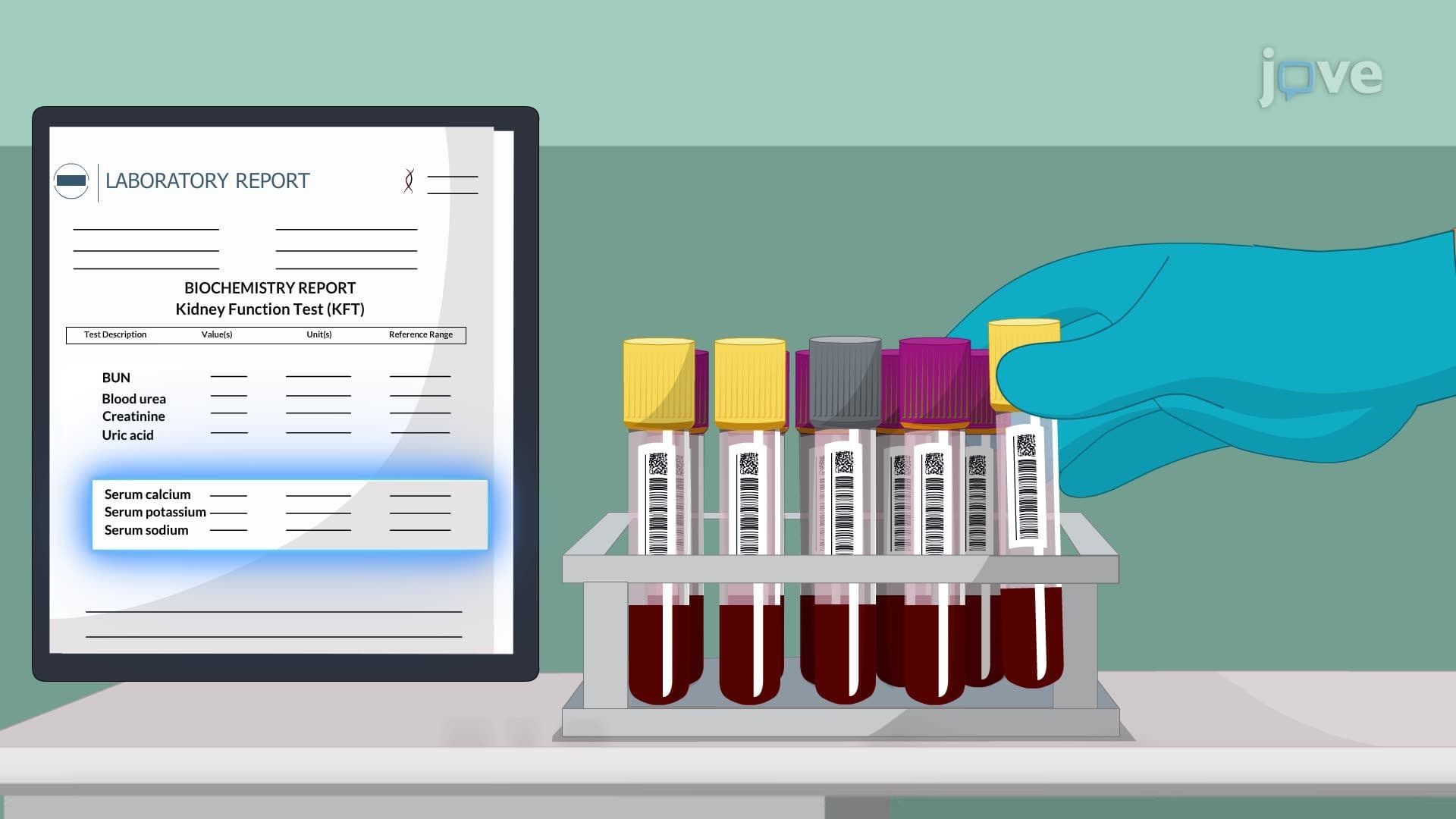
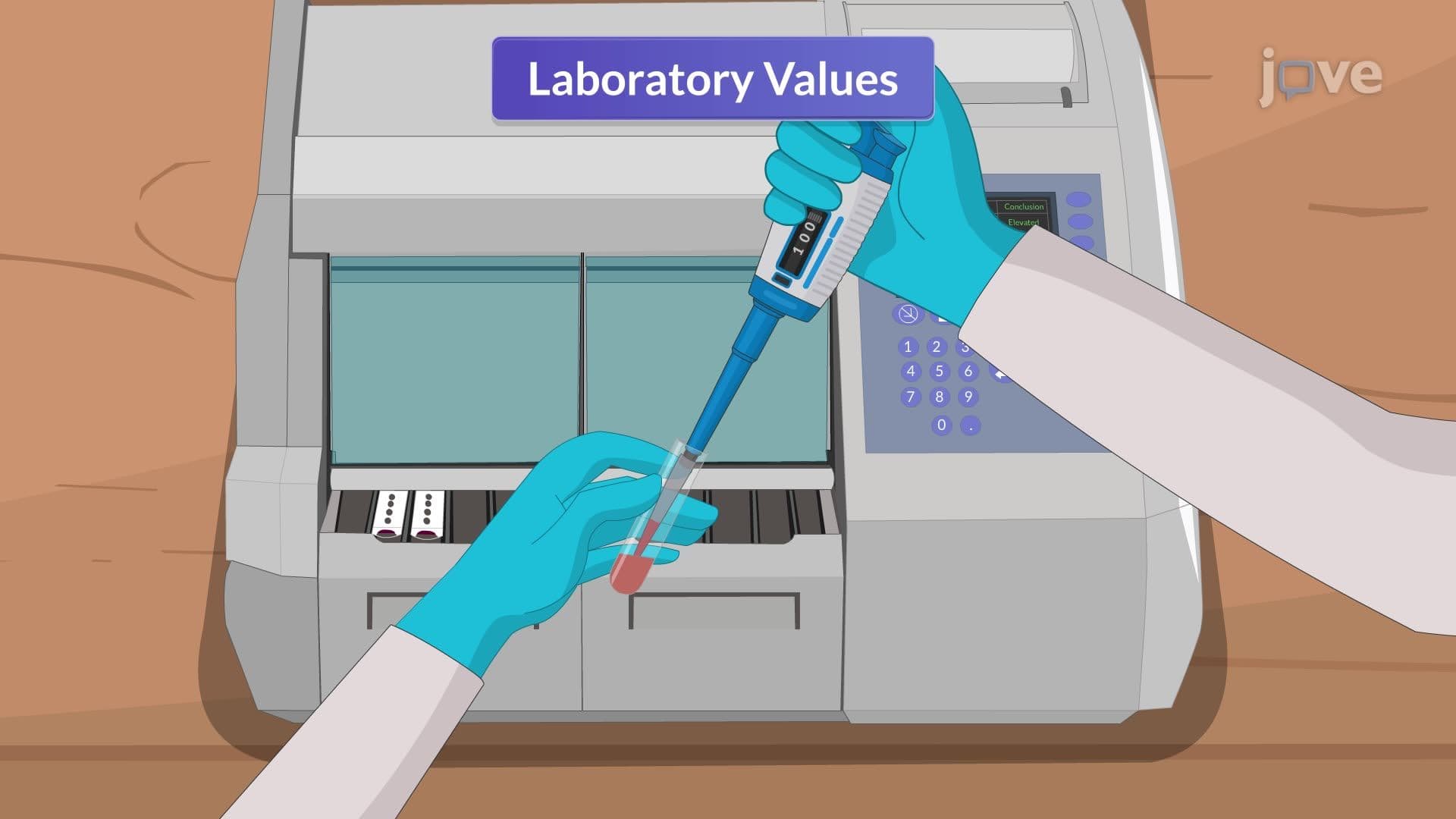
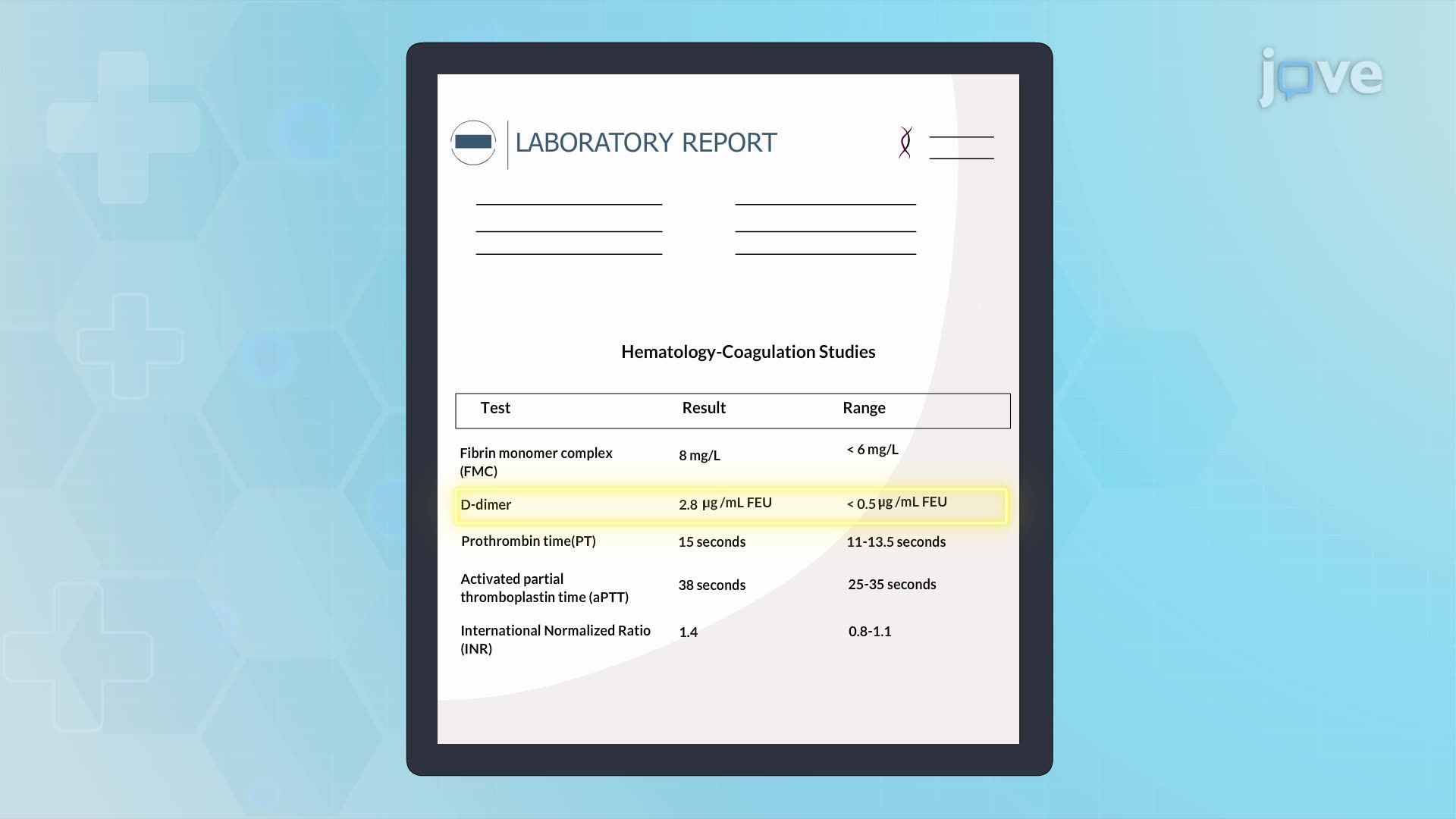
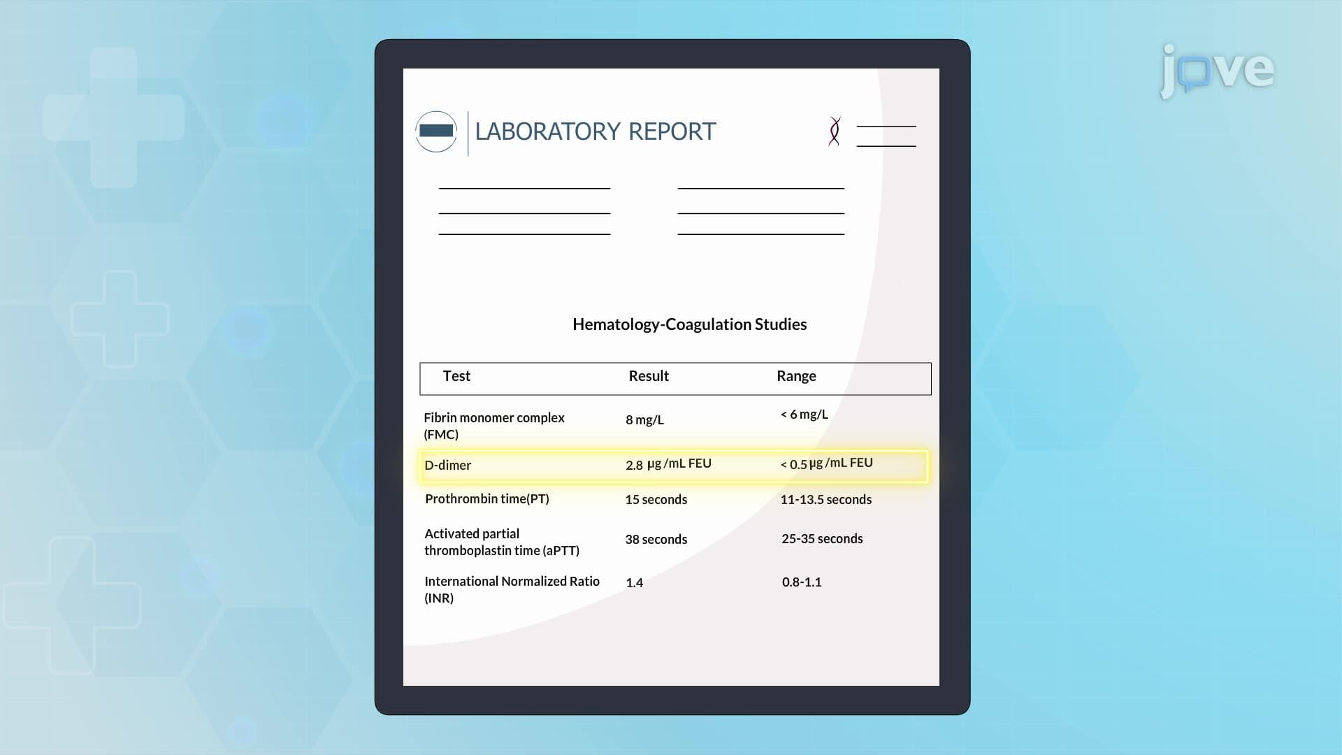
Modern diagnosis relies heavily on laboratory markers and advanced imaging. D-dimer elevation serves as a sensitive but non-specific screening tool-elevated levels suggest active clot formation but can result from many conditions including surgery, infection, or malignancy. Fibrin monomer complexes provide additional evidence of ongoing thrombosis, while coagulation studies (PT, aPTT, INR) help identify underlying bleeding disorders that predispose to clot formation.
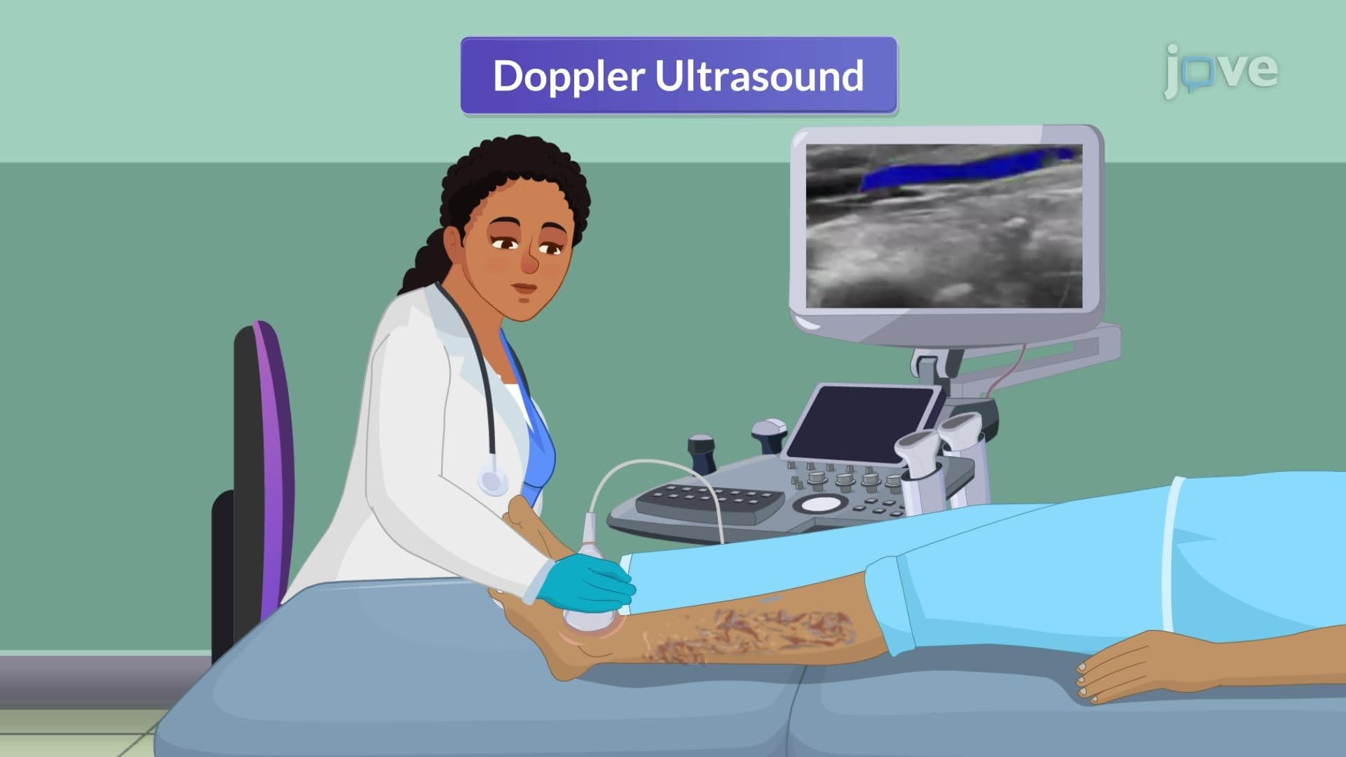
Duplex ultrasound has become the gold standard for DVT diagnosis in US hospitals, combining B-mode imaging with Doppler flow assessment. The test evaluates vein compressibility-healthy veins collapse completely under gentle pressure, while thrombosed veins remain rigid. CT venography with contrast offers superior visualization of pelvic and iliac veins, particularly important for detecting clots that might be missed on ultrasound. MR venography provides excellent soft tissue contrast without radiation exposure, making it valuable for pregnant patients or those with contrast allergies.
For students preparing for the MCAT or USMLE Step 1, understanding the diagnostic approach helps differentiate venous thrombosis from conditions like cellulitis, muscle strain, or lymphedema that can present with similar symptoms.
Related Micro-courses