Loading subject page…


112,207 views

Video Summary: What are Heart Valves
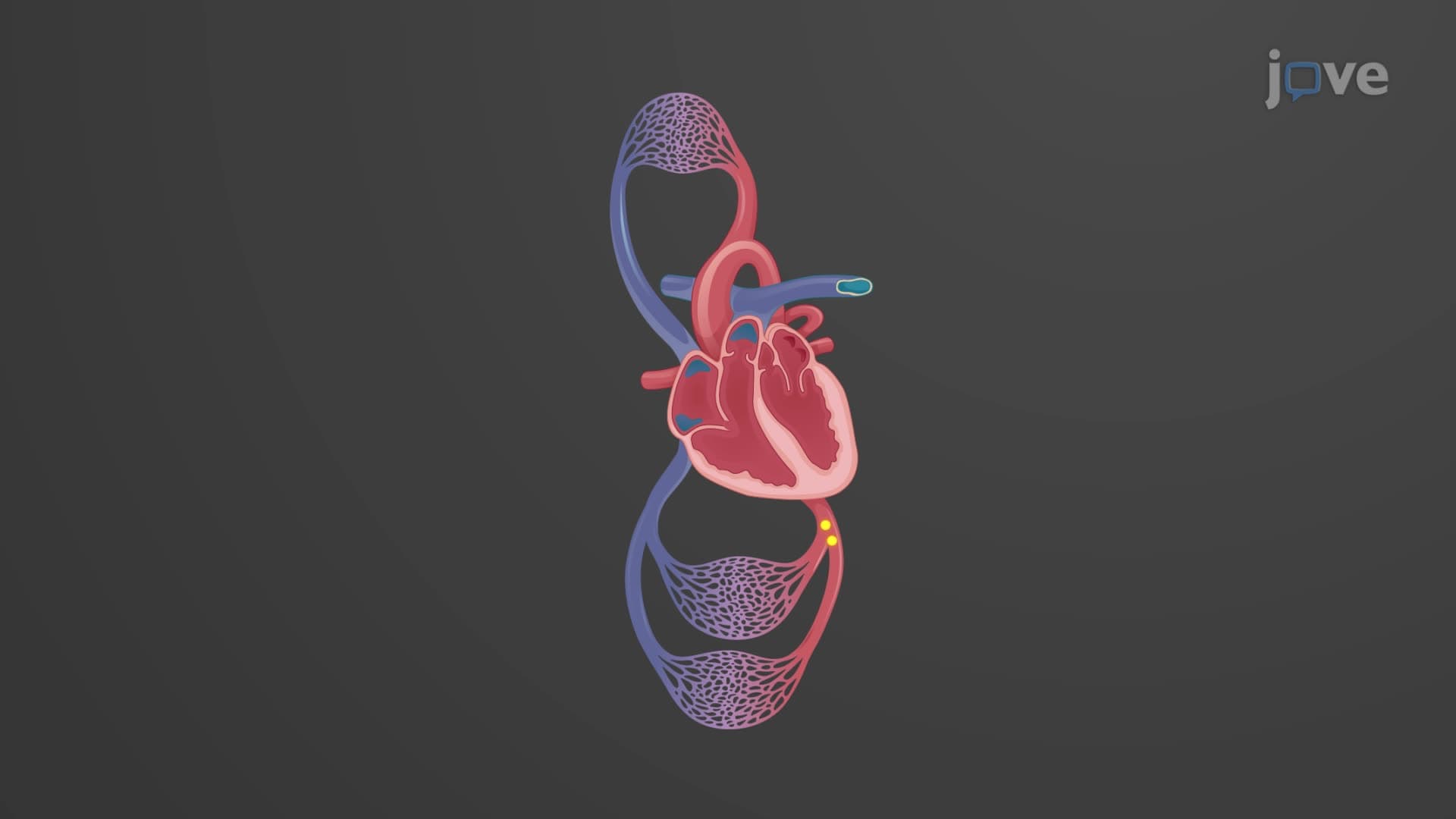
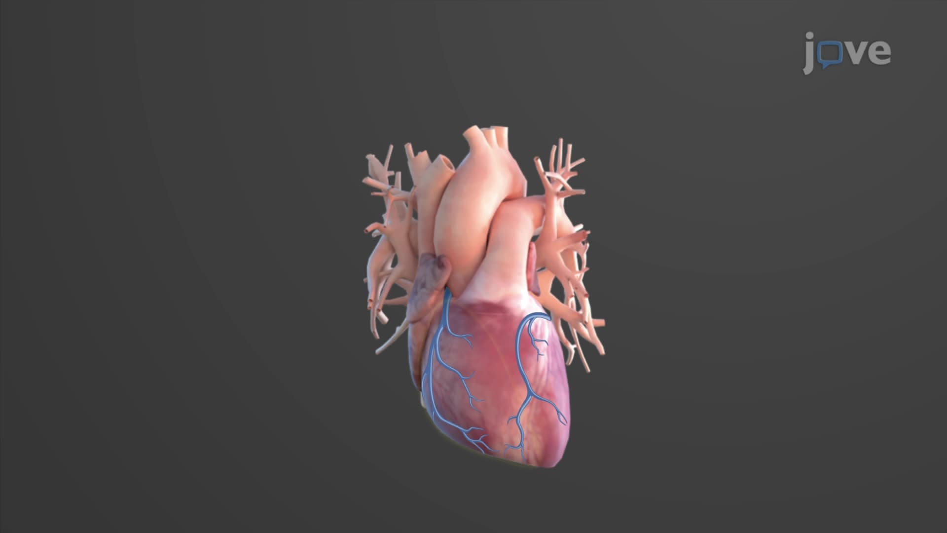
Every second of your life, four specialized structures prevent your blood from flowing backward through your heart-imagine the chaos if blood could reverse direction during each heartbeat! Understanding heart valves anatomy biology reveals how these remarkable one-way gates coordinate perfectly to maintain circulation. In the US, over 5 million Americans live with heart valve disease, making this knowledge crucial for future healthcare professionals. What are heart valves becomes clear when you see how they respond to pressure changes during each cardiac cycle. Watch the full video on JoVE Coach to master this concept with expert-led visuals and step-by-step explanations.
Heart valves anatomy biology centers on four specialized structures that function as one-way gates, ensuring blood flows in the correct direction through your cardiovascular system. These valves respond dynamically to pressure changes created by the heart's rhythmic contractions, opening and closing approximately 100,000 times daily. For students preparing for the MCAT or AP Biology exams, understanding valve mechanics provides crucial insight into cardiovascular physiology and common clinical conditions.
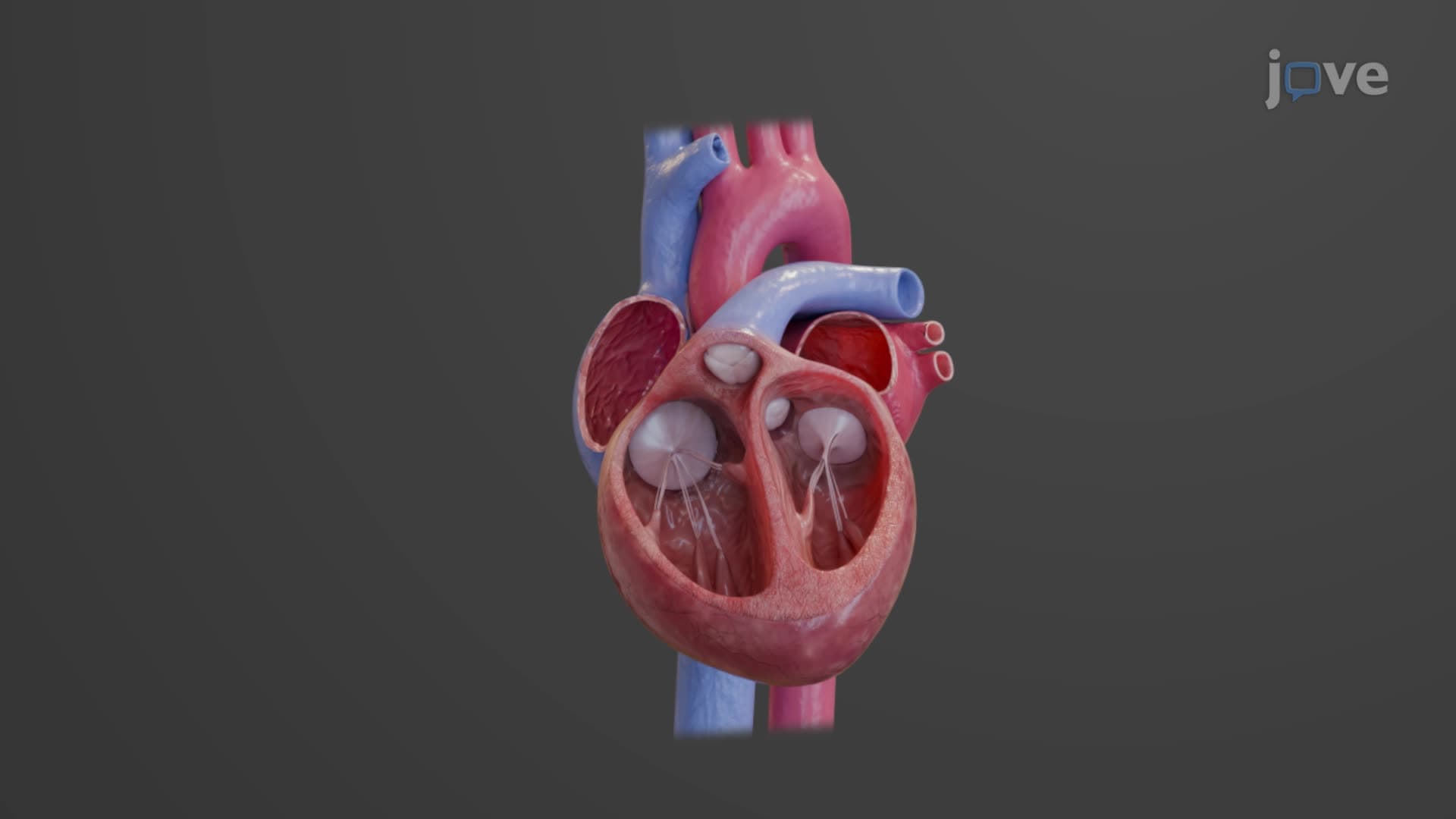
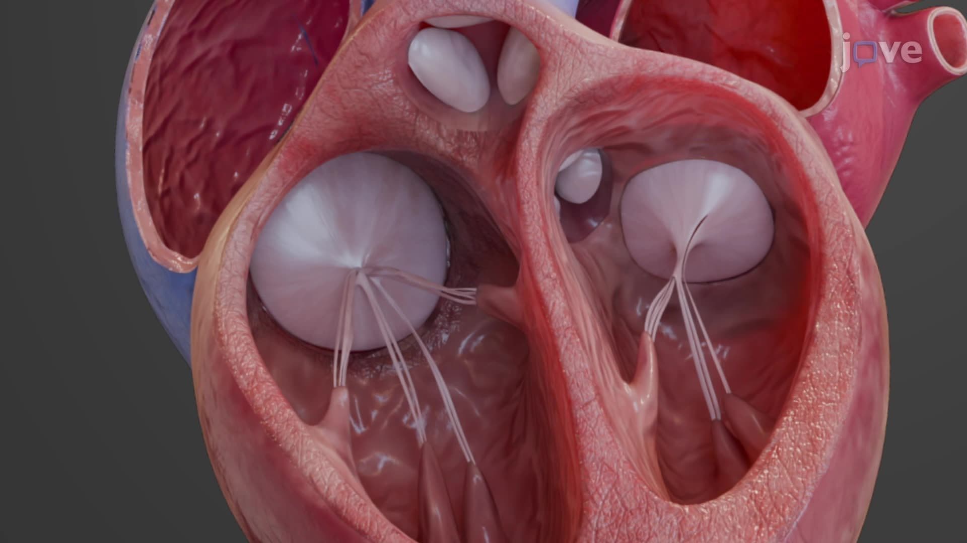
The atrioventricular (AV) valves separate the upper and lower heart chambers, preventing backflow during ventricular contraction. The tricuspid valve guards the right side of the heart, featuring three leaflets that allow blood flow from the right atrium to the right ventricle. Its counterpart, the mitral valve (also called bicuspid), has two leaflets and controls flow between the left atrium and left ventricle.
What makes AV valves particularly fascinating is their support system. Papillary muscles project from the ventricular walls, connected to valve leaflets by fibrous cords called chordae tendineae. During ventricular systole, these "heartstrings" prevent the valves from inverting into the atria-a condition called prolapse. This mechanism is frequently tested on college physiology exams and appears in USMLE Step 1 questions about cardiac pathology.
The semilunar valves, named for their half-moon shaped cusps, control blood exit from the ventricles. The aortic valve sits between the left ventricle and ascending aorta, while the pulmonary valve separates the right ventricle from the pulmonary trunk. Unlike AV valves, semilunar valves lack papillary muscle support, instead relying on their unique cusp design for proper closure.
During ventricular contraction, rising pressure forces these valves open, allowing blood to rush into the systemic and pulmonary circulations. When the ventricles relax, blood flowing backward fills the valve cusps, creating the characteristic "dub" sound of S2 heart sound that medical students learn to identify during clinical rotations at institutions like Johns Hopkins or Mayo Clinic.
Understanding normal valve function becomes critical when studying cardiovascular diseases prevalent in the US population. Aortic stenosis affects over 2.5 million Americans, while mitral valve prolapse occurs in approximately 2-3% of the population. These conditions appear frequently in NCLEX-RN questions and medical school case studies, making thorough knowledge of normal valve anatomy essential for healthcare career preparation.
Related Micro-courses