Loading subject page…


112,207 views


Video Summary: What are Layers Heart Wall
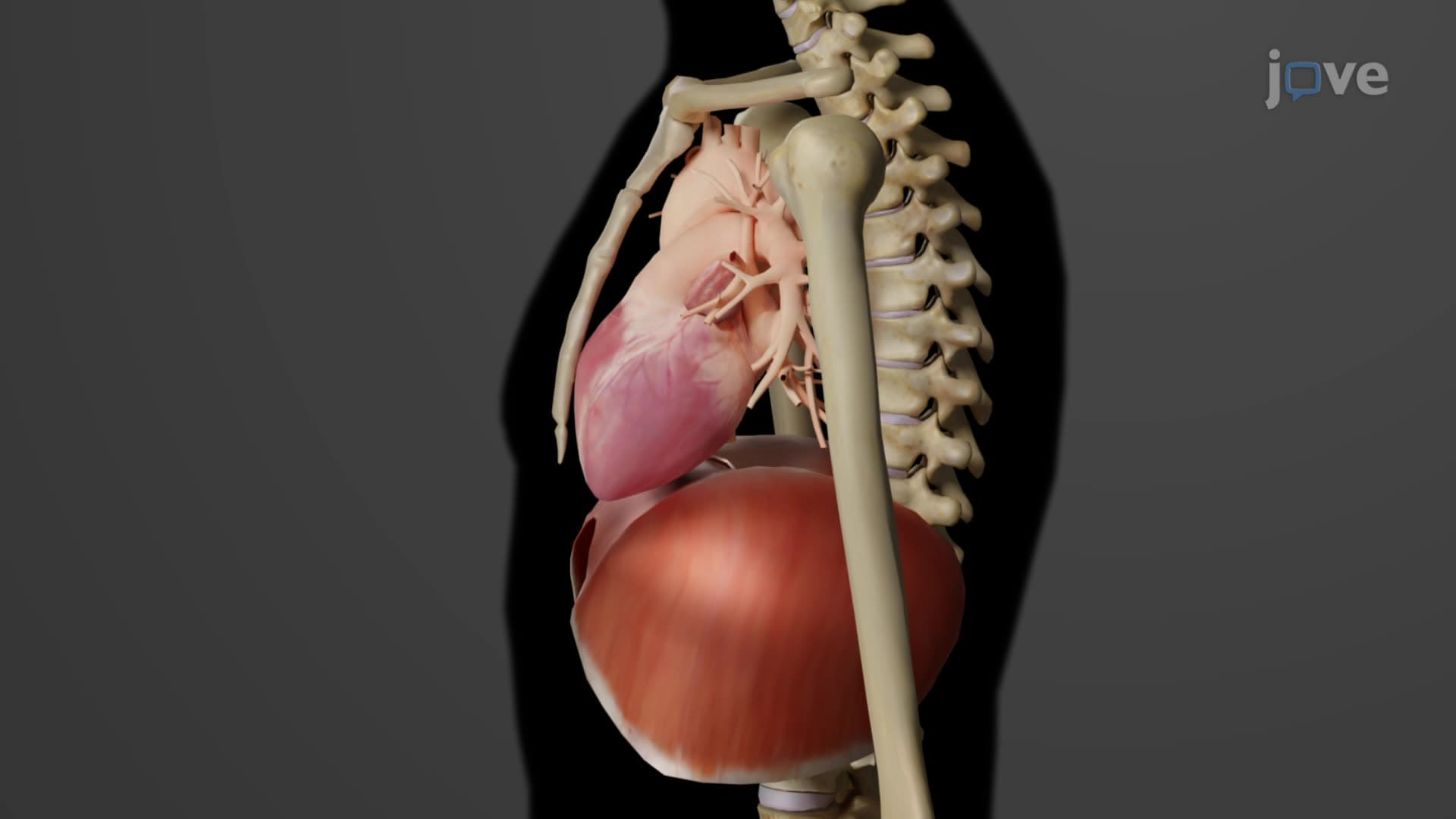
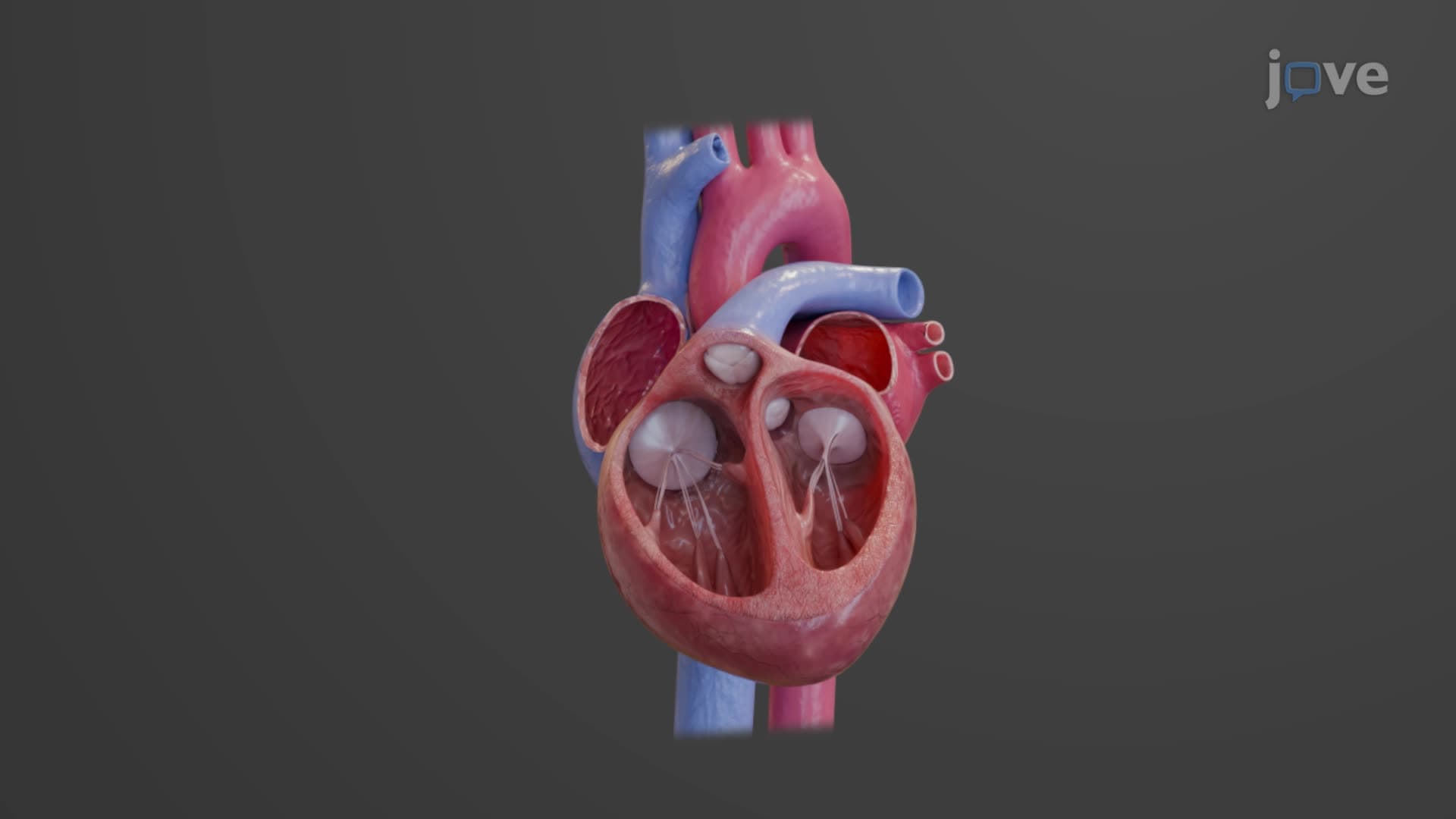
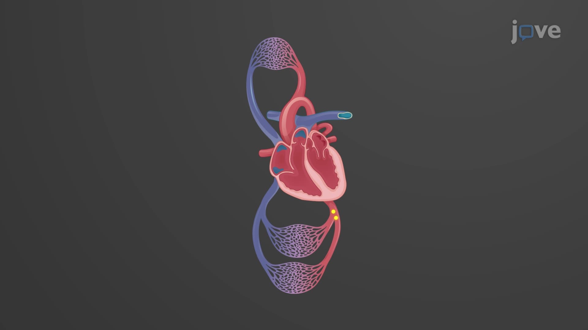
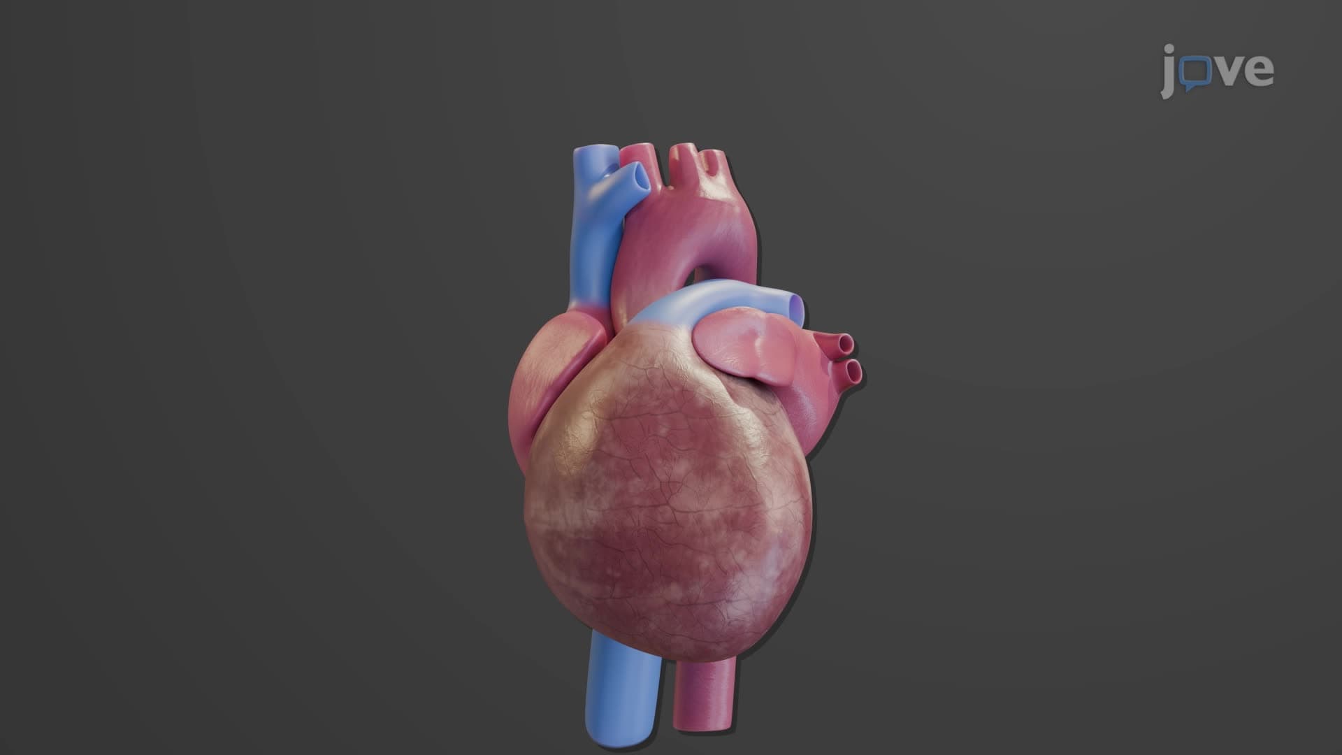
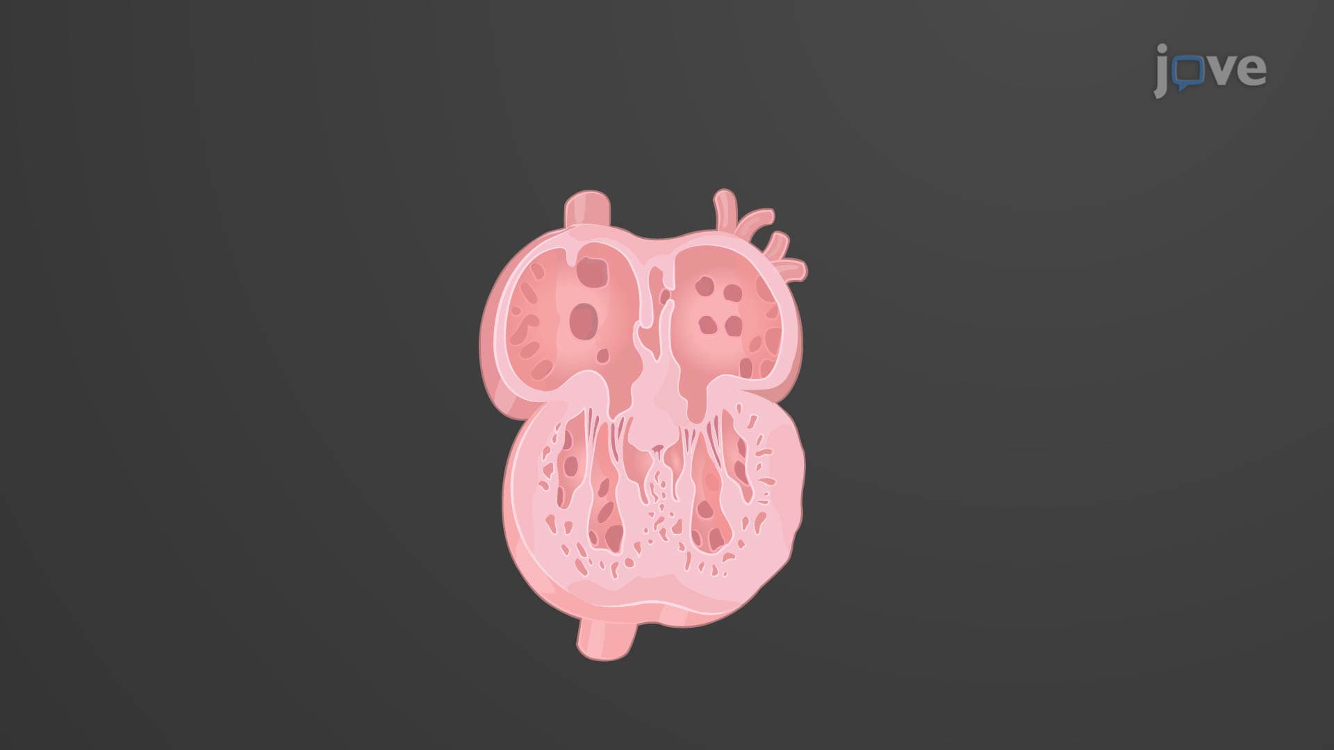
Every heartbeat you feel is powered by a three-layered muscular wall working in perfect synchrony. The heart wall layers anatomy includes the epicardium, myocardium, and endocardium-each with distinct functions that keep your cardiovascular system running efficiently. Consider that the average American heart beats over 100,000 times daily, with each layer of the what are the layers of the heart wall structure playing a critical role in this life-sustaining process. Watch the full video on JoVE Coach to master this concept with expert-led visuals and step-by-step explanations.
The human heart's remarkable ability to pump blood continuously throughout your lifetime depends on its sophisticated three-layered wall structure. Each layer of the heart wall layers anatomy serves specific physiological functions that work together to maintain cardiovascular homeostasis. Understanding this architecture is fundamental for students pursuing careers in medicine, nursing, or biomedical sciences.
The epicardium represents the heart's outermost defensive barrier, functioning as the visceral layer of the pericardium heart wall system. This layer consists of two distinct components: a thin, transparent mesothelial surface that provides a smooth gliding interface, and an underlying areolar connective tissue layer rich in adipose deposits. These fat deposits naturally increase with age, which explains why older adults often show more prominent epicardial fat on cardiac imaging studies used in US hospitals like Mayo Clinic or Johns Hopkins.
The epicardium's clinical significance becomes apparent in conditions like pericarditis, where inflammation of this layer can cause severe chest pain. Students preparing for the MCAT or nursing entrance exams should note that the epicardium's smooth surface reduces friction during cardiac contractions-a concept frequently tested in cardiovascular physiology questions.
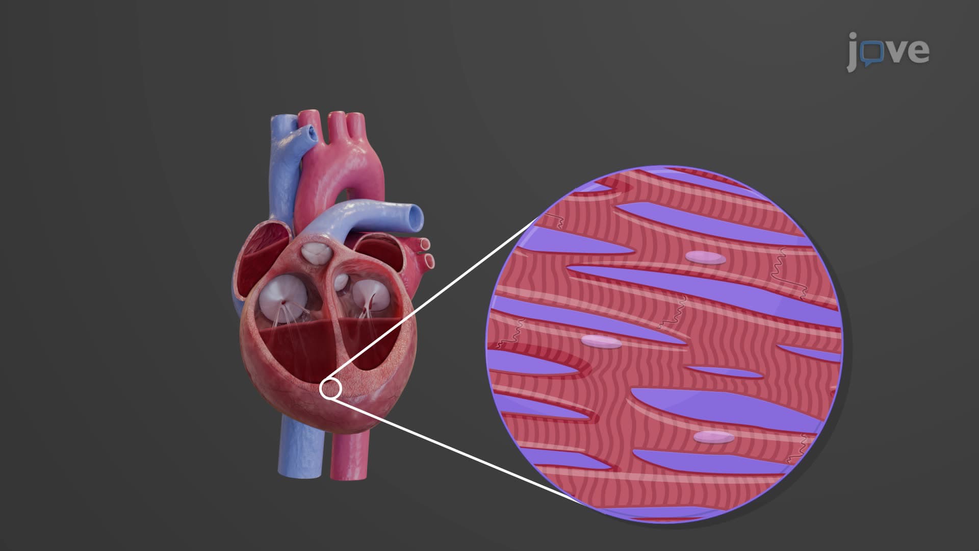
The myocardium forms the heart's thickest and most functionally critical layer, composed of specialized cardiac muscle cells called cardiomyocytes. These cells interconnect through intercalated discs, creating a syncytium that allows coordinated electrical conduction and mechanical contraction. The cardiac layer anatomy of the myocardium includes crisscrossing connective tissue fibers that provide structural integrity while allowing flexibility during the cardiac cycle.
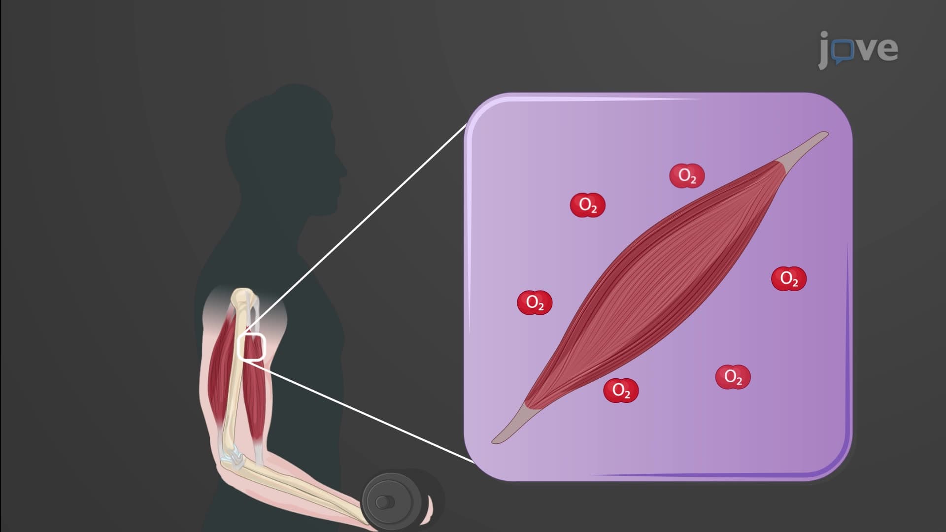
For AP Biology students, understanding myocardial function is essential for grasping concepts like cardiac output, stroke volume, and the Frank-Starling mechanism. The myocardium's unique properties-including automaticity, rhythmicity, and contractility-distinguish it from skeletal muscle tissue, making it a favorite topic for college-level physiology exams.
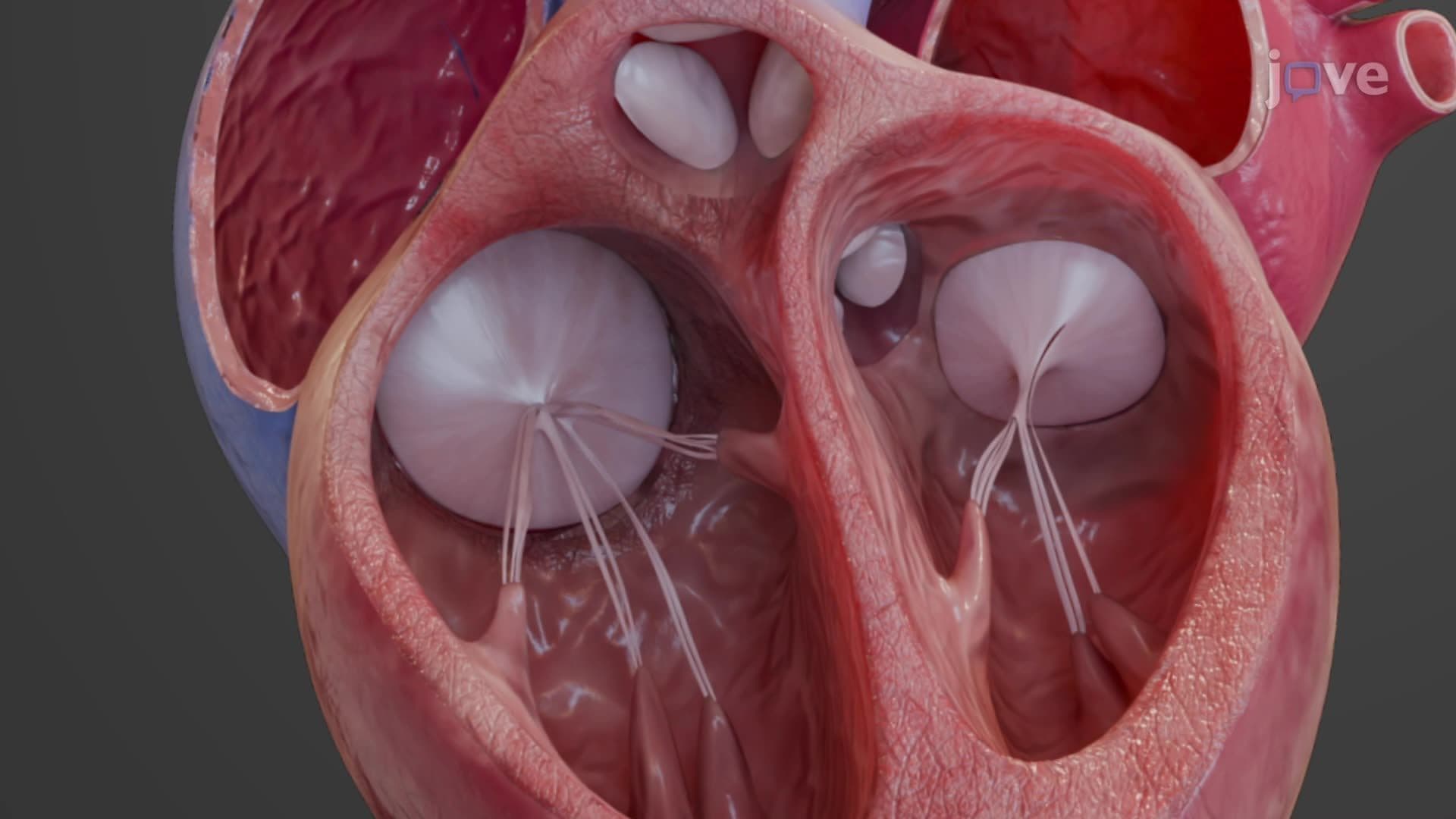
The endocardium creates the heart's smooth internal lining, consisting of endothelial cells overlying connective tissue. This specialized surface minimizes blood clot formation and reduces turbulence during blood flow-critical factors in preventing cardiovascular complications. The heart wall composition also includes the cardiac skeleton, four fibrous connective tissue rings that anchor the heart valves and provide electrical isolation between atria and ventricles.
Related Micro-courses