Loading video…


 19 Concepts
 112.2K views

Video Summary: What Is Chambers of the Heart
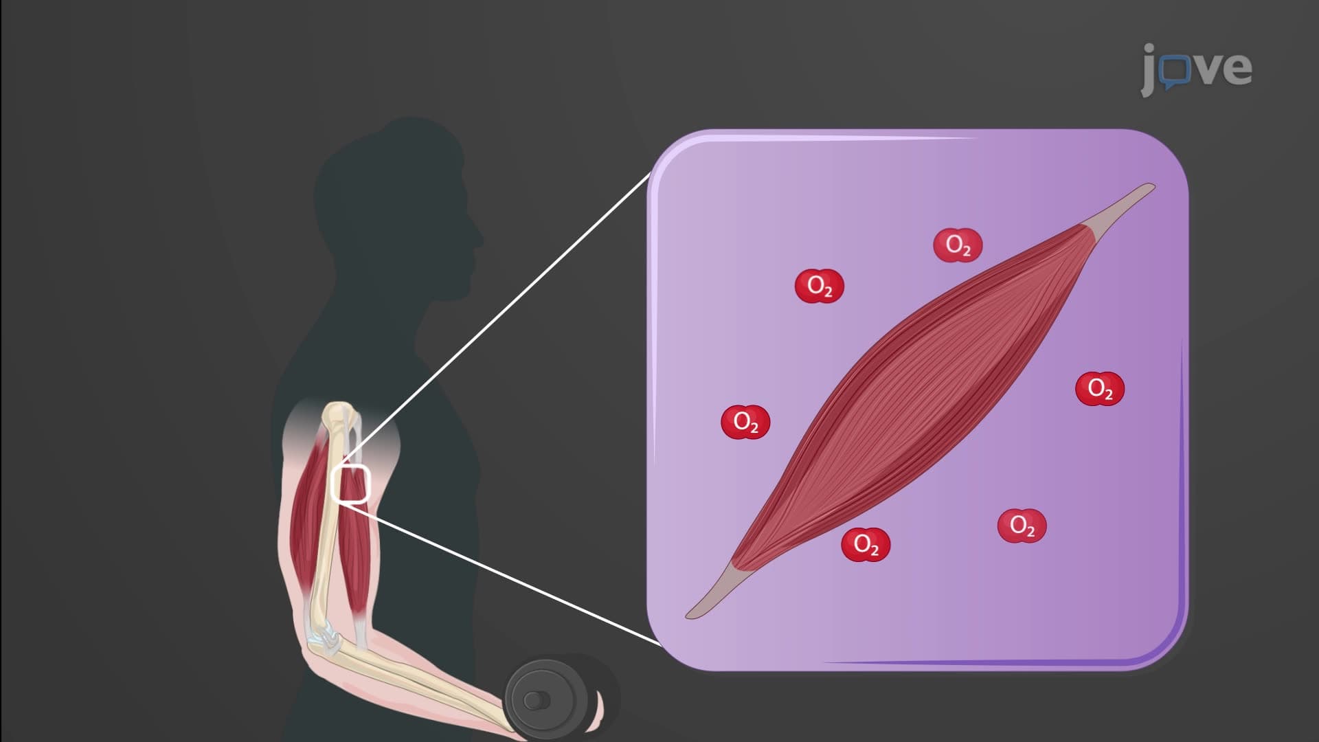
Every minute, your heart beats approximately 70 times, pumping 5 liters of blood through a four-chambered system so efficient that NASA studies it for spacecraft design. Heart chambers anatomy biology reveals how the right atrium, right ventricle, left atrium, and left ventricle work together as a dual-pump system. For instance, during a typical college basketball game at Duke University, a player's heart chambers coordinate to pump over 600 liters of blood to meet increased oxygen demands. Watch the full video on JoVE Coach to master this concept with expert-led visuals and step-by-step explanations.
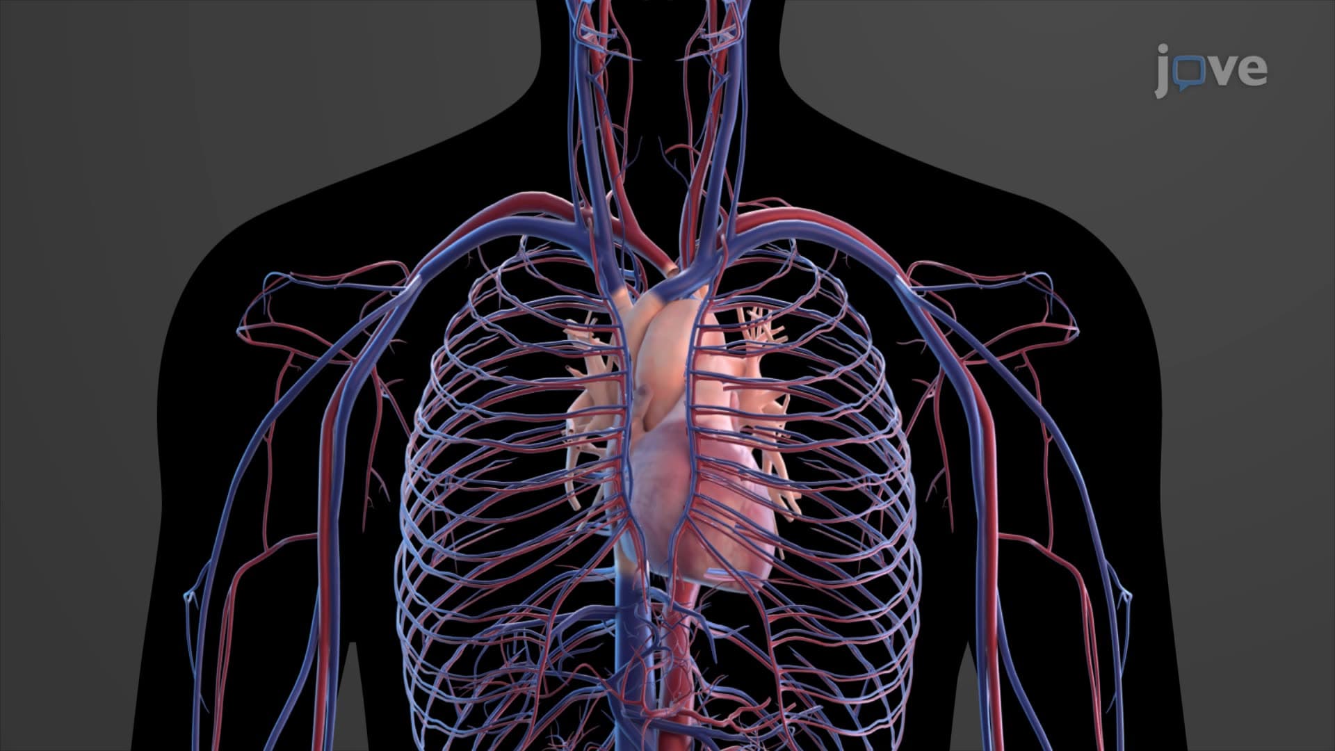
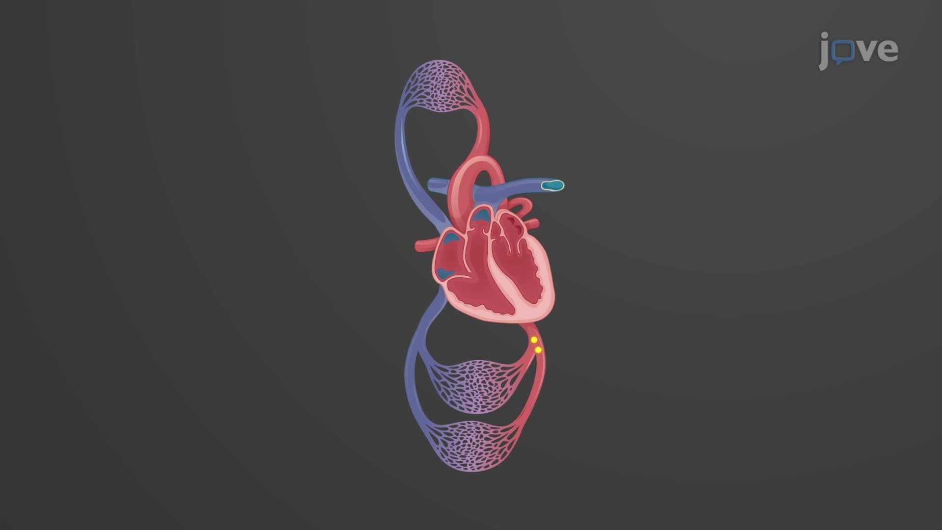
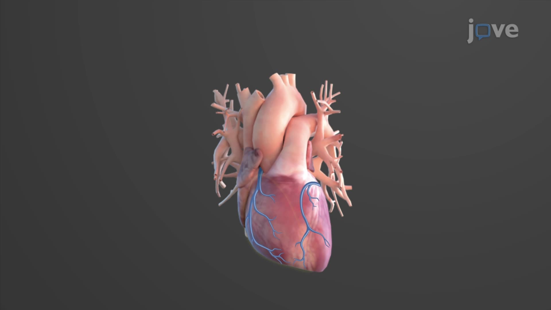
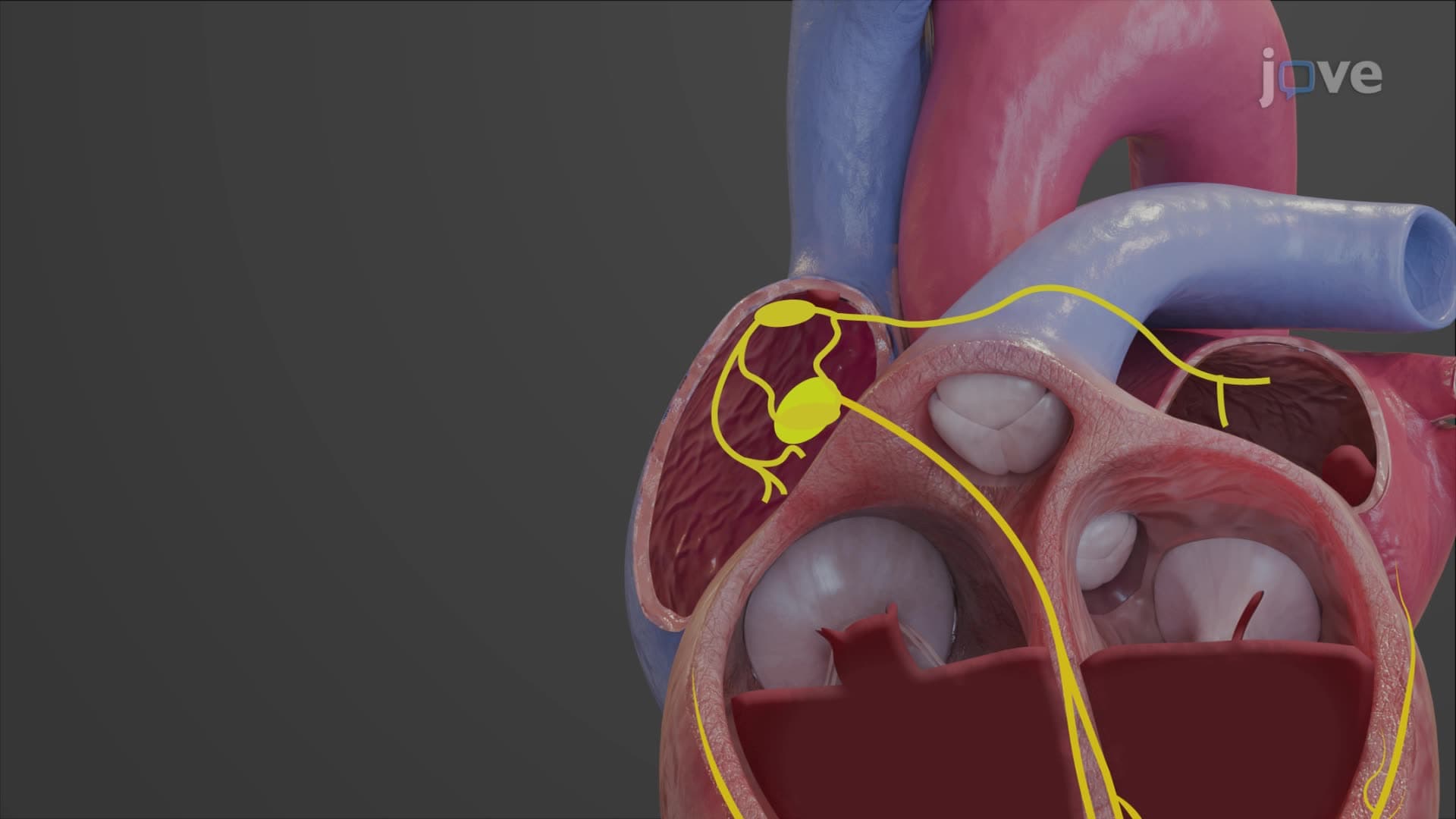
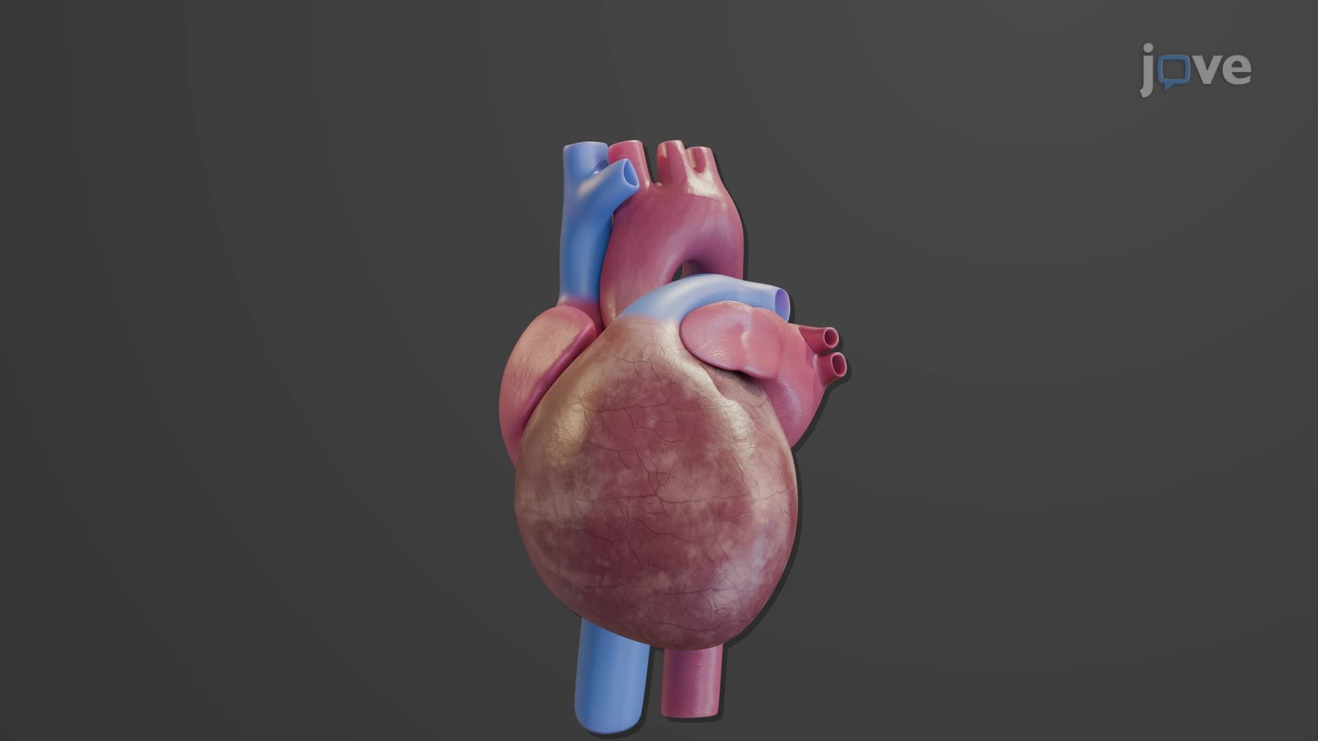
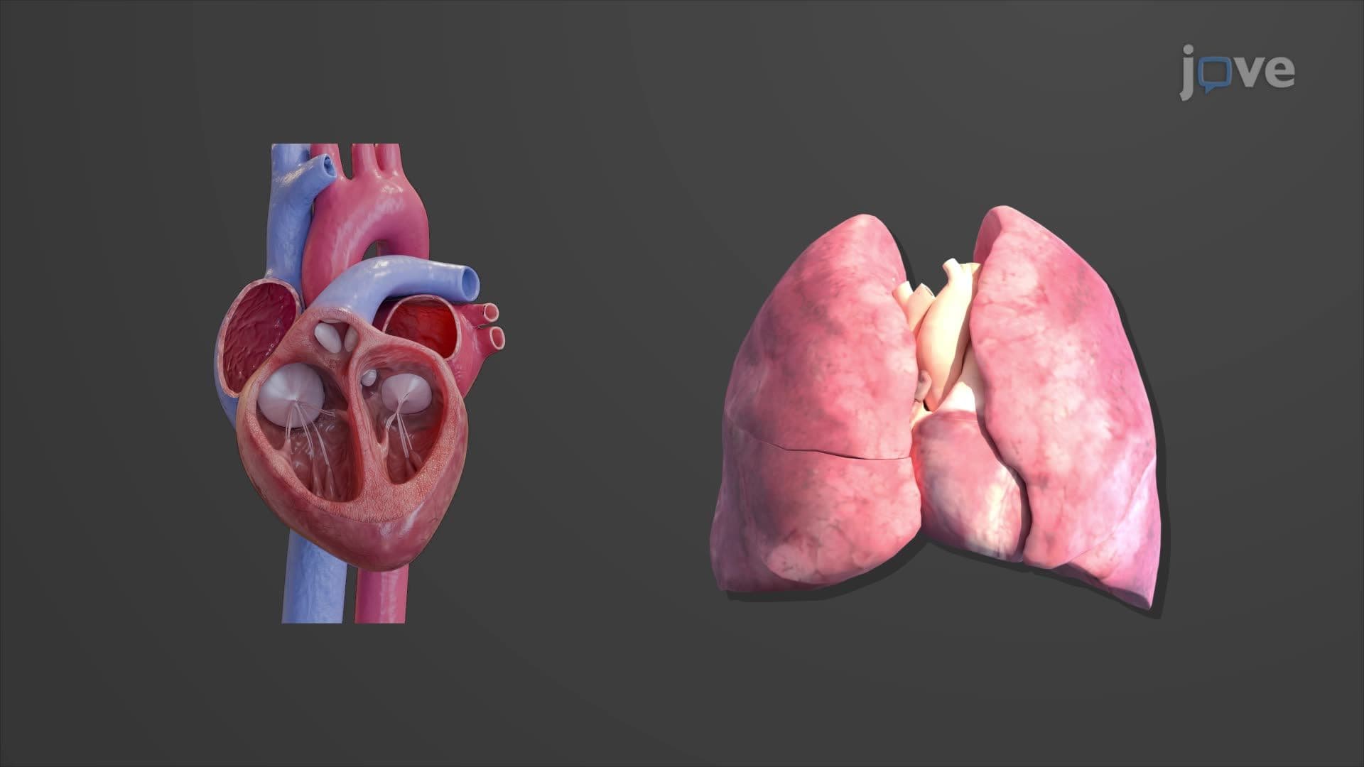
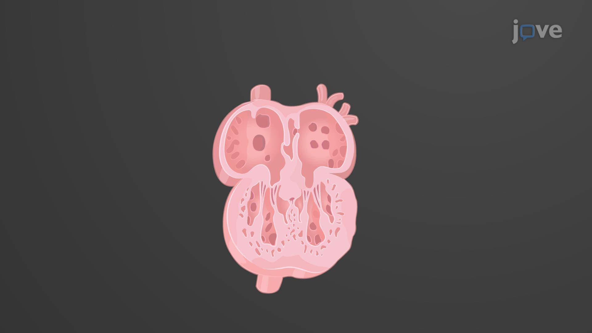
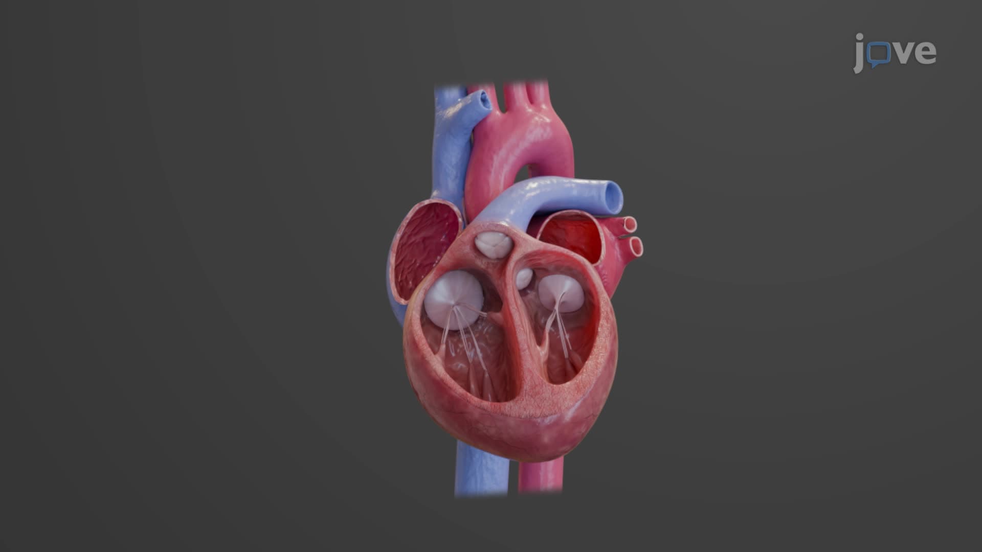
The human heart's four-chamber design represents millions of years of evolutionary refinement, creating the most efficient pump in the animal kingdom. This cardiac chamber anatomy divides into two functional sides: the right heart handles deoxygenated blood from the body and sends it to the lungs, while the left heart receives oxygenated blood from the lungs and distributes it throughout the body. Each side contains an atrium (receiving chamber) and a ventricle (pumping chamber), making four heart chambers total.
The atria serve as collection points for returning blood. The right atrium receives deoxygenated blood through the superior and inferior vena cavae, while the left atrium collects oxygenated blood from the four pulmonary veins. Both atria feature auricles-ear-like appendages that increase blood storage capacity during filling. The interatrial septum separates these chambers, and in fetal development, the foramen ovale allows blood to bypass the non-functioning lungs. After birth, this opening closes, leaving behind the fossa ovalis-a clinically important landmark that emergency physicians at hospitals like Johns Hopkins use during cardiac catheterization procedures.
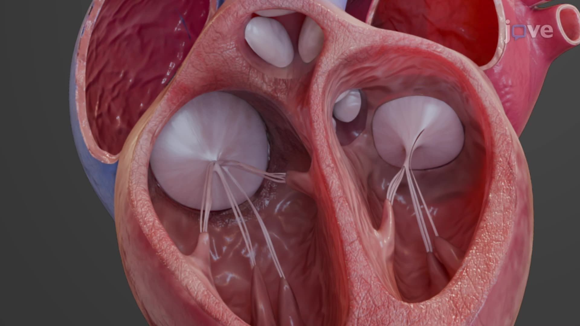
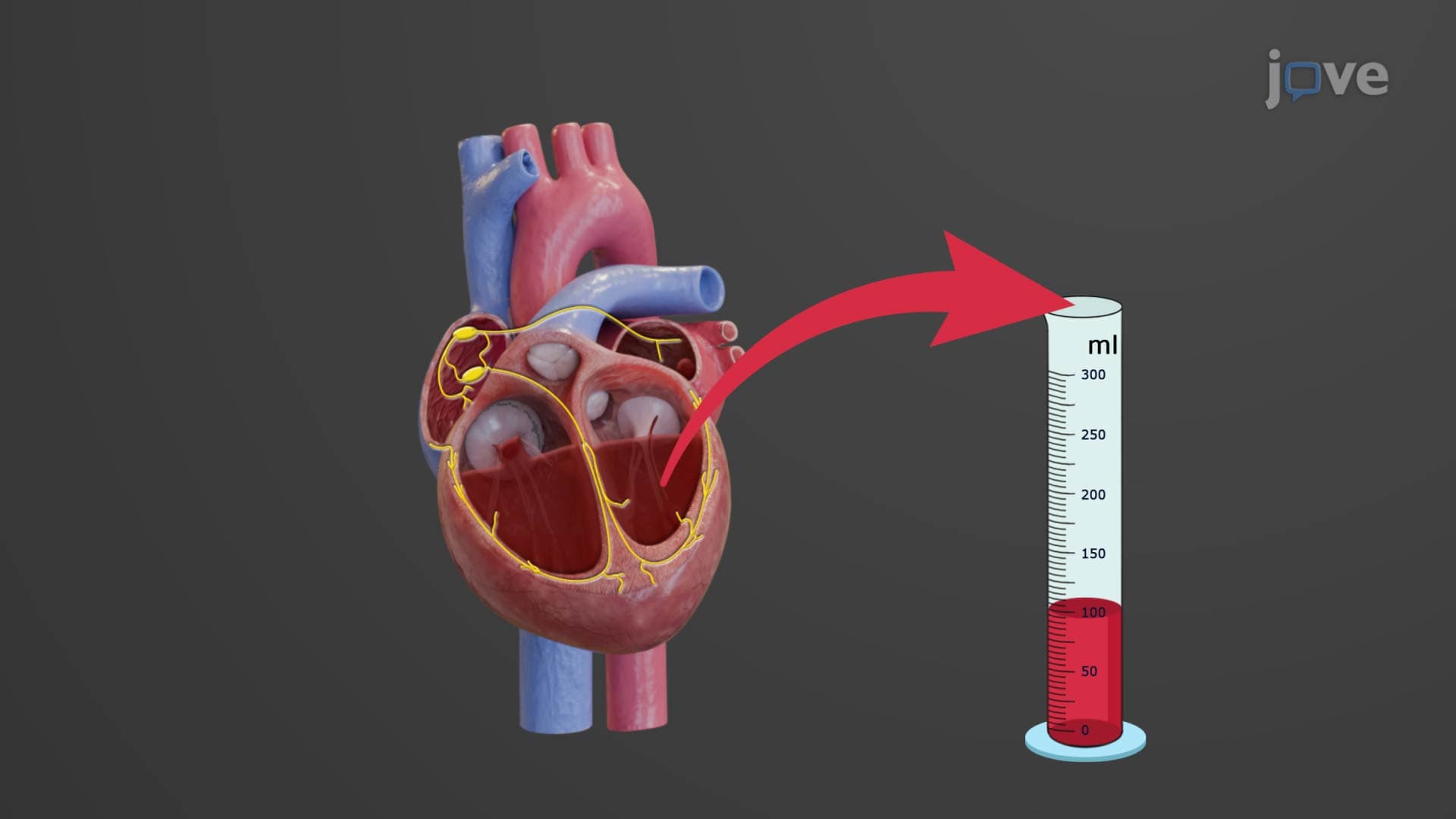
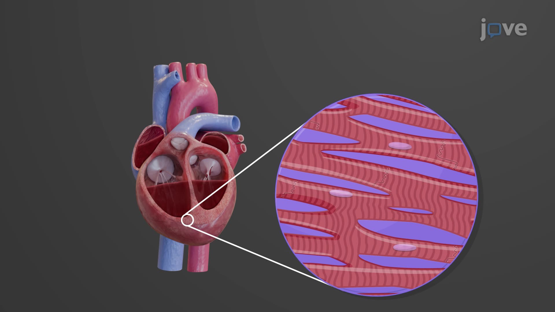
The ventricles generate the pressure needed for circulation. The right ventricle, with its thinner walls, pumps blood to the nearby lungs at relatively low pressure (25 mmHg systolic). In contrast, the left ventricle's thick muscular walls generate high pressure (120 mmHg systolic) to propel blood throughout the entire body-from your brain to your toes. Internal structures called trabeculae carneae create irregular ridges that prevent suction during contraction, while papillary muscles and their chordae tendineae act like guy-wires on a tent, preventing valve leaflets from inverting during pumping.
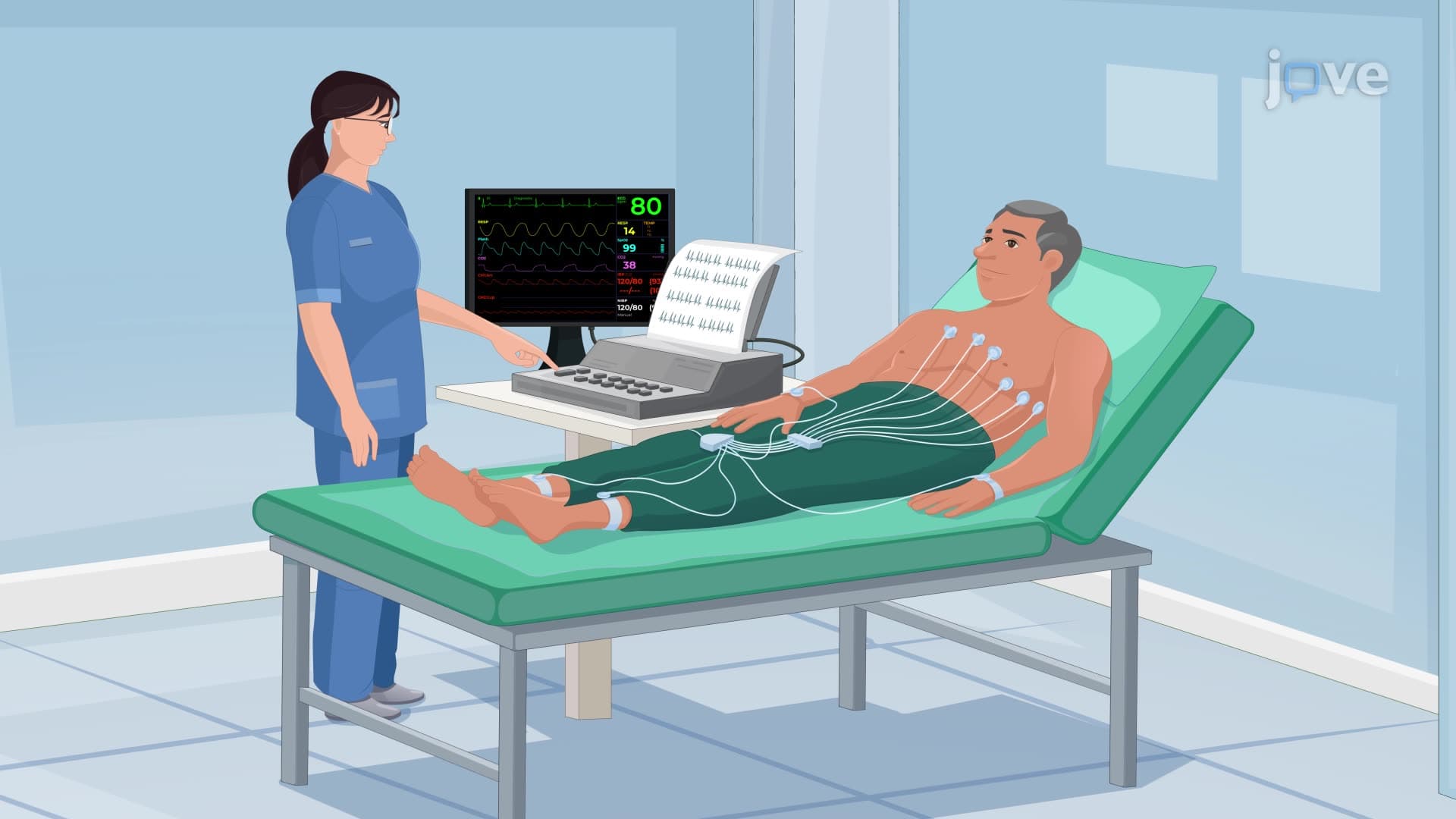
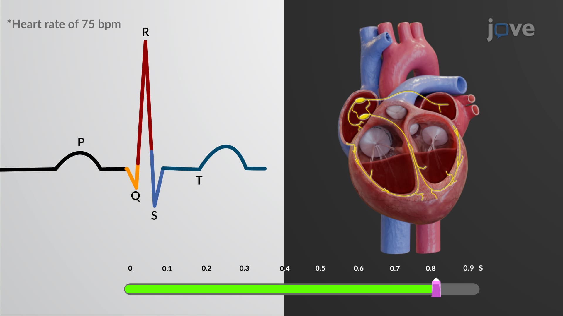
Understanding cardiac chambers proves essential for AP Biology students and pre-med students preparing for the MCAT. The interventricular septum, which separates the ventricles, becomes critically important when studying congenital heart defects like ventricular septal defects-the most common congenital heart abnormality affecting 1 in 500 births in the United States. Medical schools like Harvard and Stanford emphasize how chamber anatomy relates to electrocardiogram interpretation and cardiac catheterization procedures used in interventional cardiology.
Related Micro-courses