Loading subject page…

- Anatomy and Physiology
- The Heart
- Conduction System of the Heart
Micro-course: The Heart
112,207 views
Learn key concepts and earn your certificate.


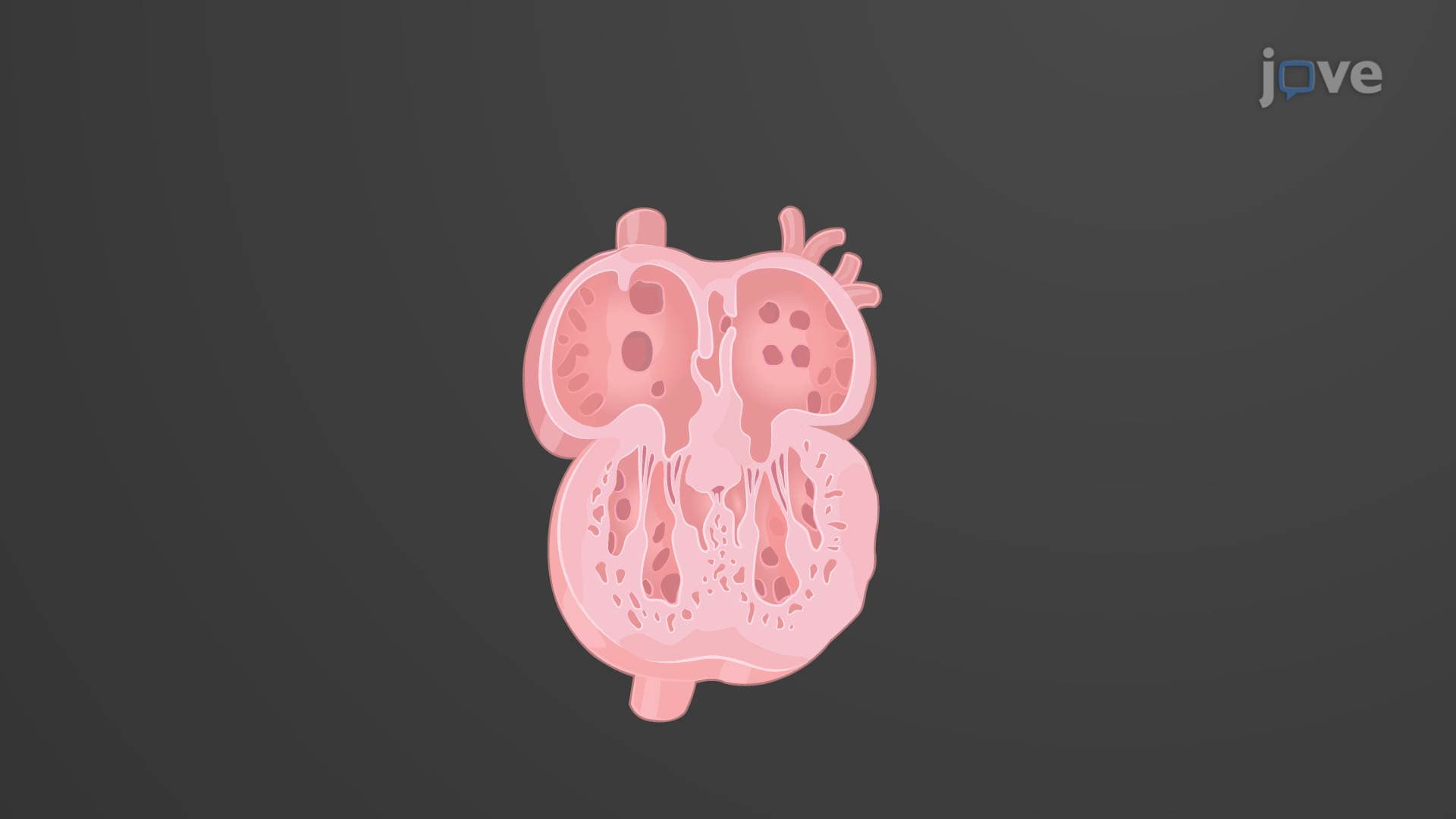
Conduction System of the Heart

00:00
01:19
Video Summary: What Is Conduction System Heart
Ever wondered why your heart beats exactly when it needs to, even during sleep? The heart conduction system anatomy works like your body's most reliable electrical network, automatically generating about 100,000 beats daily without conscious control. In the United States, understanding cardiac conduction helps explain why conditions like arrhythmias affect over 2 million Americans annually. What is conduction system heart functionality reveals how specialized cells create and conduct electrical impulses that coordinate every heartbeat through a precise pathway from the SA node to the Purkinje fibers. Watch the full video on JoVE Coach to master this concept with expert-led visuals and step-by-step explanations.
- Understand what is conduction system heart anatomy and how it generates electrical impulses automatically through specialized pacemaker cells
- Identify the five main components: SA node, internodal pathways, AV node, bundle of His, and Purkinje fiber network
- Analyze how action potentials travel from the sinoatrial node through the atrioventricular node to coordinate atrial and ventricular contractions
- Compare the intrinsic firing rates of different pacemaker regions and explain why the SA node typically controls heart rhythm
- Evaluate how the autonomic nervous system modifies heart rate through parasympathetic and sympathetic influences on pacemaker cells
- Apply knowledge of conduction timing to explain why atrial contraction precedes ventricular contraction during each cardiac cycle
- Recognize clinical implications when conduction pathway damage leads to arrhythmias or heart blocks
- Explore the relationship between electrical conduction and mechanical contraction in coordinating effective blood pumping
Understanding What is Conduction System Heart Physiology
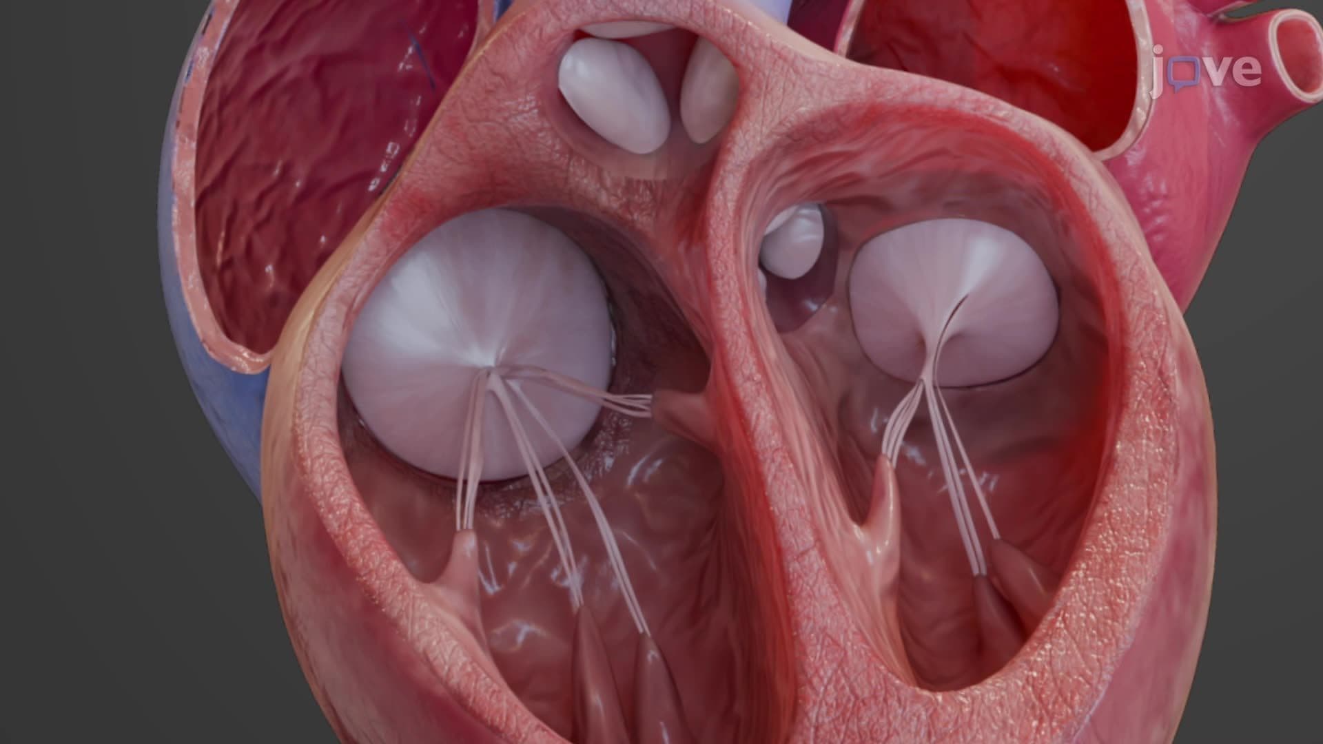
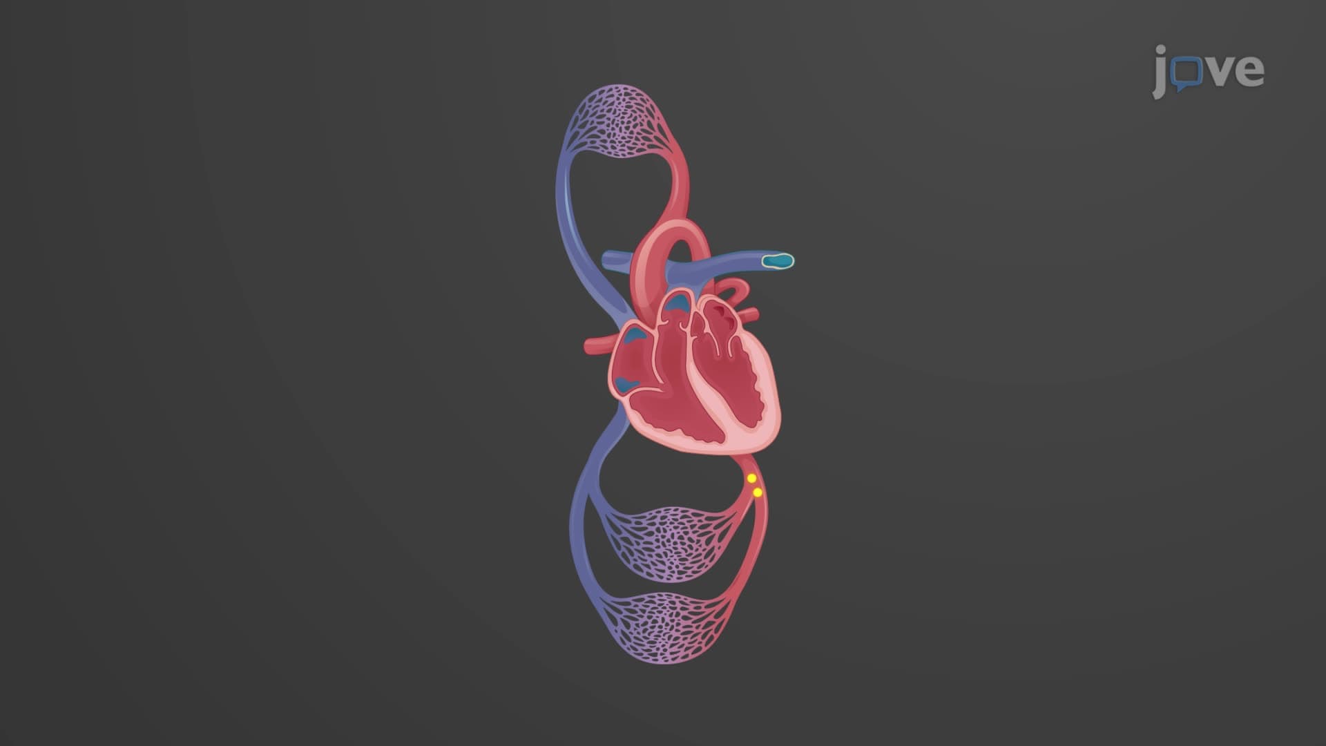
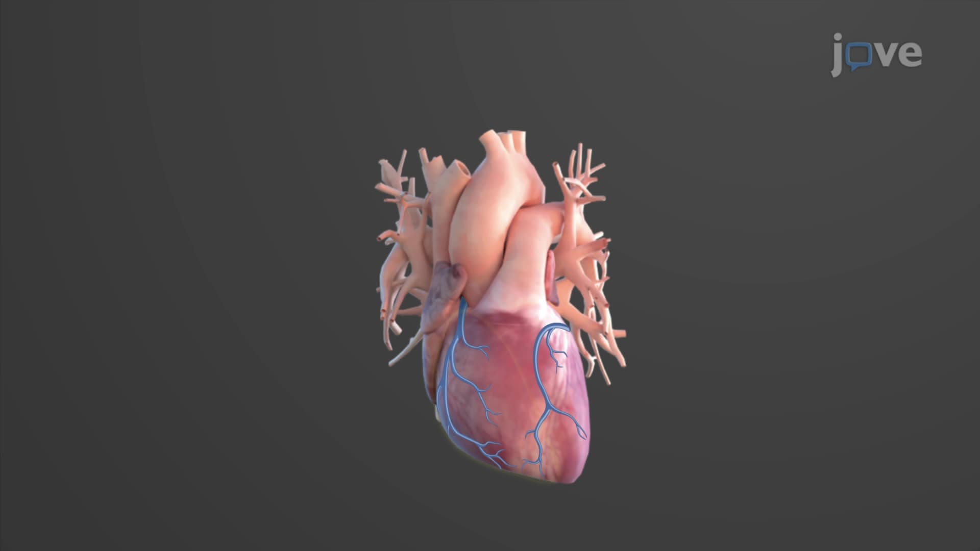
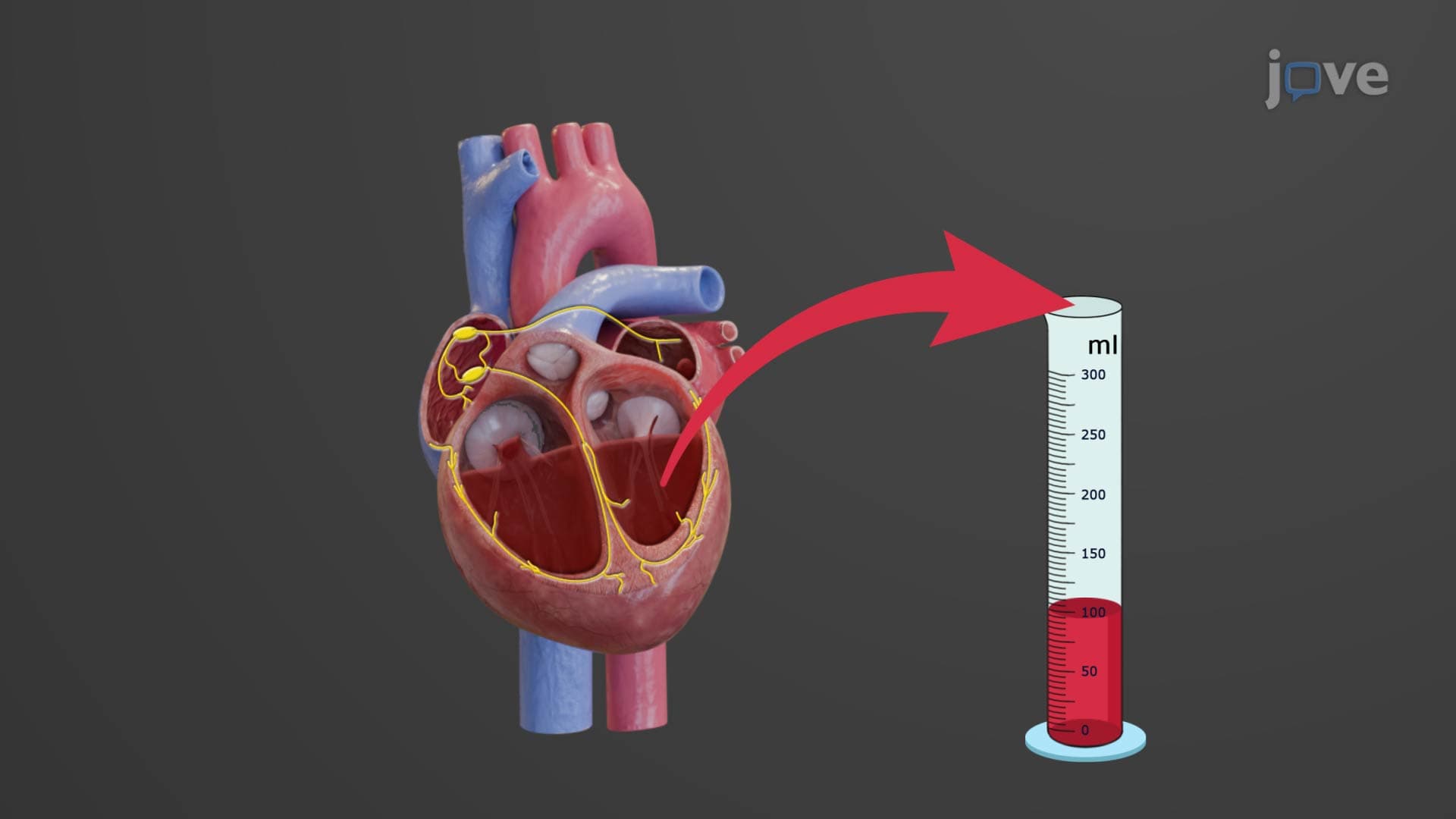
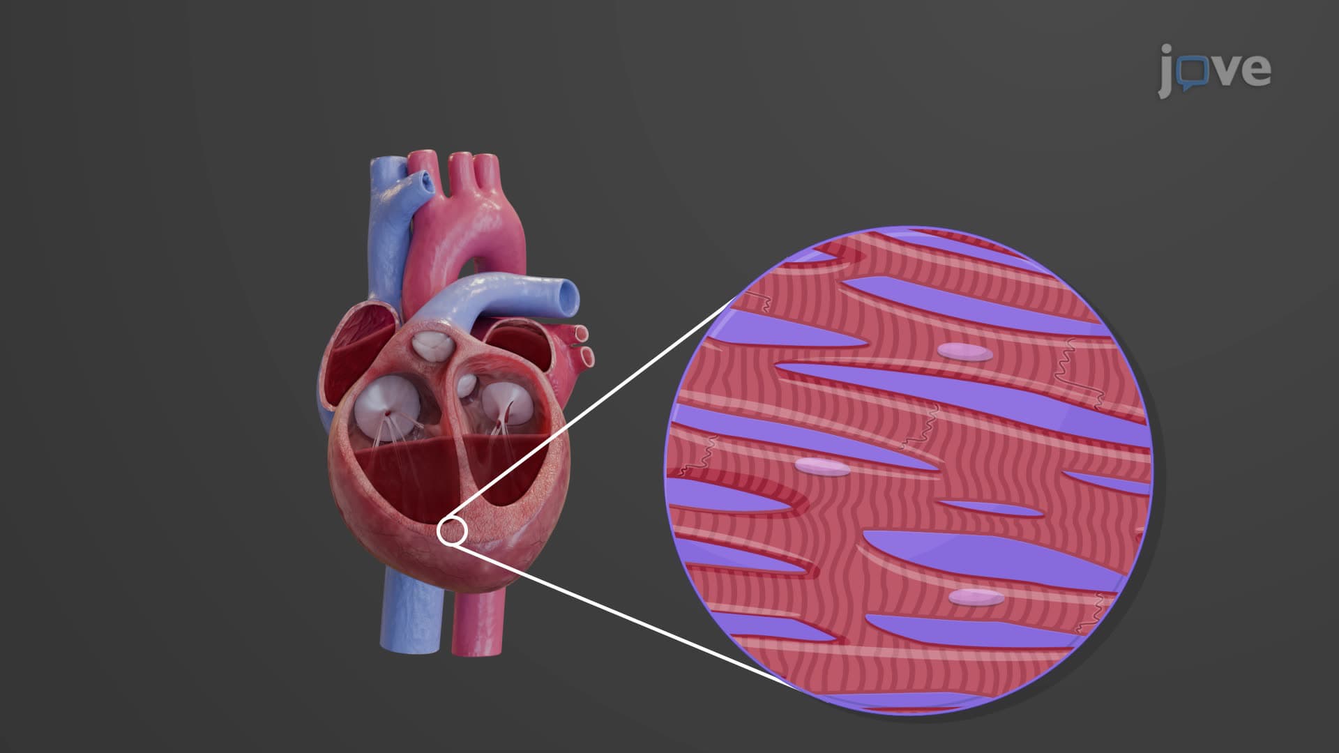
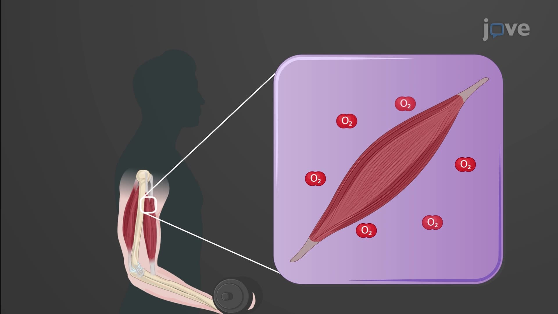
The heart conduction system anatomy represents one of the body's most sophisticated electrical networks, enabling the heart to function as both an electrical generator and mechanical pump. Unlike skeletal muscle that requires nervous system stimulation, cardiac muscle possesses autorhythmicity-the intrinsic ability to generate electrical impulses spontaneously. This remarkable property ensures continuous heart function even when disconnected from external nervous control, as demonstrated in heart transplant patients.
SA Node AV Node Conduction Pathway
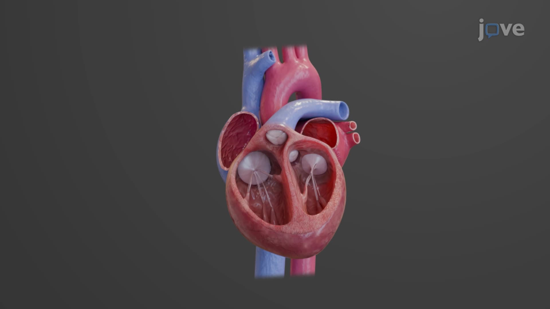
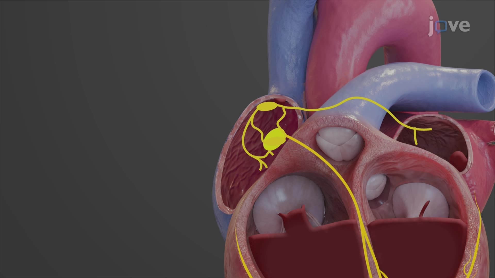
The cardiac conduction system explained begins with the sinoatrial (SA) node, often called the heart's natural pacemaker. Located in the right atrial wall near the superior vena cava, the SA node contains specialized pacemaker cells that spontaneously depolarize approximately 100 times per minute. This electrical impulse spreads through both atria via internodal pathways, causing coordinated atrial contraction that fills the ventricles with blood.
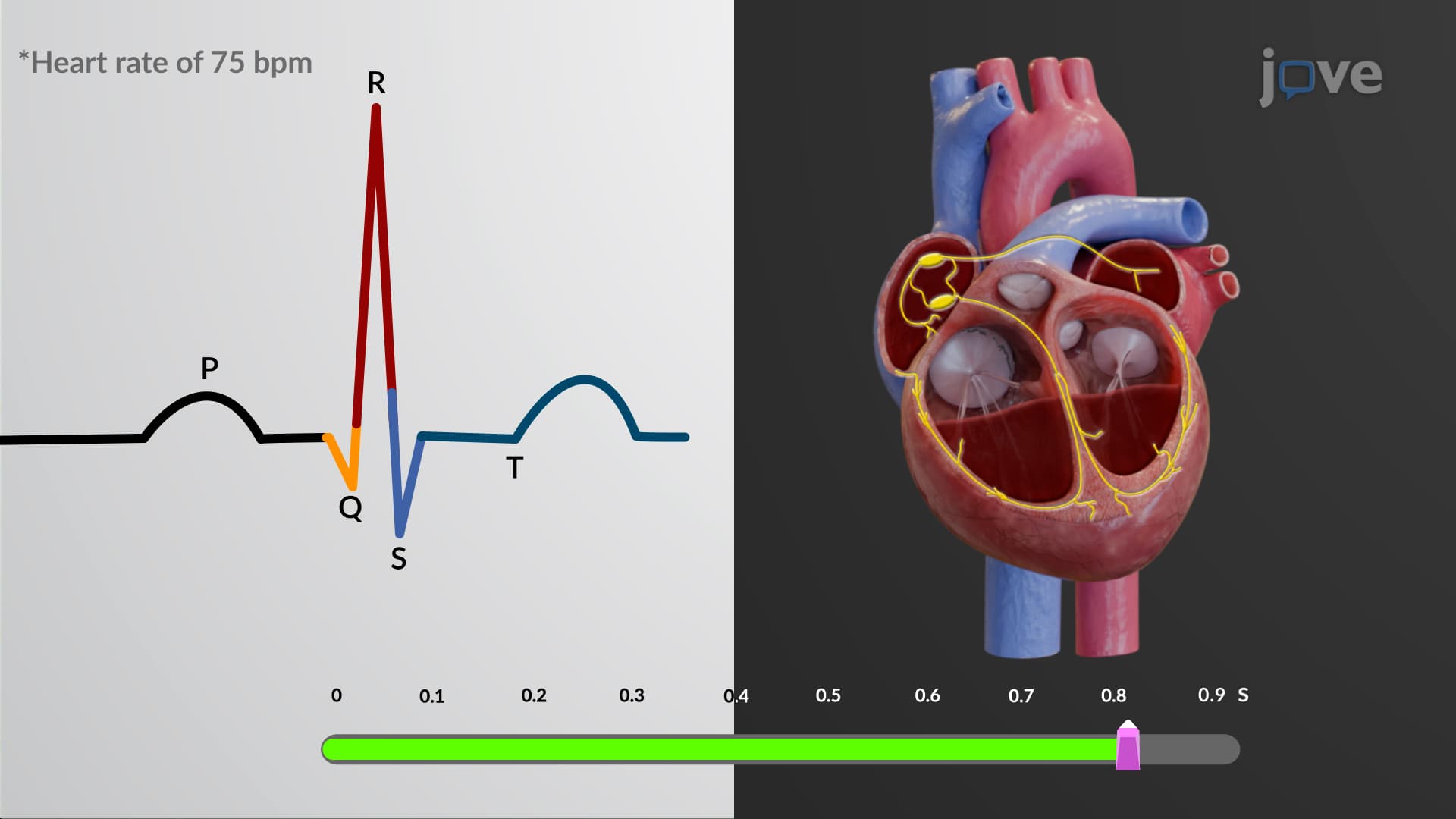
The signal then reaches the atrioventricular (AV) node, strategically positioned at the junction between atria and ventricles. The AV node introduces a crucial 0.1-second delay, allowing complete atrial emptying before ventricular contraction begins. This timing prevents the inefficient simultaneous contraction of all cardiac chambers.
Bundle of His Purkinje System Function
From the AV node, the action potential conduction heart pathway continues through the bundle of His, which penetrates the fibrous cardiac skeleton-the only electrical connection between atria and ventricles. The bundle quickly divides into left and right bundle branches, conducting impulses down the interventricular septum toward the apex.
The pathway concludes with the extensive Purkinje fiber network, which rapidly distributes electrical signals throughout the ventricular myocardium. These specialized fibers conduct impulses at approximately 4 meters per second, ensuring synchronized ventricular contraction that efficiently pumps blood to the pulmonary and systemic circulations.
Clinical Relevance and Autonomic Regulation
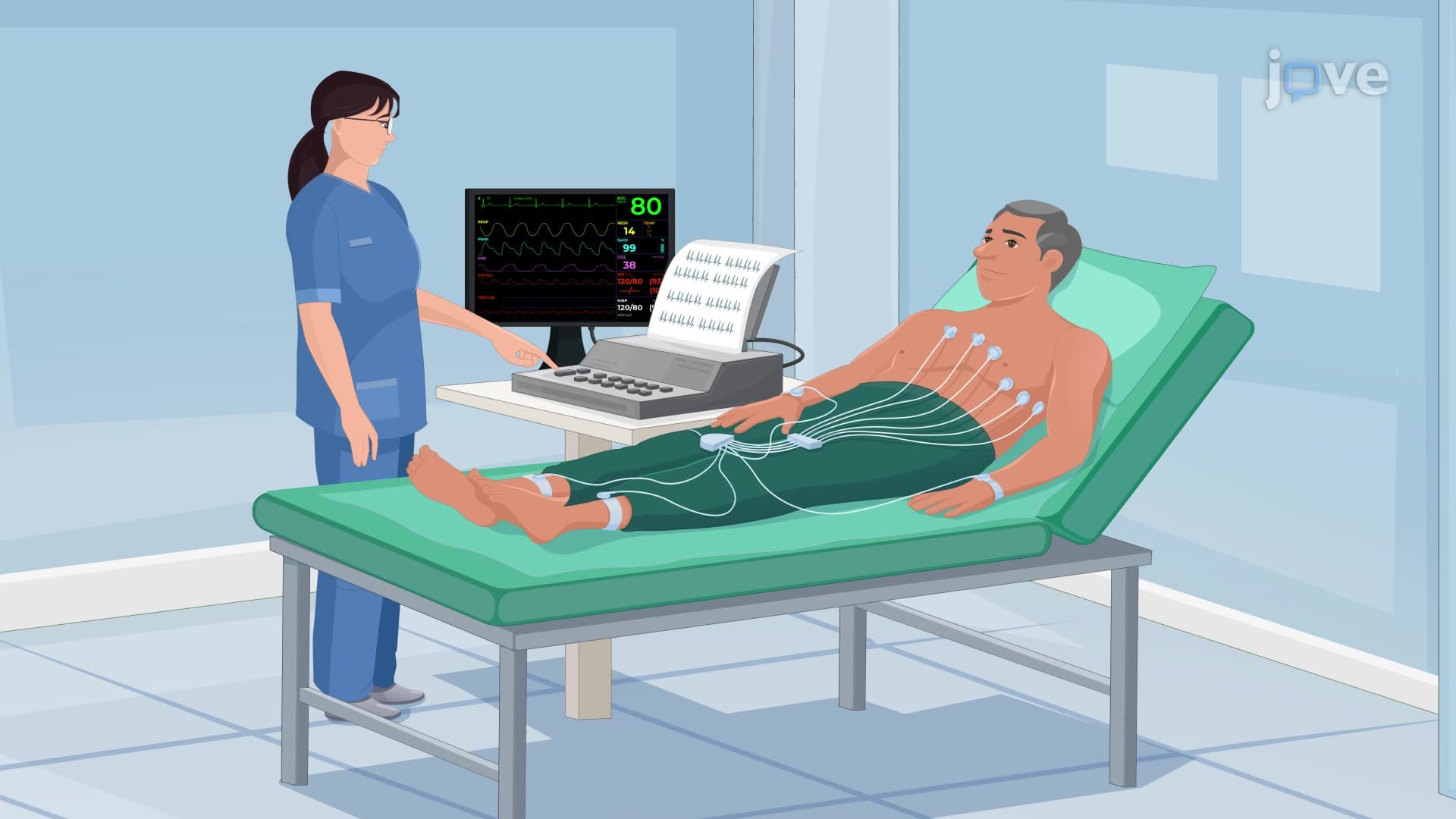
Understanding electrical conduction heart mechanisms proves essential for interpreting electrocardiograms (ECGs) on the MCAT and in clinical practice. The P wave represents atrial depolarization, the QRS complex shows ventricular depolarization, and the T wave indicates ventricular repolarization. Healthcare professionals use this knowledge to diagnose conditions like heart blocks, where conduction between chambers becomes impaired.
The autonomic nervous system continuously fine-tunes heart rate based on physiological demands. Parasympathetic stimulation through the vagus nerve slows SA node firing to about 75 beats per minute at rest, while sympathetic activation can increase rates to over 180 beats per minute during exercise or stress. This regulation explains how trained athletes often display resting heart rates below 60 beats per minute due to enhanced parasympathetic tone.
Frequently Asked Questions
The heart conduction system is your body's internal electrical network that automatically generates and coordinates heartbeats through specialized cells. This system ensures your heart beats approximately 100,000 times daily without conscious control, pumping blood efficiently throughout your body. Understanding this system helps explain how conditions like arrhythmias develop and why maintaining cardiovascular health is crucial for overall wellbeing.
MCAT questions frequently test conduction pathway sequences, timing delays, and autonomic regulation of heart rate. AP Biology exams emphasize the relationship between structure and function in cardiac cells, particularly autorhythmicity and action potential propagation. Focus on understanding why the AV node delay is necessary and how sympathetic versus parasympathetic stimulation affects pacemaker cell activity.
USMLE heavily emphasizes ECG interpretation, arrhythmia mechanisms, and pharmacological effects on conduction. Key topics include understanding how different drugs affect SA and AV node function, recognizing heart block patterns, and correlating conduction defects with clinical presentations. Practice linking anatomical conduction pathways to ECG waveform changes and pathophysiological conditions.
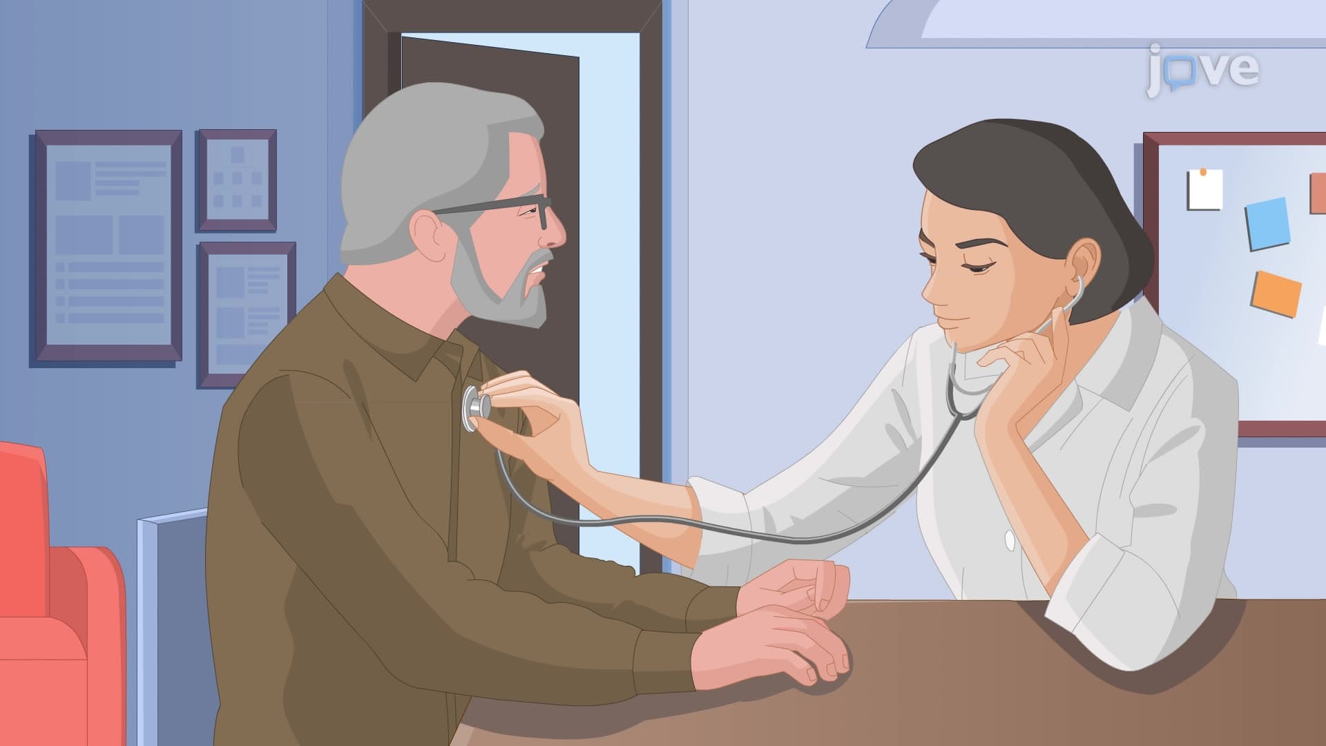
American cardiologists rely on conduction system understanding to interpret thousands of ECGs, diagnose arrhythmias, and plan treatments like pacemaker implantations. When patients present with chest pain at hospitals like Mayo Clinic or Johns Hopkins, physicians immediately assess conduction system function to rule out heart attacks or dangerous rhythm disturbances. This knowledge guides life-saving interventions and long-term cardiac care strategies.
While the topic involves multiple components, the conduction system follows a logical sequence that's easier to master than many students expect. Start by learning the five main structures in order, then understand why each timing delay serves a purpose. Visual aids and practice with ECG patterns make the electrical concepts more concrete and memorable for most high school learners.
Use the acronym "Some Athletes Need Better Performance" (SA node, Atrial pathways, AV Node, Bundle of His, Purkinje fibers) to remember the sequence. Create a flow chart showing timing delays and draw the pathway multiple times while explaining each step aloud. Practice correlating each anatomical structure with its corresponding ECG component to strengthen long-term retention.
Mastering basic conduction principles provides the foundation for understanding complex arrhythmias, electrophysiology procedures, and cardiac pharmacology in medical school. This knowledge directly applies to interpreting stress tests, understanding pacemaker programming, and grasping how medications like beta-blockers or calcium channel blockers affect different parts of the conduction system.
Disease, aging, or medications can damage the SA node or disrupt conduction pathways, causing dangerously slow heart rates or complete heart blocks. Artificial pacemakers restore normal rhythm by electrically stimulating the heart when natural conduction fails. Over 200,000 Americans receive pacemakers annually, highlighting how understanding normal conduction helps recognize when artificial intervention becomes necessary.
Related Micro-courses

38 Concepts

38 Concepts

25 Concepts