Loading subject page…


112,207 views

Video Summary: What Is Development of the Heart
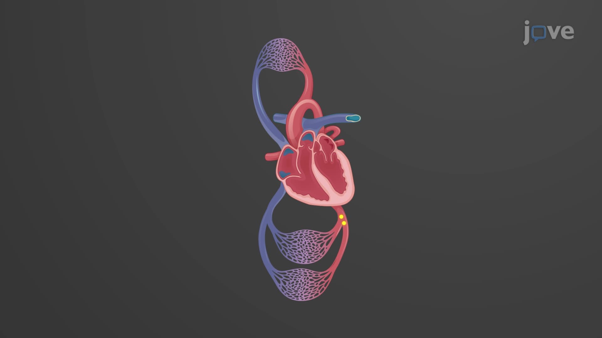
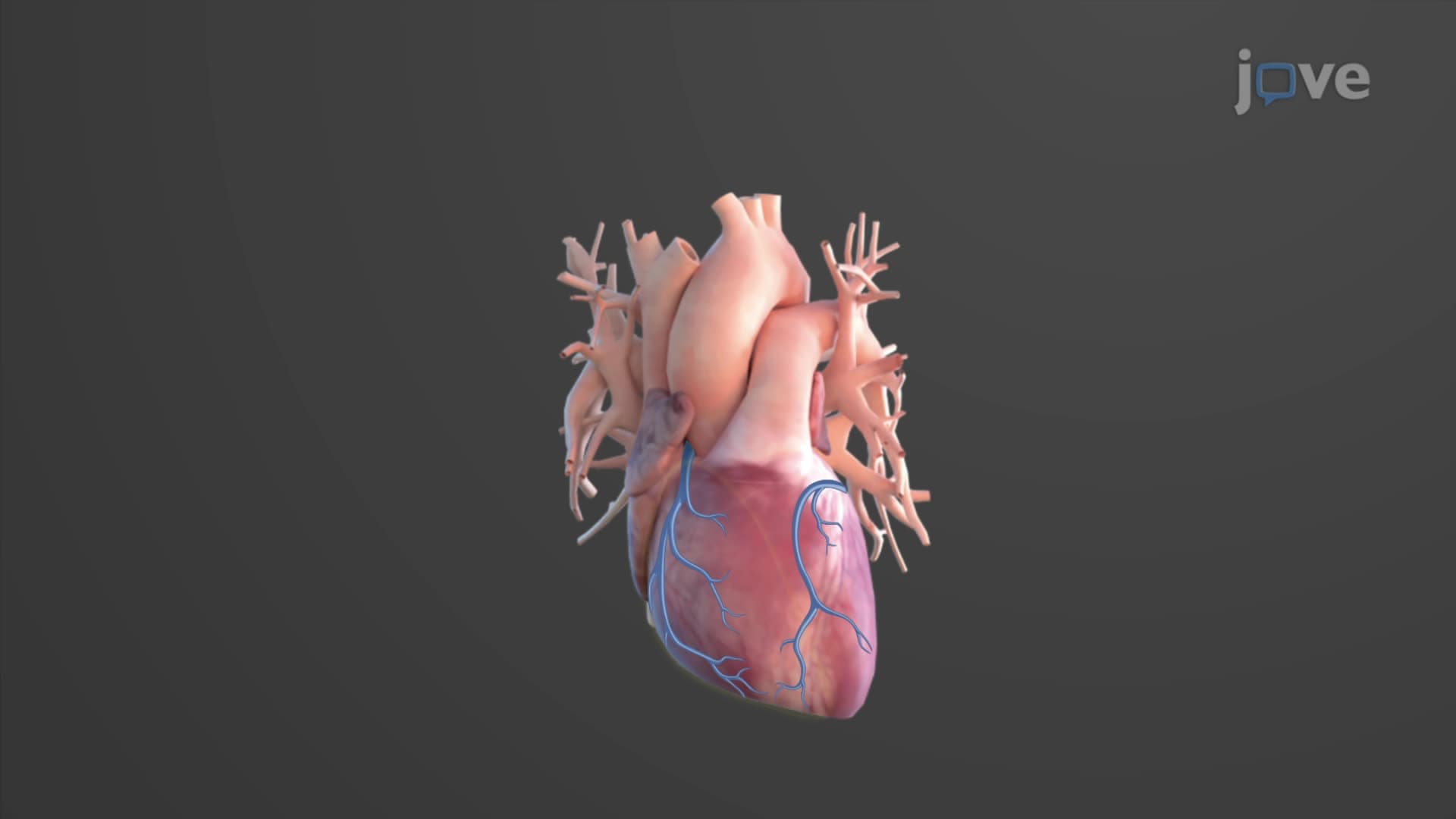
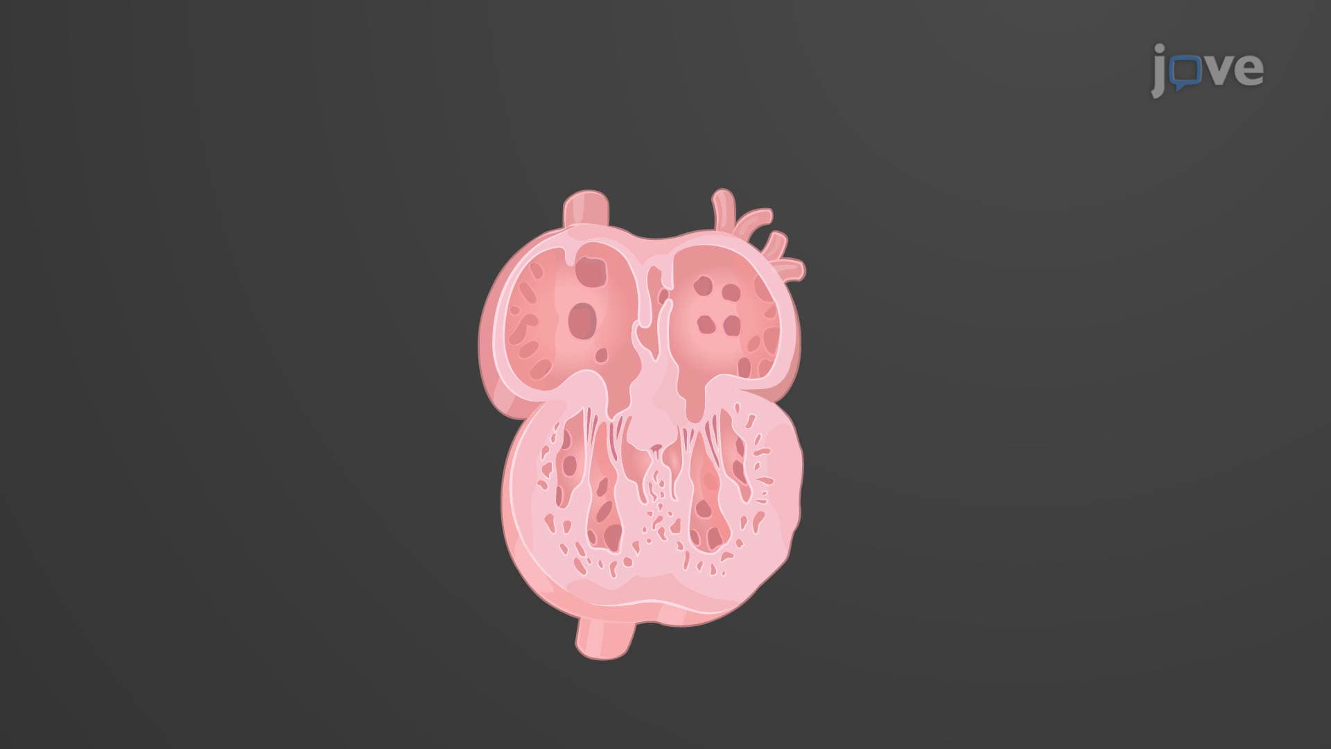
Every minute, your heart beats roughly 60-100 times-but did you know this incredible organ begins developing just 18 days after conception? Heart development embryology follows a precise timeline where mesodermal cells transform into a functioning four-chambered heart by week 9. Consider how congenital heart defects affect approximately 40,000 babies born annually in the United States, making understanding what is the development of the heart in embryology crucial for future healthcare professionals. Watch the full video on JoVE Coach to master this concept with expert-led visuals and step-by-step explanations.
Heart development embryology represents one of the most precisely orchestrated processes in human development. Unlike other organ systems that develop gradually, the cardiovascular system must function almost immediately to support the growing embryo. This developmental urgency explains why cardiac malformations represent the most common type of birth defect, affecting nearly 1% of all live births in the United States.
The process begins remarkably early-before many women even realize they're pregnant. By day 18-19 post-fertilization, specialized mesodermal cells aggregate in the cardiogenic area heart development, forming the embryonic heart field. This timing coincides with critical neural tube closure, highlighting why prenatal vitamins containing folic acid are recommended even before conception.
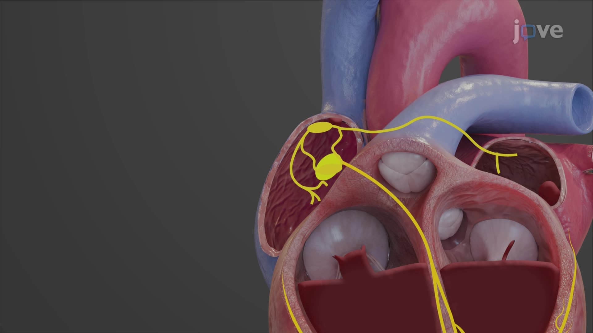
Cardiac development biology explained reveals how two separate endocardial tubes fuse by day 20 to create a single, linear heart tube. This primitive structure contains five distinct regions that will eventually become recognizable cardiac structures. The sinus venosus transforms into the right atrial wall, coronary sinus, and sinoatrial (SA) node-the heart's natural pacemaker. Students preparing for the MCAT or AP Biology exams should remember this developmental origin when studying cardiac conduction systems.
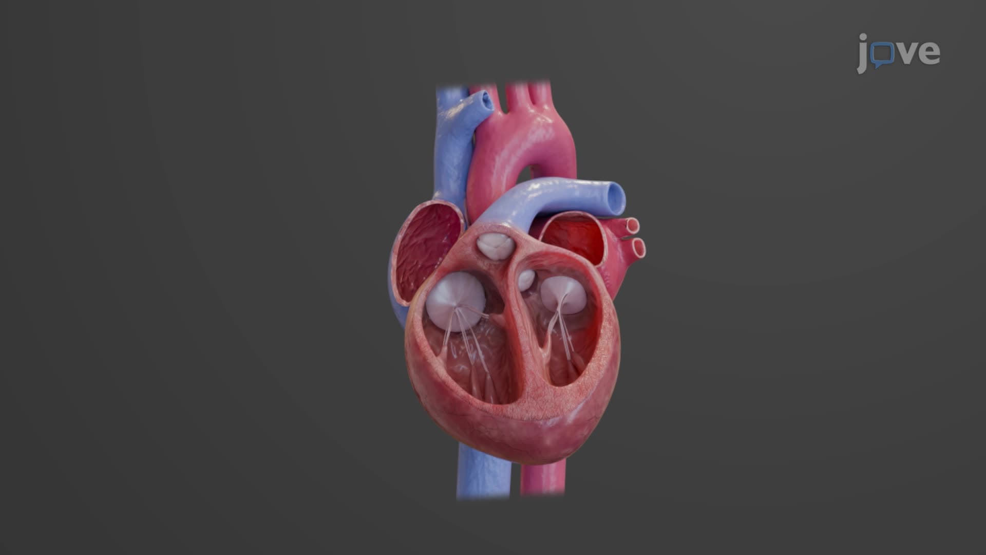
The primitive atrium and ventricle give rise to the muscular walls we recognize in adult hearts, while the bulbus cordis and truncus arteriosus form the great vessels. This developmental sequence explains why certain congenital defects, like transposition of the great arteries, occur when normal rotation and septation fail during weeks 5-8.
Heart tube looping begins around day 23, transforming the linear tube into the familiar heart shape. This rightward looping positions the future ventricles below the atria-a critical step that can go wrong in conditions like dextrocardia. For students taking the USMLE Step 1, understanding this looping process helps explain why the right ventricle wraps around the left ventricle in normal cardiac anatomy.
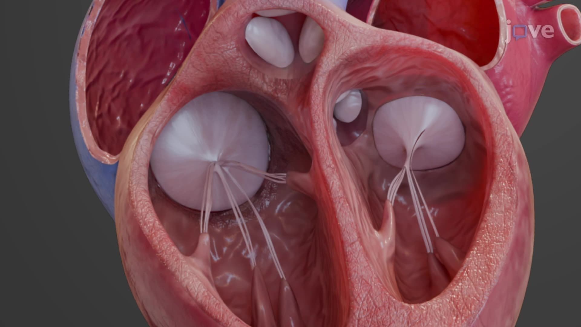
Septum heart formation occurs through coordinated growth of endocardial cushions around day 28. These tissue masses not only form the atrioventricular and semilunar valves but also contribute to the interatrial and interventricular septa. The foramen ovale, a temporary opening between fetal atria, exemplifies how fetal circulation bypasses non-functioning lungs. Nursing students preparing for NCLEX should understand that foramen ovale closure failure results in patent foramen ovale, present in approximately 25% of adults.
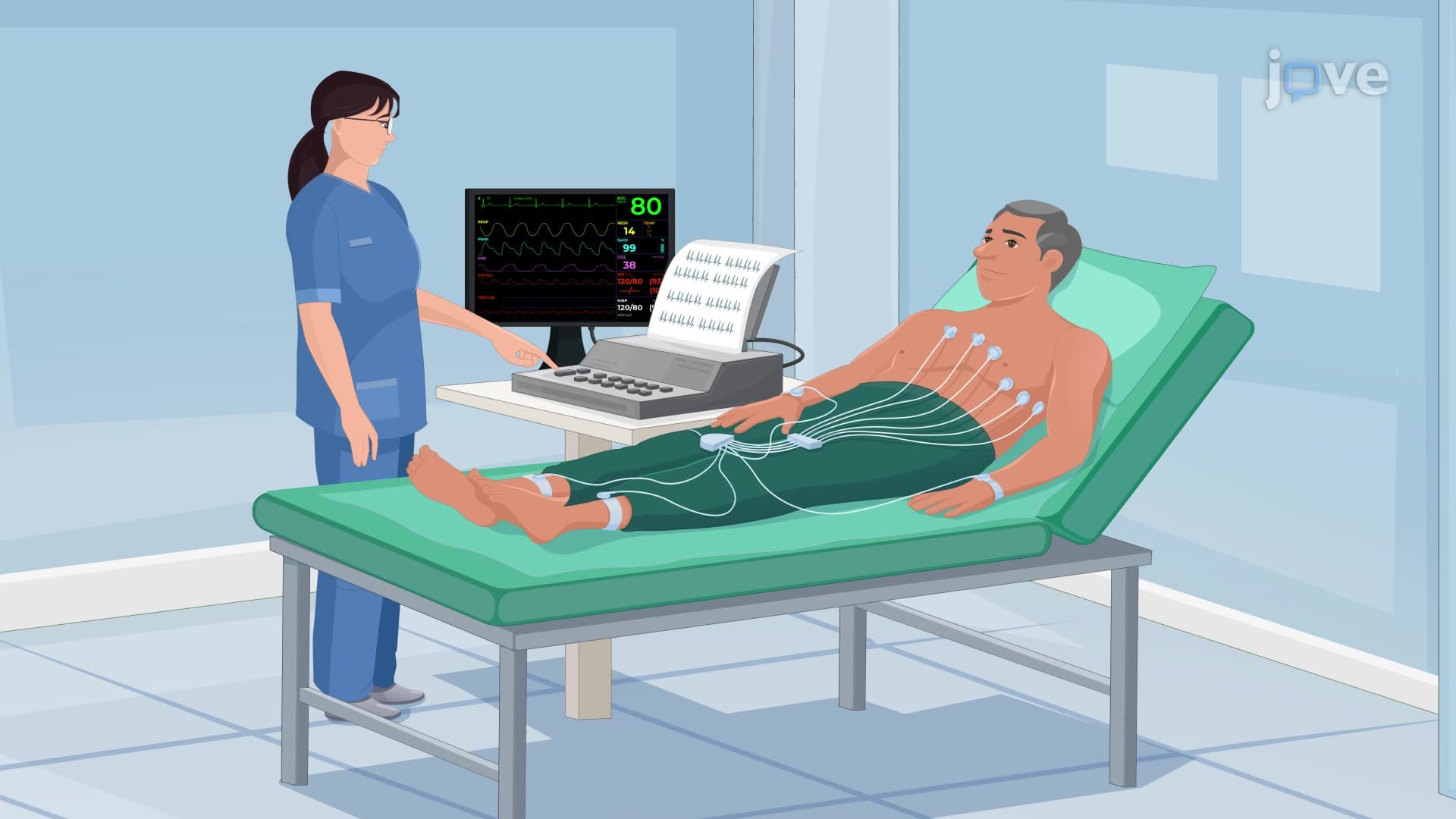
Understanding normal cardiac embryology development proves essential for interpreting echocardiograms and cardiac catheterizations. Pediatric cardiologists at institutions like Boston Children's Hospital and Texas Children's Hospital rely on embryological knowledge to predict surgical outcomes for complex congenital heart disease. Students planning medical careers should recognize that advances in fetal cardiac intervention now allow surgeons to repair certain heart defects before birth, demonstrating how embryological understanding directly improves patient outcomes.
Related Micro-courses