Loading video…


 19 Concepts
 112.2K views

Video Summary: What Is Disturbances in Heart Rhythm
Did you know that approximately 6.1 million Americans live with atrial fibrillation, making heart rhythm disturbances biology a critical health topic? When the heart's electrical system malfunctions, it creates cardiac arrhythmia explained through irregular beating patterns that can range from harmless to life-threatening. Understanding what are disturbances in heart rhythm helps explain why someone might experience sudden dizziness during exercise or require a pacemaker like former Vice President Dick Cheney. Watch the full video on JoVE Coach to master this concept with expert-led visuals and step-by-step explanations.
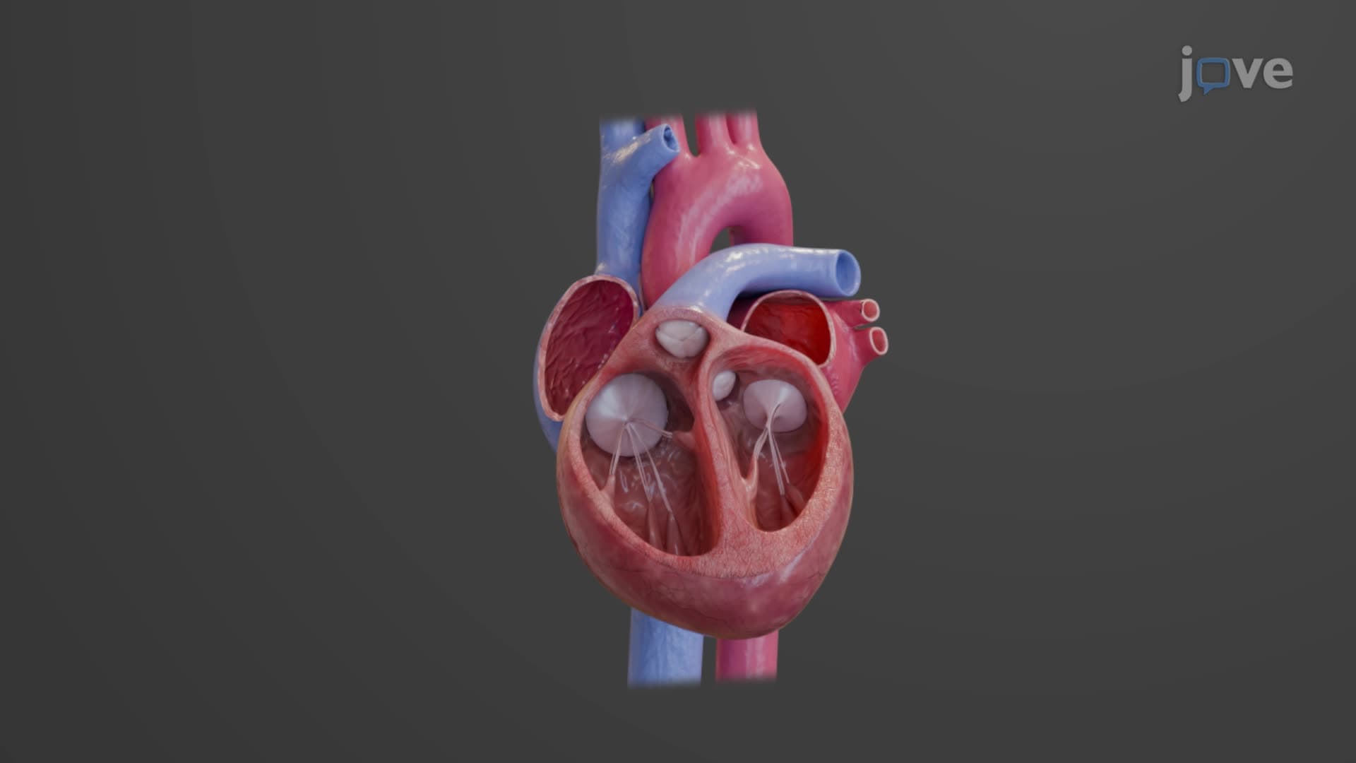
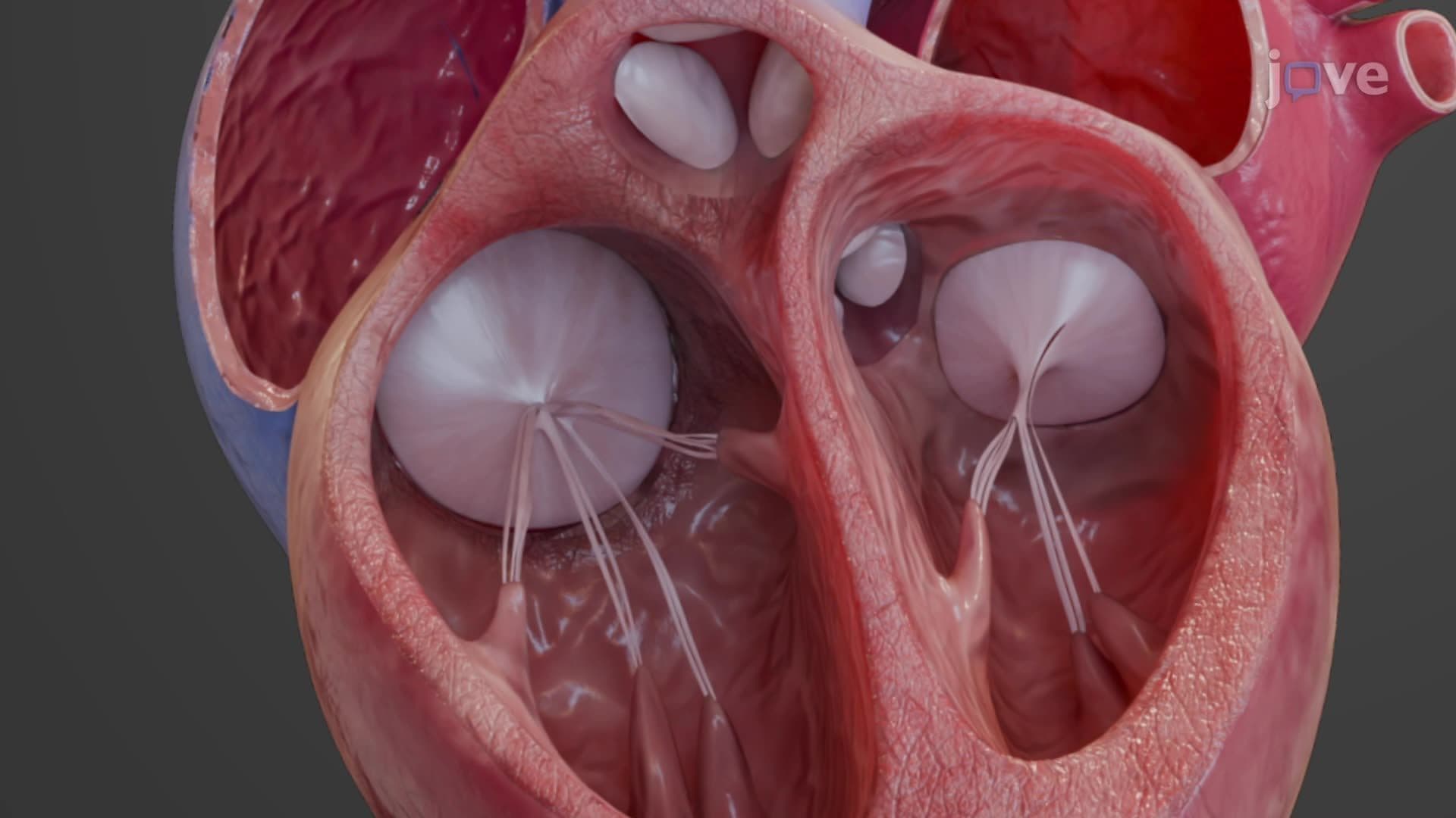
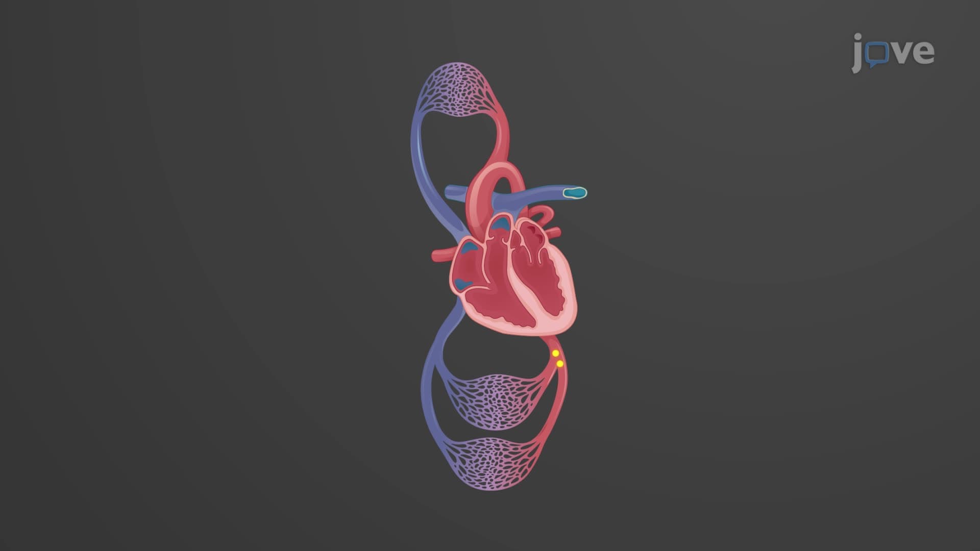
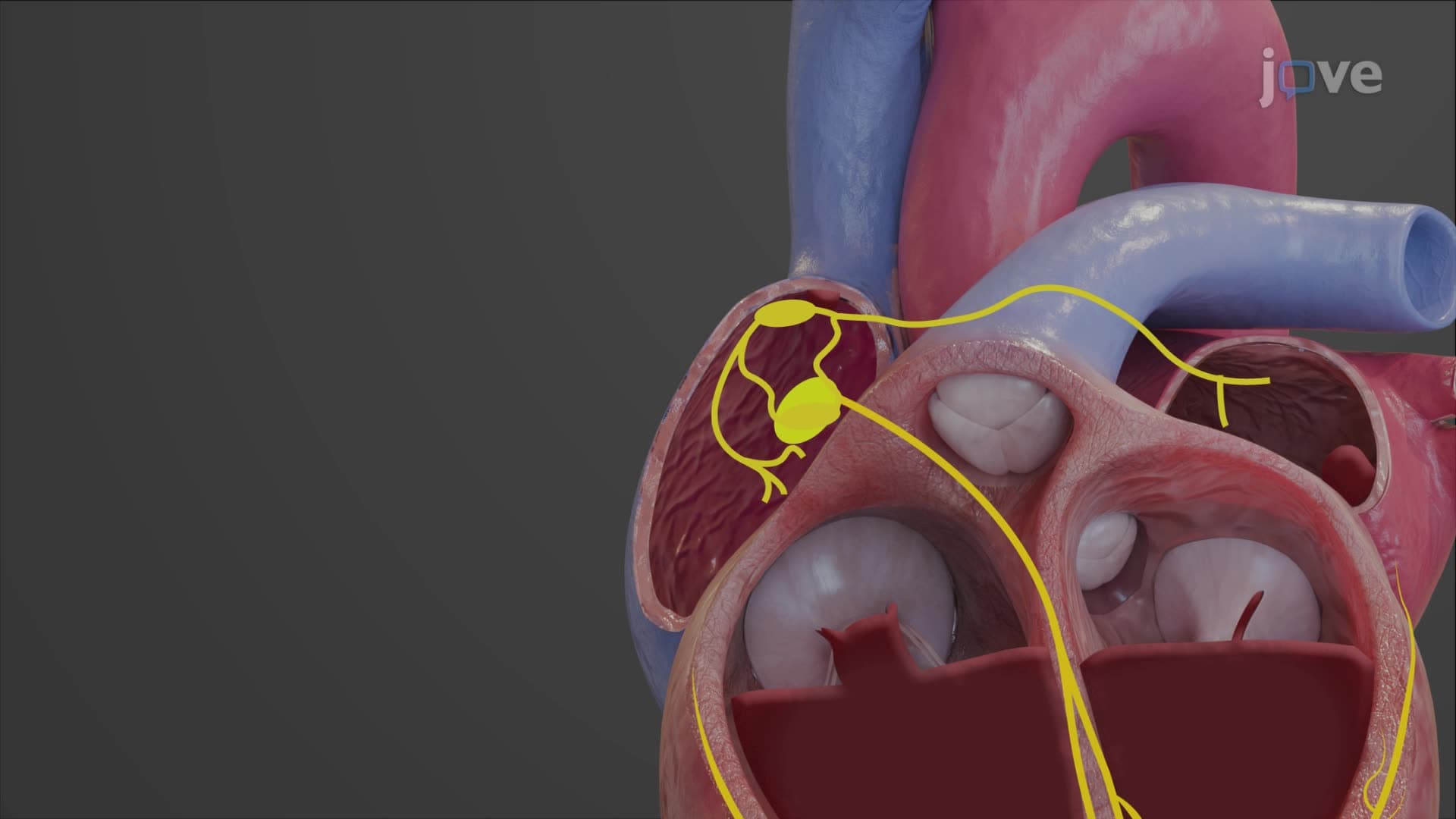
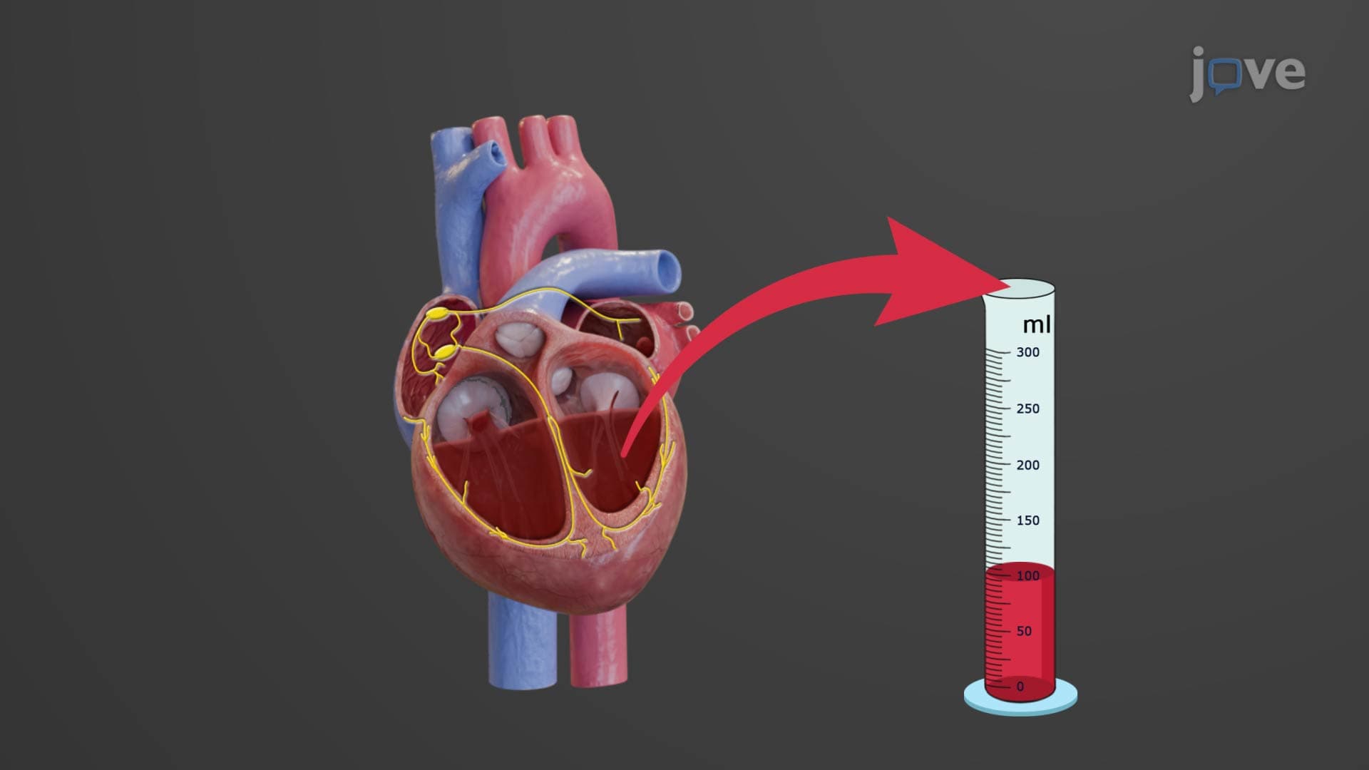
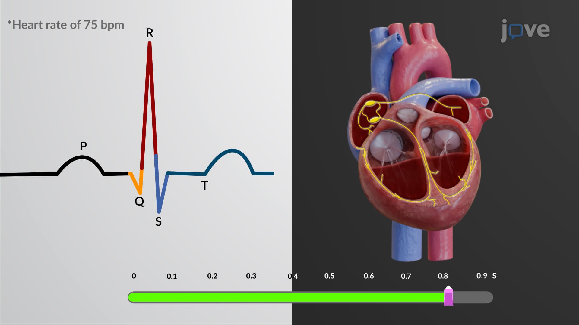
The heart's electrical system orchestrates every heartbeat through a precise sequence of electrical impulses. When this system malfunctions, cardiac arrhythmia explained becomes essential for understanding cardiovascular health. The sinoatrial (SA) node, located in the right atrium, serves as the heart's natural pacemaker, generating electrical impulses at approximately 60-100 beats per minute. These impulses travel through specialized conduction pathways, including the atrioventricular (AV) node and bundle of His, coordinating the contraction of atria and ventricles.
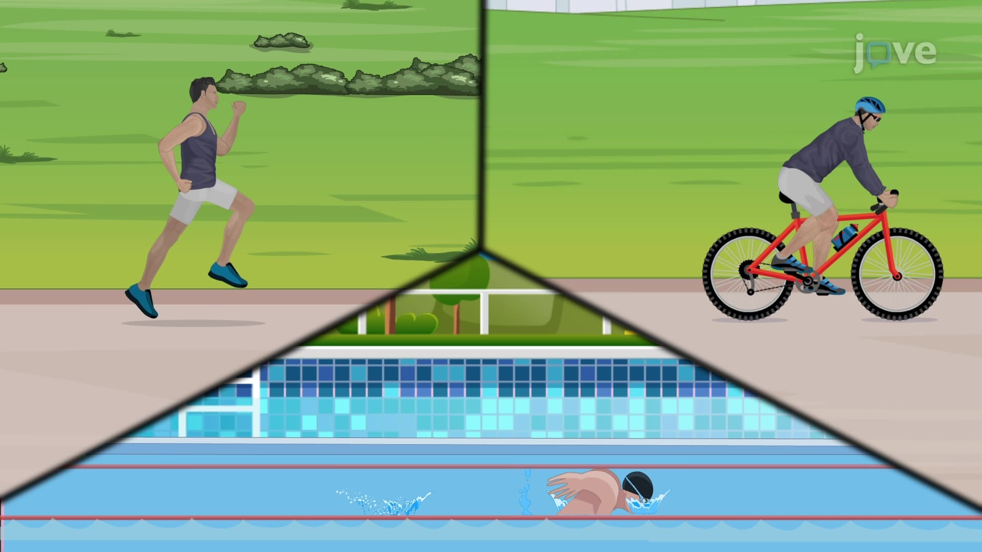
Arrhythmia dysrhythmia type disorders fall into distinct categories based on heart rate and origin. Bradycardia occurs when the heart rate drops below 60 BPM, often seen in well-conditioned athletes or elderly patients with degenerative conduction system disease. Conversely, tachycardia involves rates exceeding 100 BPM and can originate from atrial or ventricular tissue. Ventricular tachycardia fibrillation represents particularly dangerous rhythms where rapid, uncoordinated electrical activity prevents effective blood pumping.
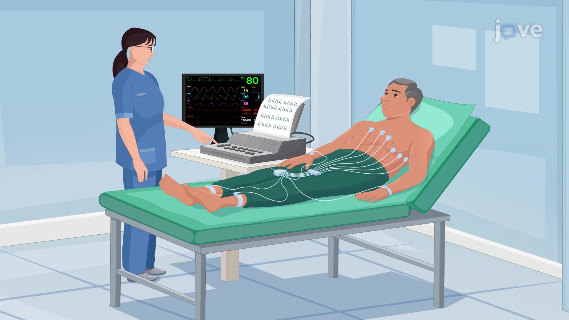
Atrial fibrillation arrhythmia affects millions of Americans and increases stroke risk due to blood stasis in the atria. During atrial fibrillation, chaotic electrical signals cause the atria to quiver instead of contracting effectively. On ECG analysis, this appears as irregularly spaced QRS complexes with absent or poorly defined P waves. The condition often develops in patients with hypertension, diabetes, or structural heart disease, making it a frequent topic in MCAT cardiovascular sections and nursing school pathophysiology courses.
Heart block conduction disorders occur when electrical impulses traveling from atria to ventricles encounter delays or complete obstruction. First-degree AV block shows prolonged P-Q intervals (over 200 milliseconds) but maintains 1:1 conduction. Second-degree blocks demonstrate intermittent conduction failure, with some P waves not followed by QRS complexes. Third-degree or complete heart block represents total AV dissociation, forcing ventricles to rely on escape rhythms typically firing at 20-40 BPM. These concepts frequently appear in AP Biology cardiac physiology units and NCLEX-RN questions about ECG interpretation.
Understanding these cardiac rhythm disorder mechanisms proves essential for healthcare careers and provides foundational knowledge for advanced studies in cardiology, emergency medicine, and critical care nursing.
Related Micro-courses