Loading subject page…

- Anatomy and Physiology
- The Heart
- Electrocardiogram
Micro-course: The Heart
112,207 views
Learn key concepts and earn your certificate.

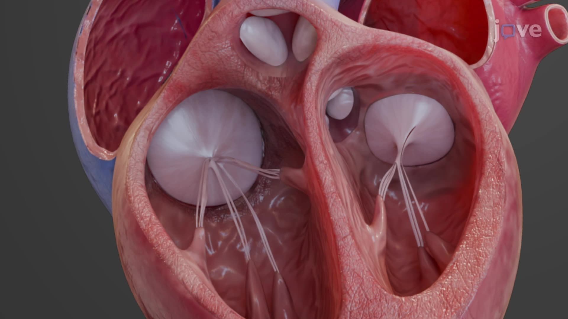
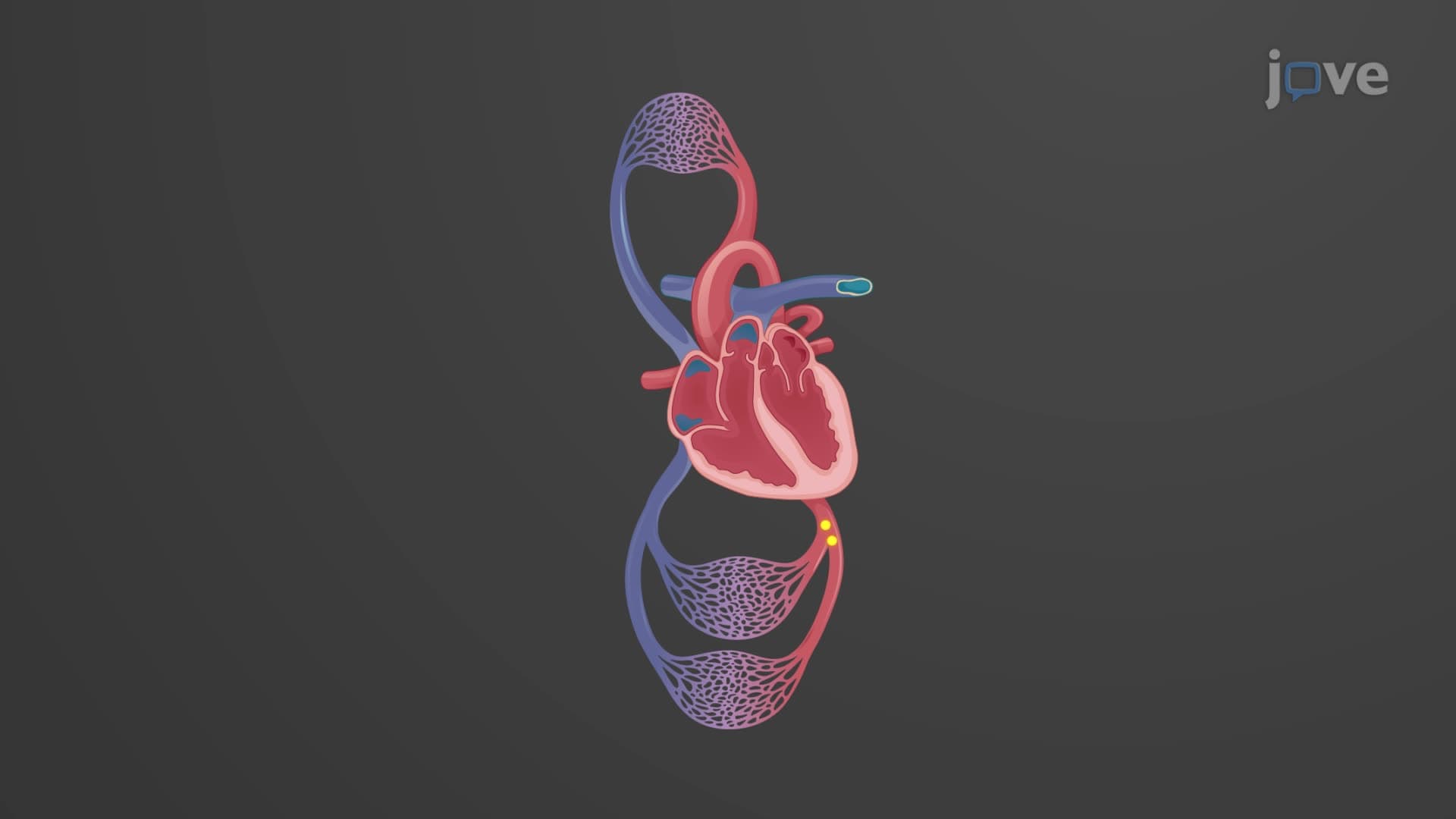
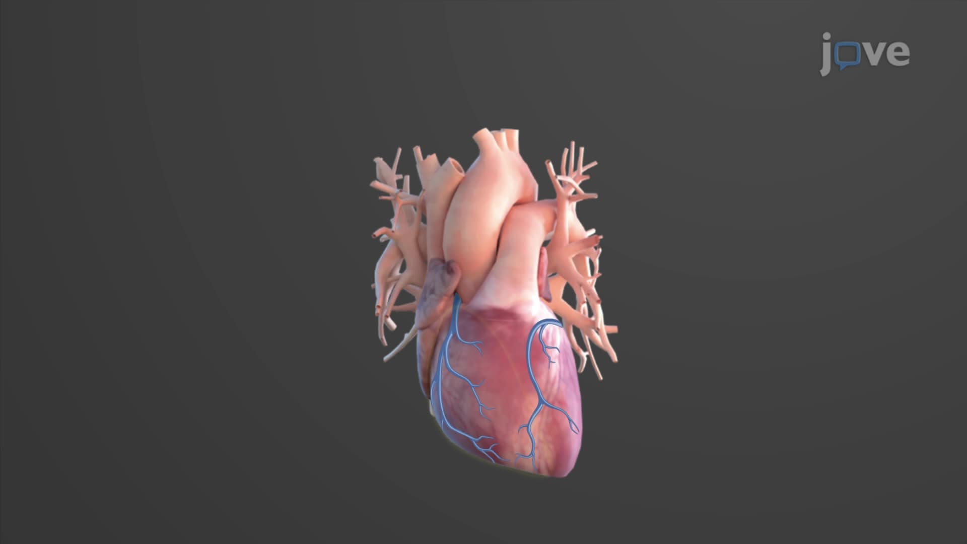
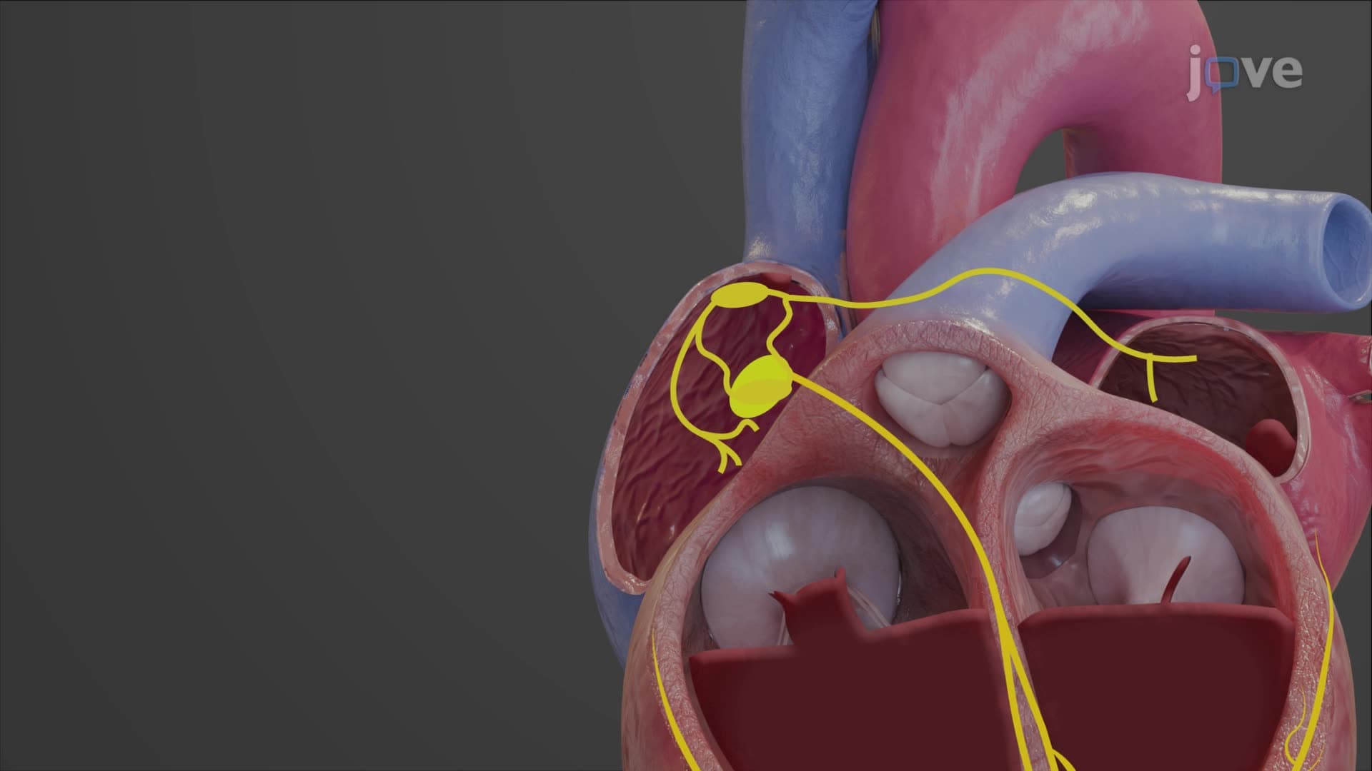
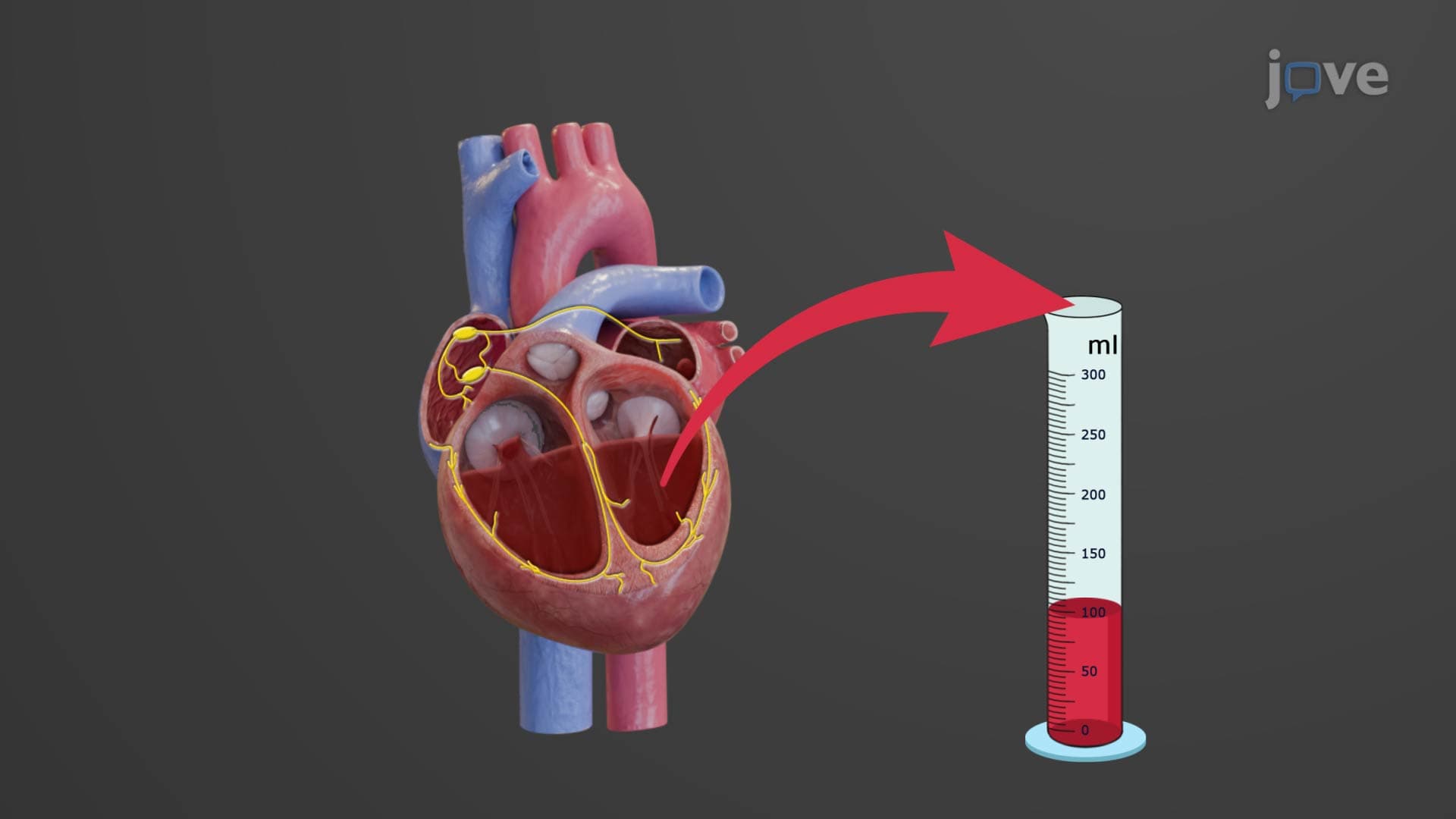
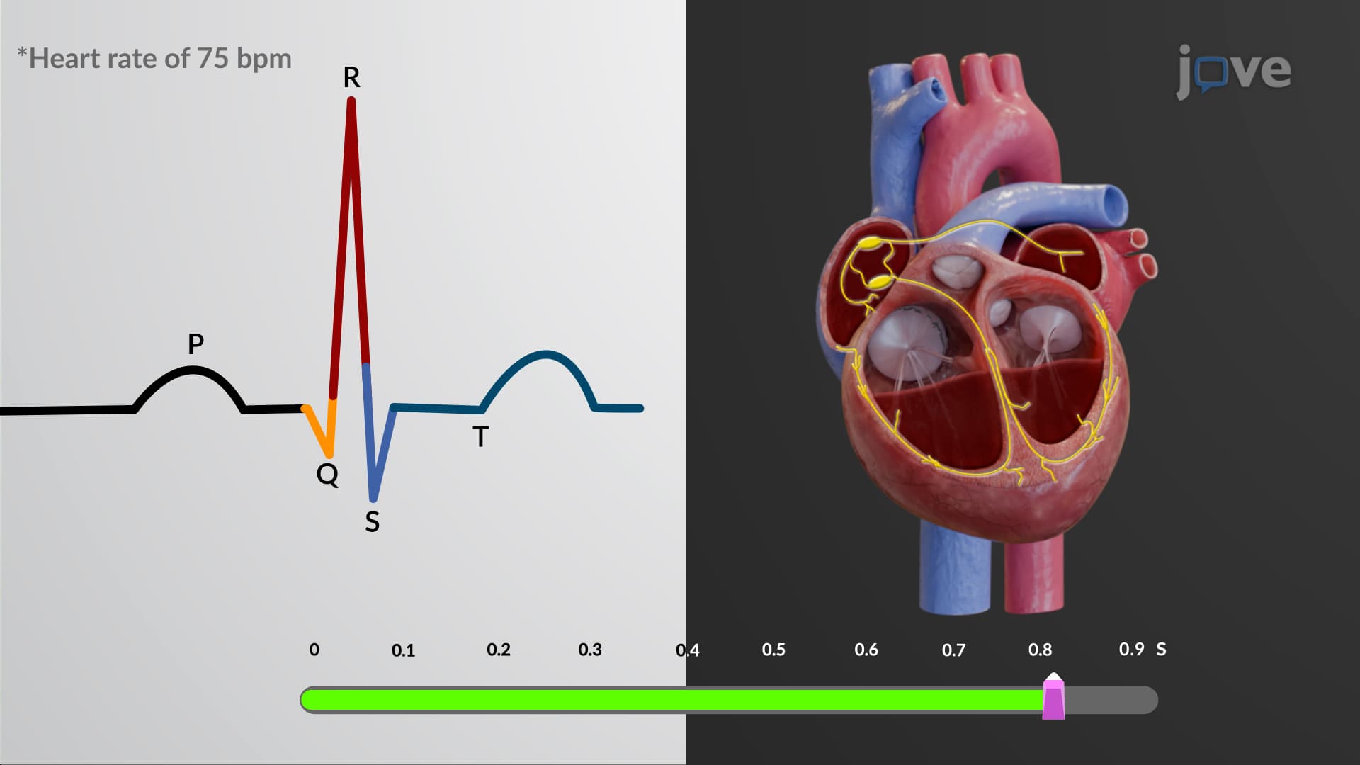
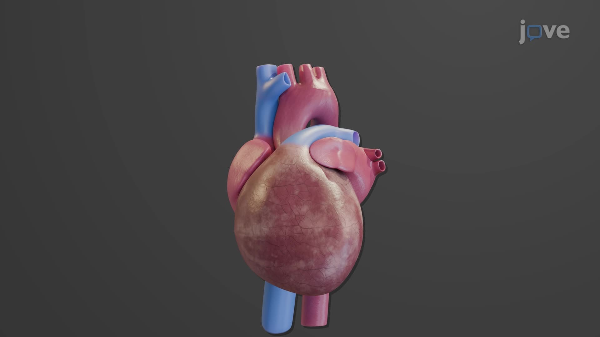
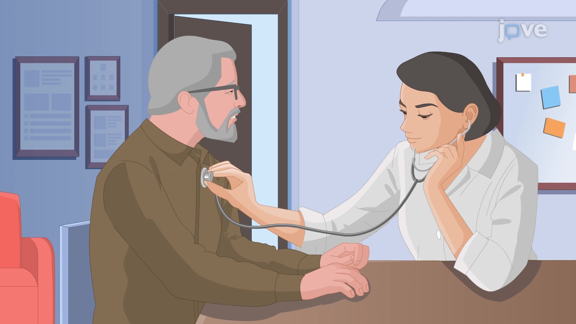

Electrocardiogram

00:00
01:29
Video Summary: What Is Electrocardiogram
Did you know that every heartbeat in your body generates enough electrical activity to be detected through your skin? An ECG electrocardiogram biology test captures these tiny electrical signals using electrodes placed on your chest, arms, and legs. When patients arrive at Mayo Clinic's emergency department with chest pain, doctors immediately order an electrocardiogram to assess heart function within minutes. This non-invasive cardiac diagnostic tool reveals critical information through distinctive waveforms including P waves, QRS complexes, and T waves. Watch the full video on JoVE Coach to master this concept with expert-led visuals and step-by-step explanations.
- Understand what is electrocardiogram technology and how it measures cardiac electrical activity through skin-surface electrodes
- Identify the three main ECG waveform components: P wave (atrial depolarization), QRS complex (ventricular depolarization), and T wave (ventricular repolarization)
- Analyze normal ECG intervals including P-R, S-T, and Q-T segments and their physiological significance
- Compare atrial versus ventricular electrical events as they appear on standard 12-lead ECG recordings
- Recognize abnormal ECG patterns that may indicate cardiac enlargement, myocardial infarction, or repolarization disorders
- Apply ECG interpretation skills to distinguish between normal and pathological cardiac rhythms
- Evaluate the clinical importance of ECG timing intervals in diagnosing cardiovascular conditions
- Explore the relationship between cardiac action potentials and their corresponding ECG waveform manifestations
Understanding Electrocardiogram Fundamentals
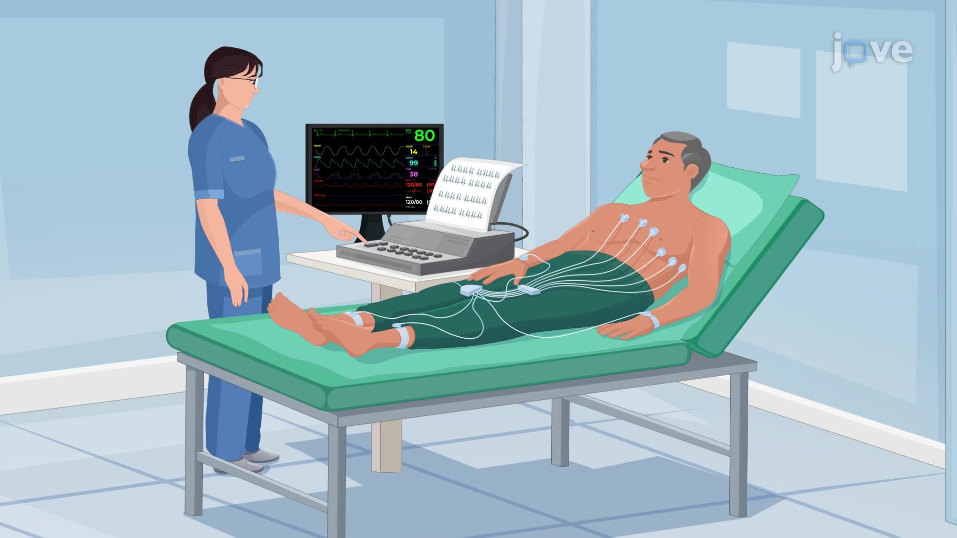
An electrocardiogram represents one of the most essential diagnostic tools in modern cardiology, providing a non-invasive window into cardiac electrical activity. This technology works by detecting the small electrical changes that occur when heart muscle depolarizes and repolarizes during each cardiac cycle. The ECG recording electrode system typically uses 10-12 electrodes strategically placed on the patient's limbs and chest to capture electrical signals from multiple cardiac perspectives simultaneously.
ECG Waveform Component Analysis
The P wave QRS complex T wave sequence represents the fundamental pattern of normal ECG interpretation. The P wave, typically lasting 80-100 milliseconds, reflects atrial depolarization as electrical impulses spread from the sinoatrial (SA) node throughout both atria. This small, rounded deflection precedes atrial contraction and normally measures less than 2.5mm in height. The QRS complex follows, representing ventricular depolarization and appearing as a sharp, narrow spike lasting 80-120 milliseconds. This complex consists of three components: the initial Q wave (downward), the prominent R wave (upward), and the final S wave (downward). The T wave completes the cycle, showing ventricular repolarization as a broader, gentler curve.
Clinical Significance and Diagnostic Applications
Healthcare professionals at institutions like Johns Hopkins Hospital and Cleveland Clinic rely on EKG explained cardiology principles to diagnose various cardiac conditions. Enlarged P waves may indicate atrial hypertrophy, while abnormally tall QRS complexes suggest ventricular enlargement. The S-T segment, normally flat and level with the baseline, becomes elevated during acute myocardial infarction, providing crucial diagnostic information for emergency physicians. Changes in Q-T interval duration can indicate dangerous repolarization abnormalities that predispose patients to life-threatening arrhythmias.
Educational and Examination Context
Students preparing for the MCAT, AP Biology, or college-level anatomy and physiology courses frequently encounter cardiac electrical ECG concepts. The American Heart Association emphasizes ECG literacy as fundamental knowledge for healthcare professionals. Pre-med students at universities like Harvard and Stanford learn to correlate ECG findings with underlying cardiac pathophysiology, understanding how ion channel function, action potential propagation, and cardiac muscle contraction create the characteristic waveforms visible on every electrocardiogram.
Frequently Asked Questions
An electrocardiogram (ECG or EKG) is a diagnostic test that records the heart's electrical activity by detecting signals through electrodes placed on the skin. The heart's electrical impulses, generated by specialized cardiac cells, spread through the heart muscle and can be measured from the body's surface. This creates a characteristic waveform pattern showing P waves (atrial activity), QRS complexes (ventricular activity), and T waves (heart muscle recovery). The test is completely painless and provides immediate information about heart rhythm, rate, and potential abnormalities.
MCAT questions often focus on correlating ECG waveforms with underlying cardiac physiology, particularly action potentials and ion movements. Students must identify normal versus abnormal patterns and explain the cellular mechanisms behind each wave. AP Biology exams typically test basic ECG component recognition and the relationship between electrical conduction and heart contraction. Practice identifying P-R intervals, QRS duration, and S-T segment changes, as these commonly appear in multiple-choice and free-response questions about cardiovascular system function.
Abnormal ECG patterns help students understand disease processes and normal cardiac function by contrast. Elevated S-T segments indicate heart attack, while irregular rhythms show conduction system problems. Learning abnormal patterns reinforces normal physiology concepts and prepares pre-health students for clinical applications. Students should focus on major abnormalities like atrial fibrillation, ventricular tachycardia, and myocardial infarction patterns that commonly appear in textbooks and exams.
At leading medical centers like Mayo Clinic and Massachusetts General Hospital, cardiologists use 12-lead ECGs to diagnose heart attacks, arrhythmias, and structural heart problems within minutes of patient arrival. Emergency physicians rely on ECG findings to make critical treatment decisions, while cardiac surgeons use continuous ECG monitoring during operations. Ambulance paramedics transmit ECG data to hospitals, allowing physicians to prepare for incoming cardiac patients before they arrive.
No advanced mathematics is required to master basic ECG interpretation for high school or college courses. The key concepts involve pattern recognition, timing measurements, and understanding the relationship between electrical activity and muscle contraction. Students with basic biology knowledge of cell membranes and action potentials can successfully learn ECG fundamentals. Focus on memorizing normal waveform characteristics and common abnormal patterns rather than complex electrical calculations.
Create flashcards showing normal ECG waveforms with labels for P waves, QRS complexes, and T waves, including their normal durations and what each represents physiologically. Practice identifying basic rhythm strips and common abnormalities like atrial fibrillation or heart blocks. Use mnemonics to remember interval measurements and focus on clinically relevant patterns that nurses encounter frequently. Many nursing programs emphasize practical ECG interpretation over detailed electrophysiology mechanisms.
Students should progress to studying cardiac arrhythmias, heart blocks, and 12-lead ECG interpretation from different anatomical perspectives. Advanced topics include bundle branch blocks, axis deviation, and complex arrhythmia mechanisms. Many pre-med students benefit from learning about cardiac catheterization, echocardiography, and stress testing as complementary diagnostic tools. Consider exploring cardiac pharmacology to understand how medications affect ECG patterns and heart rhythm management.
Related Micro-courses

38 Concepts

38 Concepts

25 Concepts