Loading subject page…


112,207 views

Video Summary: What Is Location and Orientation Heart
Ever wonder why chest compressions during CPR target a specific spot on the sternum? Understanding heart location orientation anatomy is crucial because the heart sits in a precise position within your chest cavity. In US emergency medicine, paramedics must know exactly where the heart is located to perform life-saving procedures effectively. What is the location and orientation of the heart becomes critical knowledge when every second counts in cardiac emergencies. Watch the full video on JoVE Coach to master this concept with expert-led visuals and step-by-step explanations.
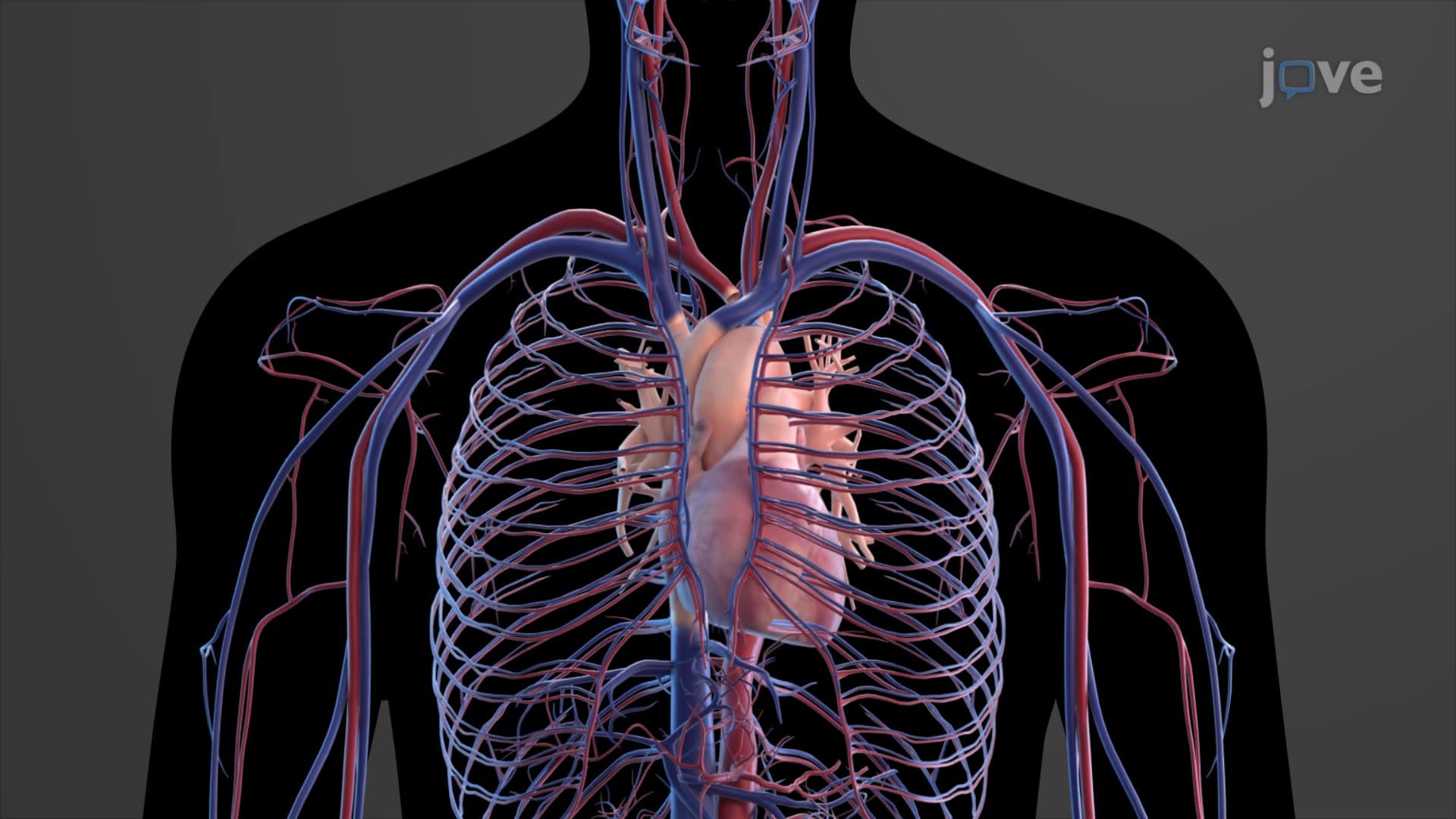
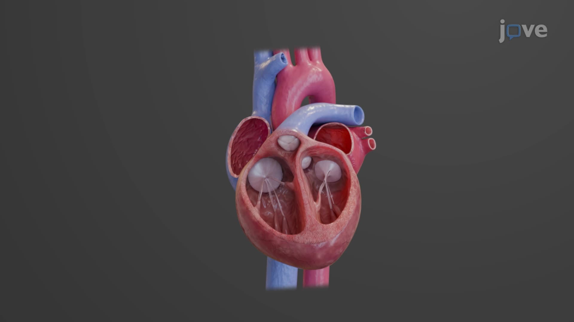
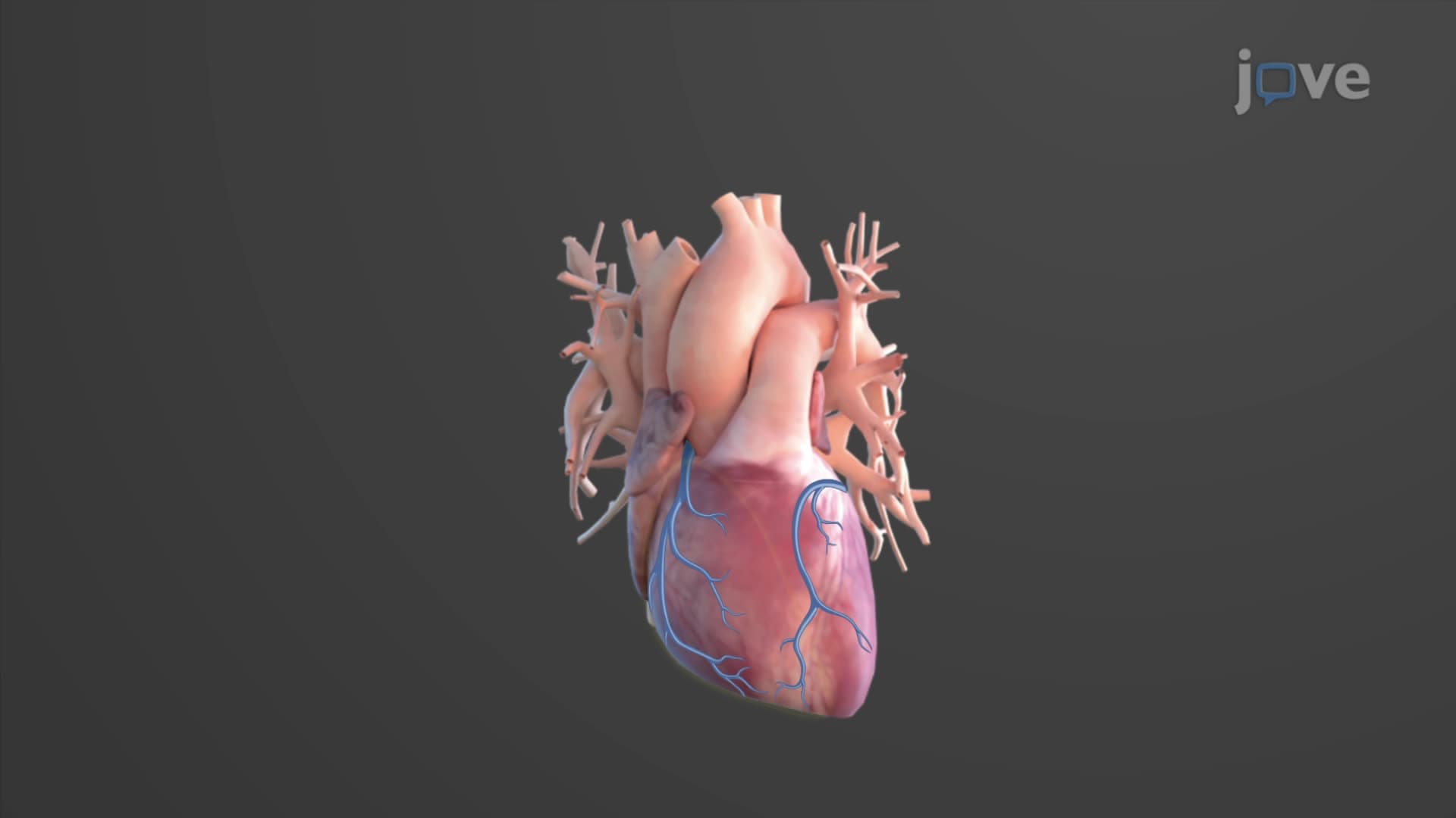
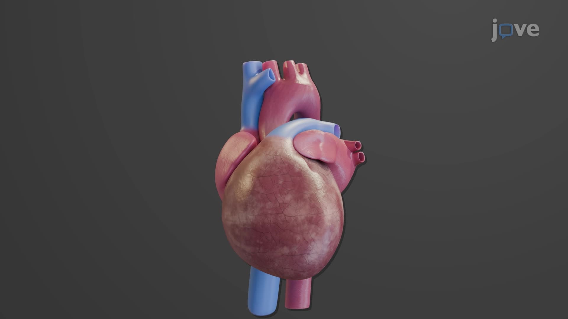
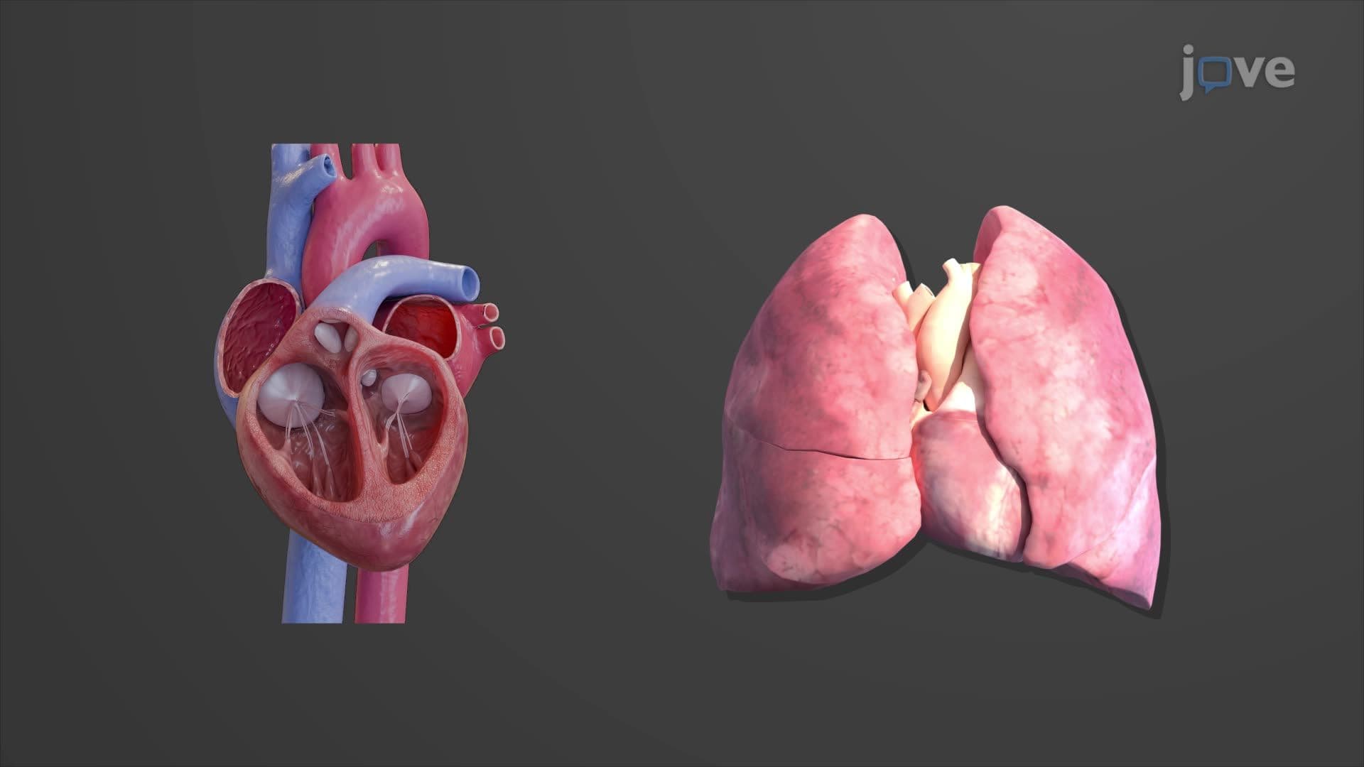
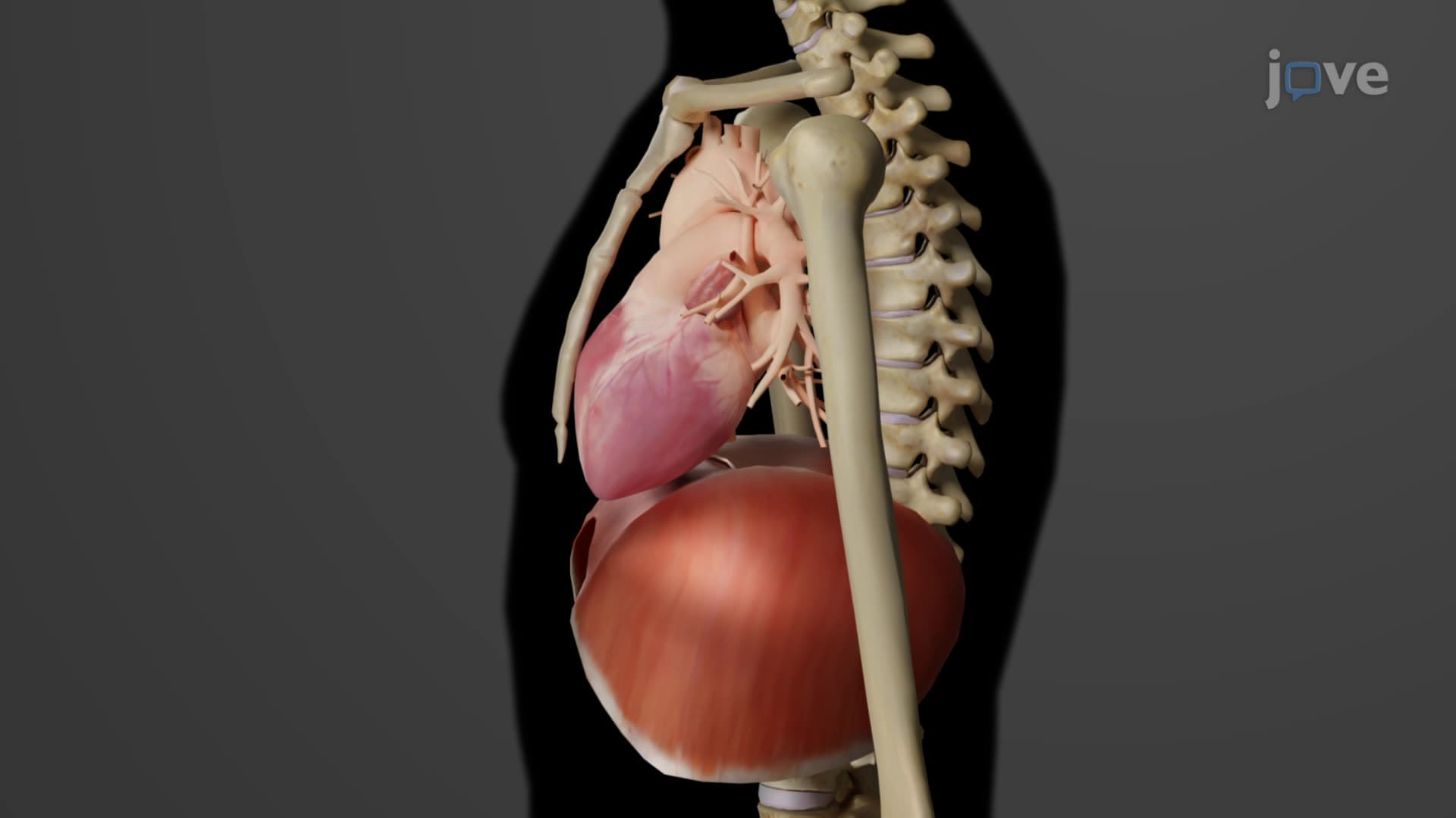
The heart location orientation anatomy represents one of the most critical concepts in cardiovascular physiology. The heart occupies a specific region called the mediastinum, which serves as the central compartment of the thoracic cavity. This mediastinum heart location positions the organ strategically between the lungs, anterior to the vertebral column, and posterior to the sternum, creating optimal protection while maintaining accessibility for medical procedures.
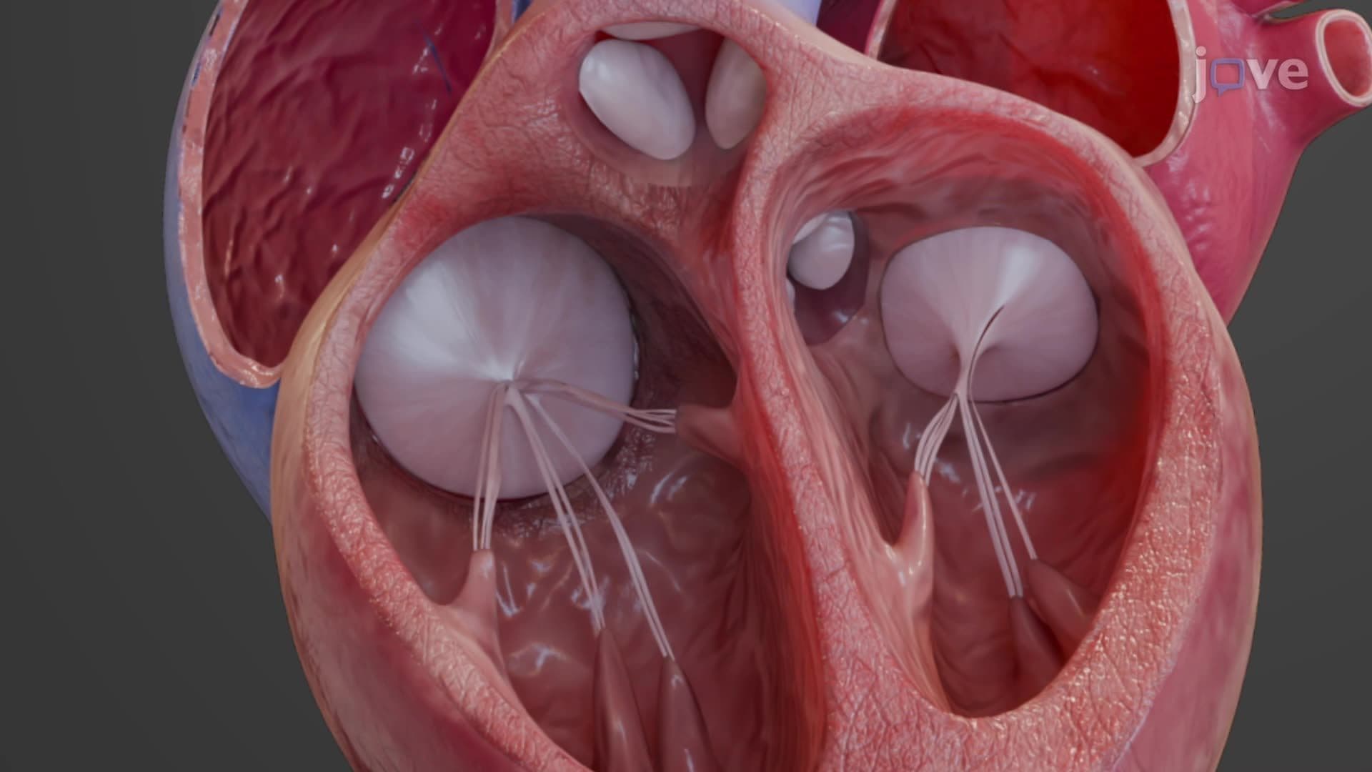
The heart's orientation follows a distinctive pattern that medical professionals must master. The heart apex base orientation shows the heart tilted at approximately 45 degrees to the left of the midline. The base, formed by the atria, points toward the right shoulder and serves as the primary entry and exit point for major blood vessels including the superior and inferior vena cavae, pulmonary veins, and great arteries. Conversely, the apex consists of the left ventricle's tip, angling toward the left hip and resting directly on the diaphragm.
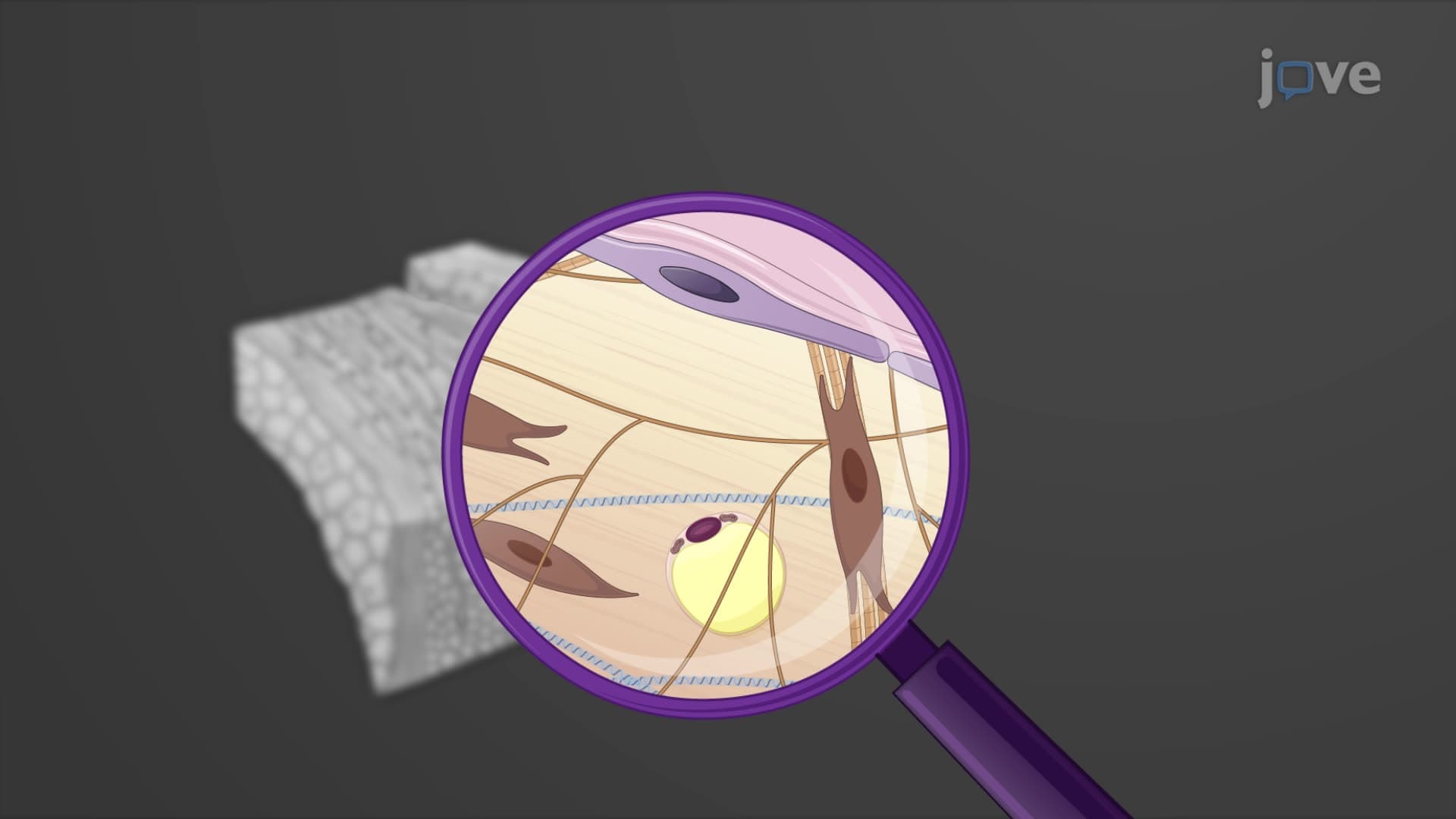
The pericardial sac location encompasses the heart within a sophisticated double-layered protective system. The fibrous pericardium forms the tough outer layer, preventing excessive cardiac expansion and anchoring the heart within the mediastinum. The serous pericardium consists of two components: the outer parietal layer lining the fibrous pericardium and the inner visceral layer (epicardium) directly covering the heart muscle. The pericardial cavity between these layers contains lubricating fluid essential for smooth cardiac contractions.
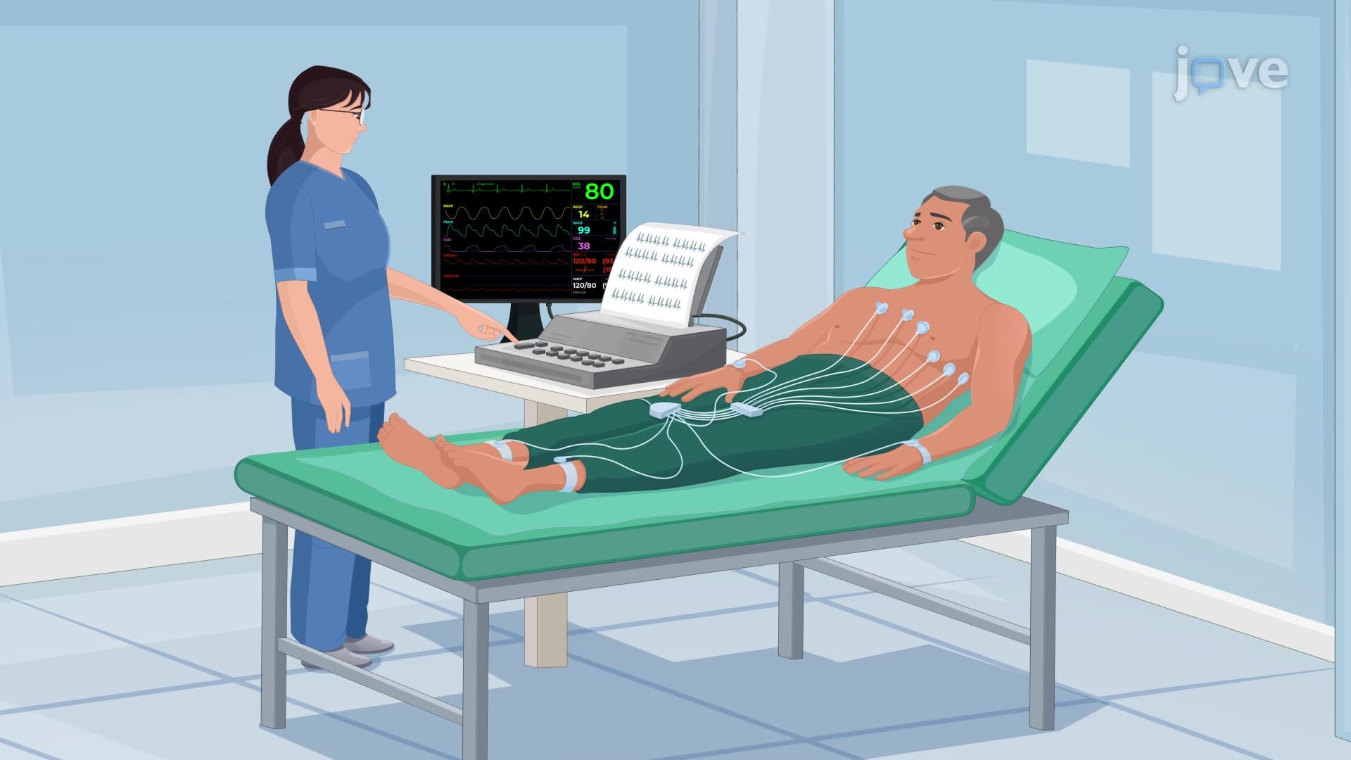
Understanding where the heart is located proves essential for US medical professionals across multiple specialties. Emergency medicine physicians use anatomical landmarks when performing procedures like pericardiocentesis or interpreting electrocardiograms. The left lateral heart position influences how cardiologists position echocardiogram transducers for optimal imaging. Medical students preparing for the USMLE Step 1 must demonstrate comprehensive knowledge of cardiac anatomy, while nursing students taking the NCLEX-RN encounter questions about heart positioning related to patient assessment and care planning.
This anatomical knowledge also supports understanding of pathological conditions. Cardiac tamponade affects the pericardial space, while conditions like dextrocardia involve abnormal heart positioning that impacts diagnostic approaches and surgical planning.
Related Micro-courses