Loading video…


 19 Concepts
 112.2K views

Video Summary: What Is Overview of the Heart
Did you know your heart anatomy overview biology reveals an organ that beats over 100,000 times daily, pumping 2,000 gallons of blood? This remarkable cardiac muscle organ weighs just 10 ounces yet powers your entire circulatory system through four specialized chambers. Understanding what is an overview of the heart in anatomy helps explain how this cone-shaped powerhouse maintains life in every American from newborns in Boston Children's Hospital to athletes training at the U.S. Olympic Training Center. Watch the full video on JoVE Coach to master this concept with expert-led visuals and step-by-step explanations.
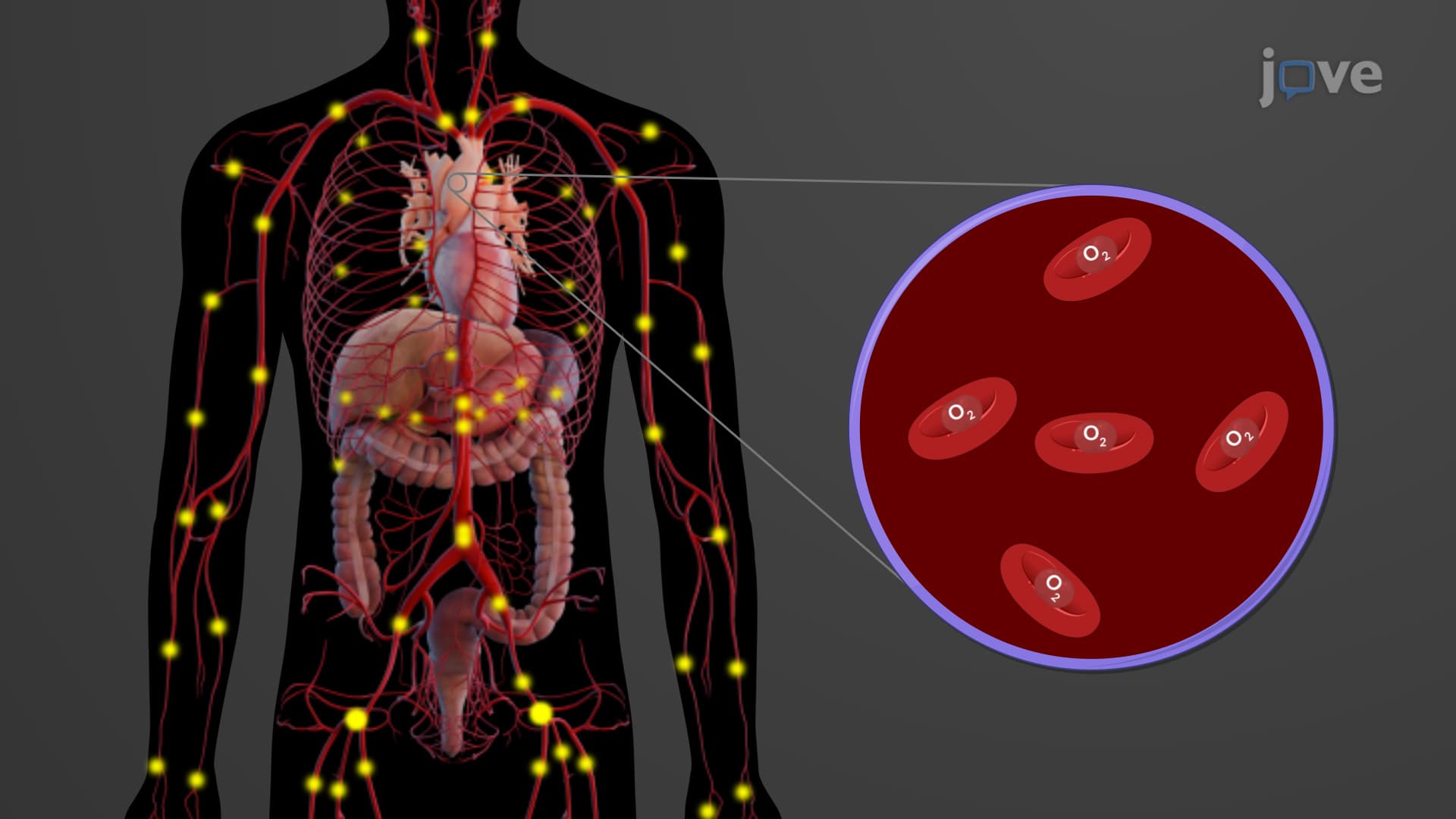
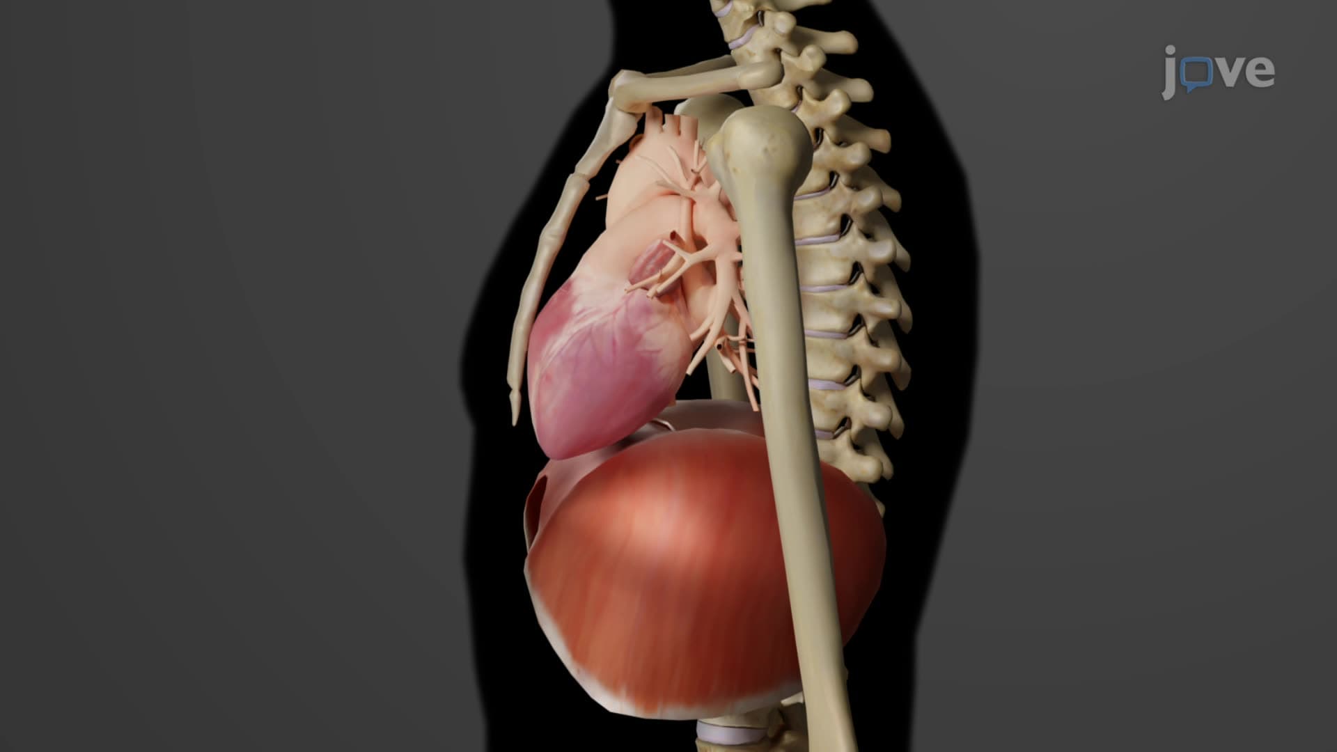
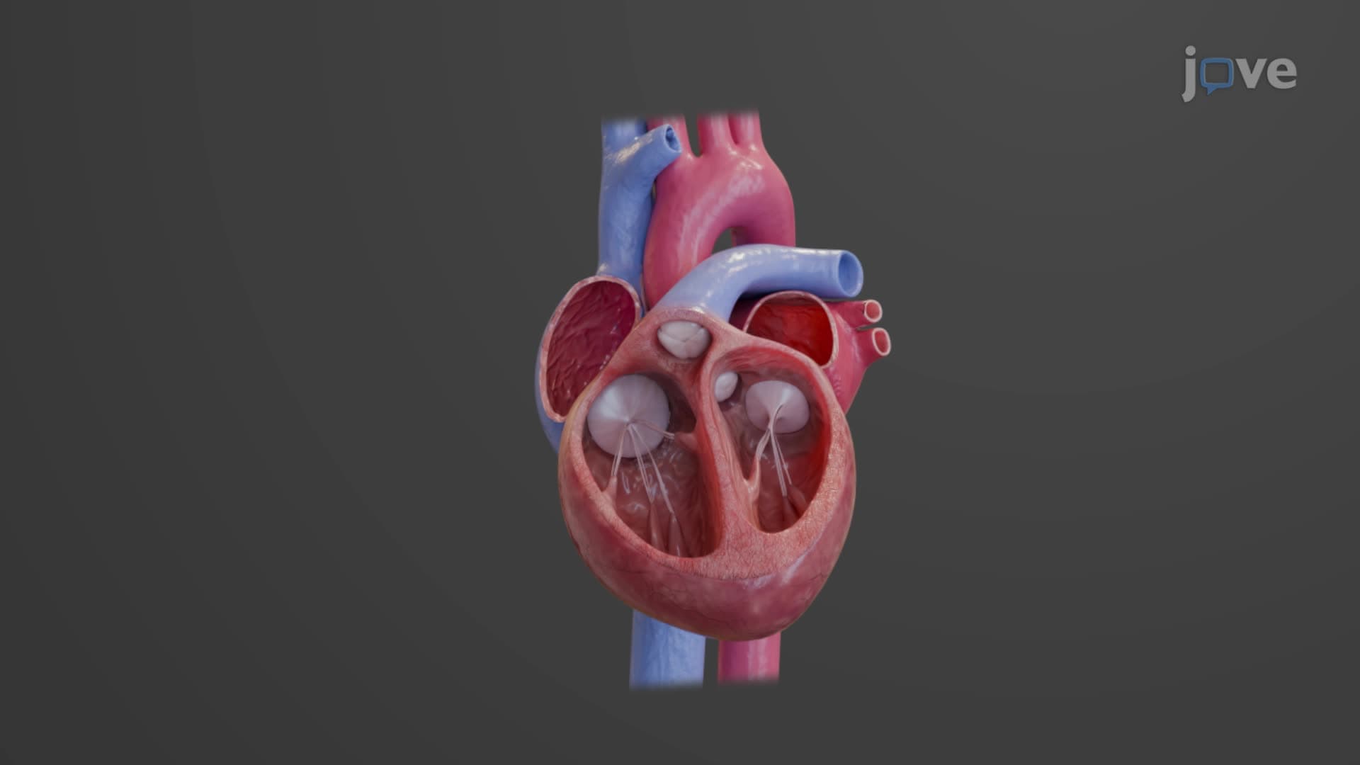
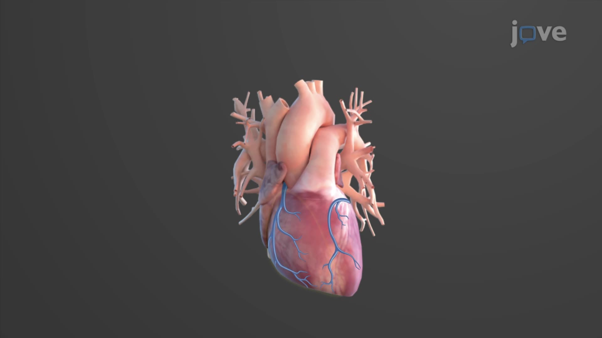
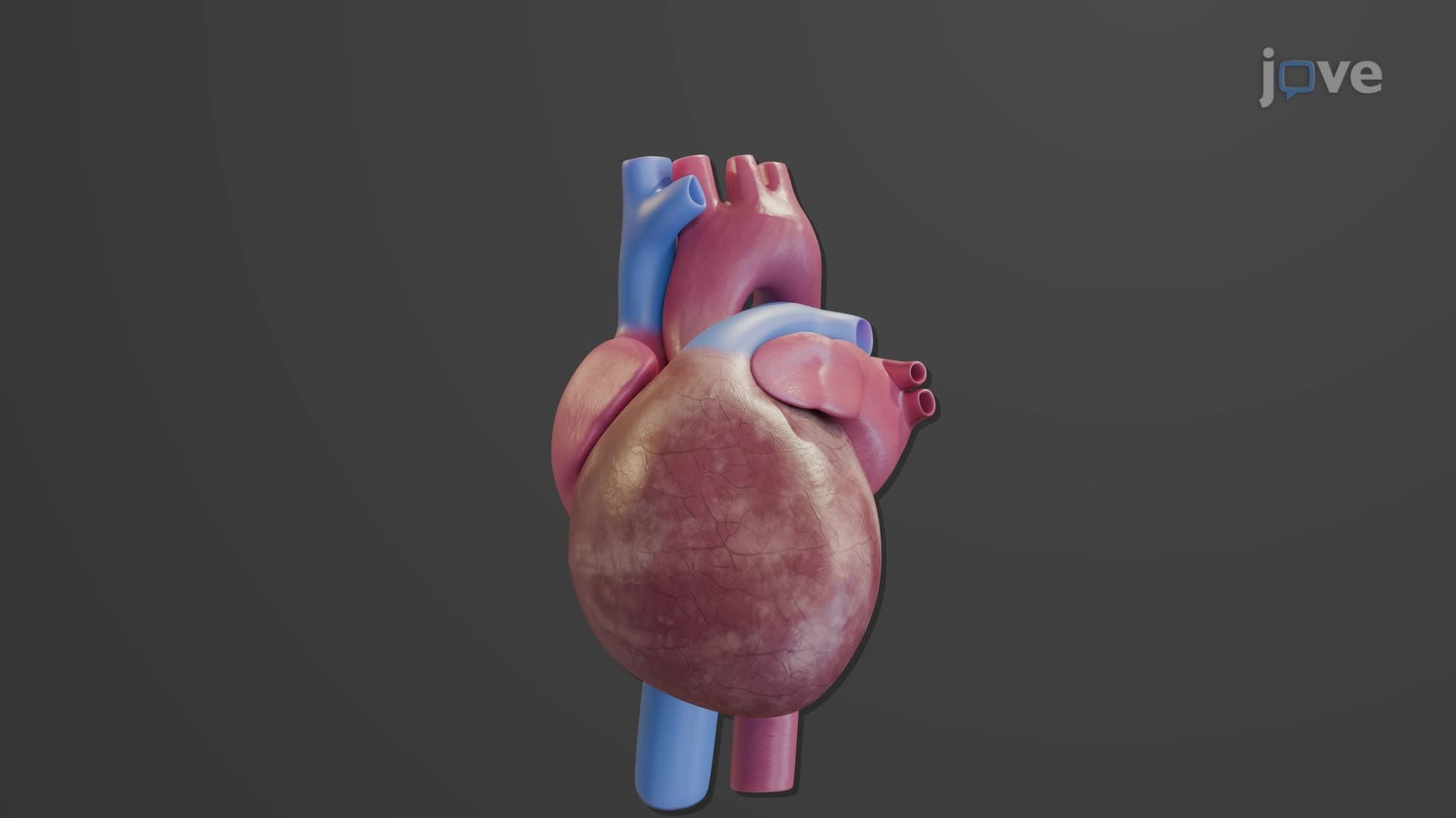
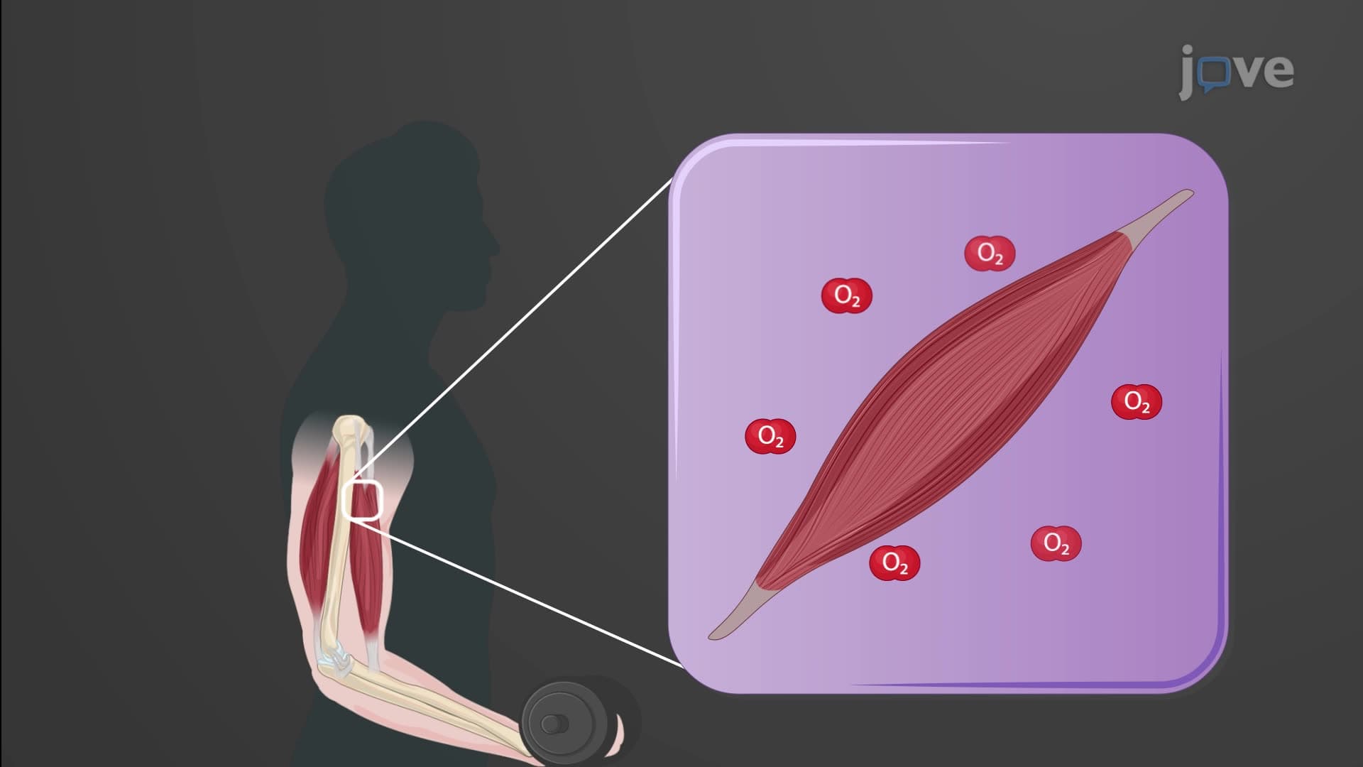
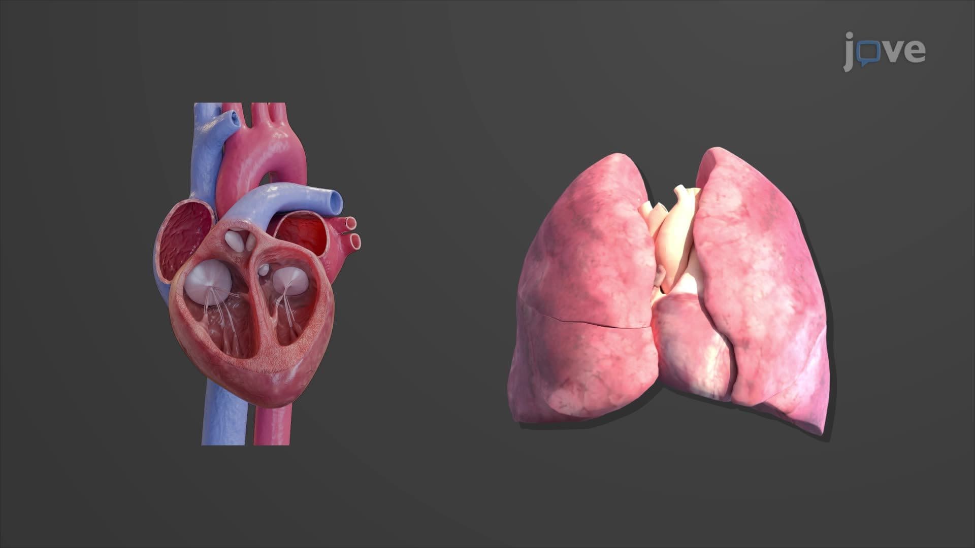
The heart anatomy overview biology reveals one of nature's most efficient mechanical pumps. This hollow, muscular organ serves as the central component of the cardiovascular system, continuously circulating blood to sustain cellular metabolism. Located in the mediastinum between the lungs, the heart's strategic positioning allows optimal blood distribution throughout the body.
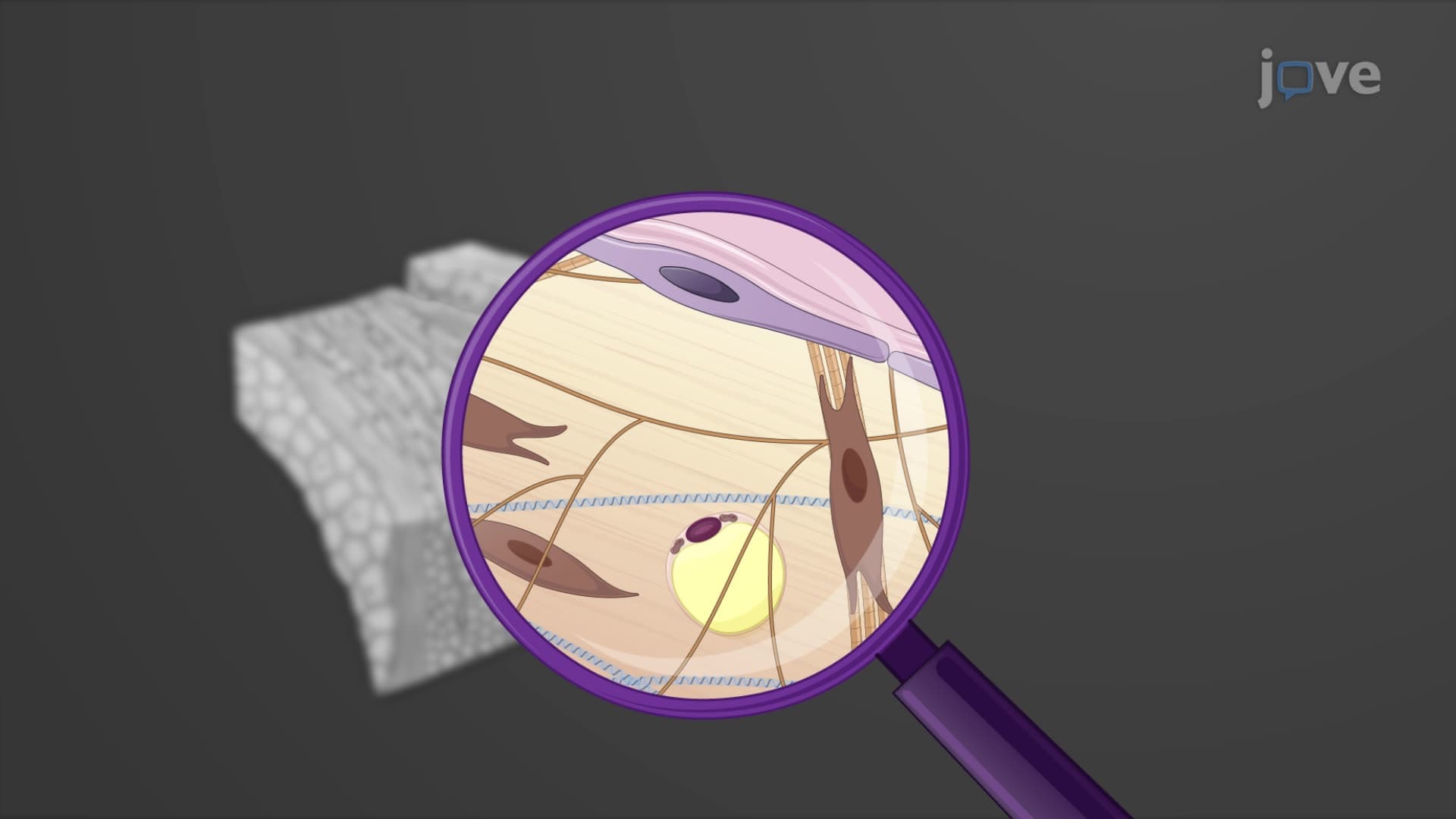
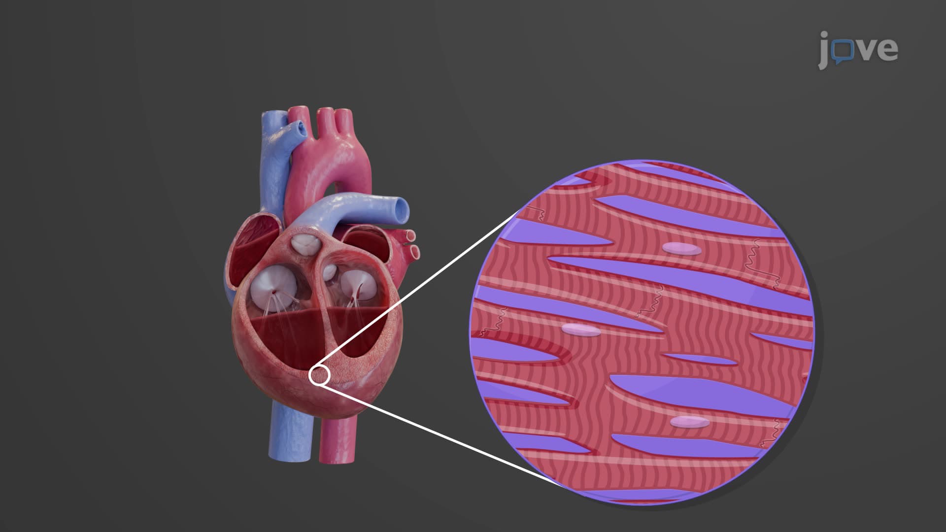
The heart's distinctive cone shape maximizes pumping efficiency while fitting within the chest cavity's constraints. Weighing approximately 250-300 grams (roughly 10-11 ounces), this cardiac muscle organ measures about 12 cm in length, 9 cm in width, and 6 cm in thickness-roughly the size of a clenched fist. The heart wall consists of three layers: the epicardium (outer layer), myocardium (muscular middle layer), and endocardium (smooth inner lining). The pericardium heart covering provides additional protection and reduces friction during cardiac contractions.
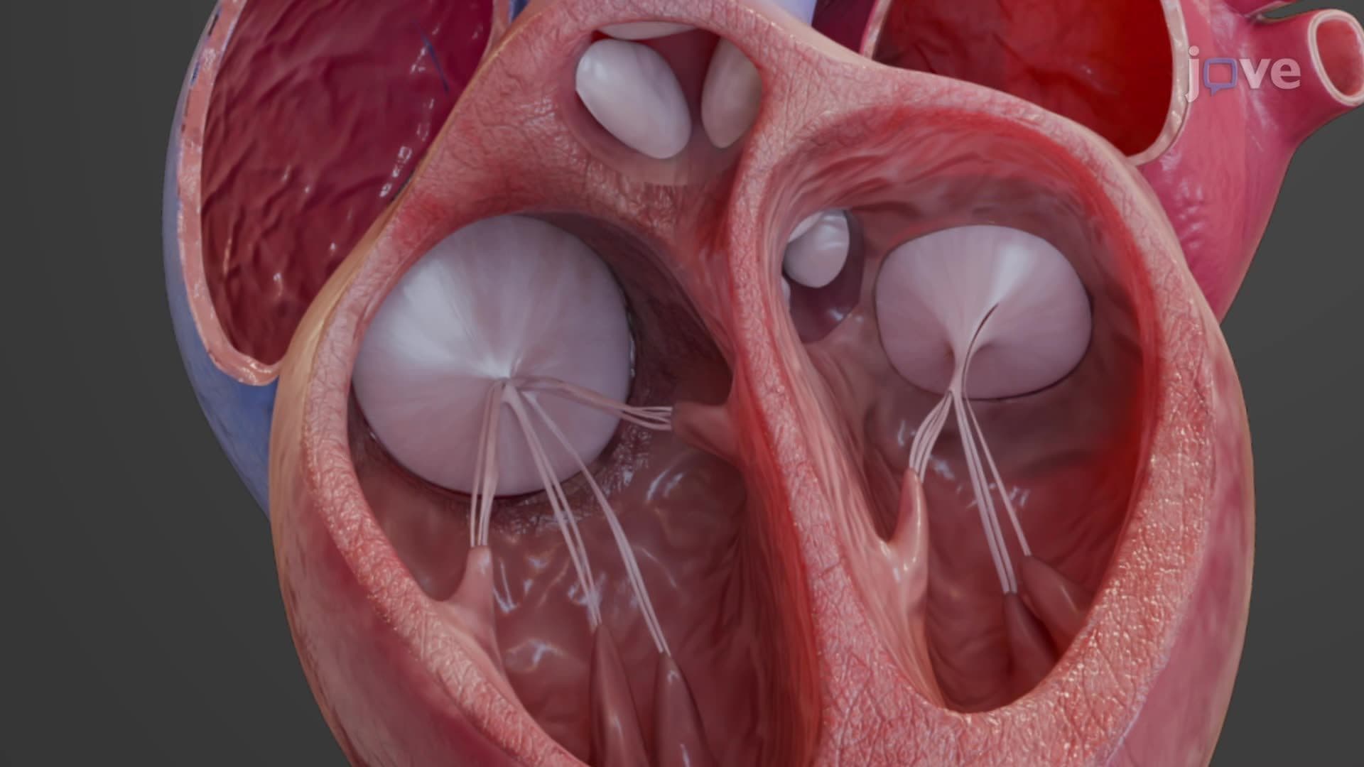
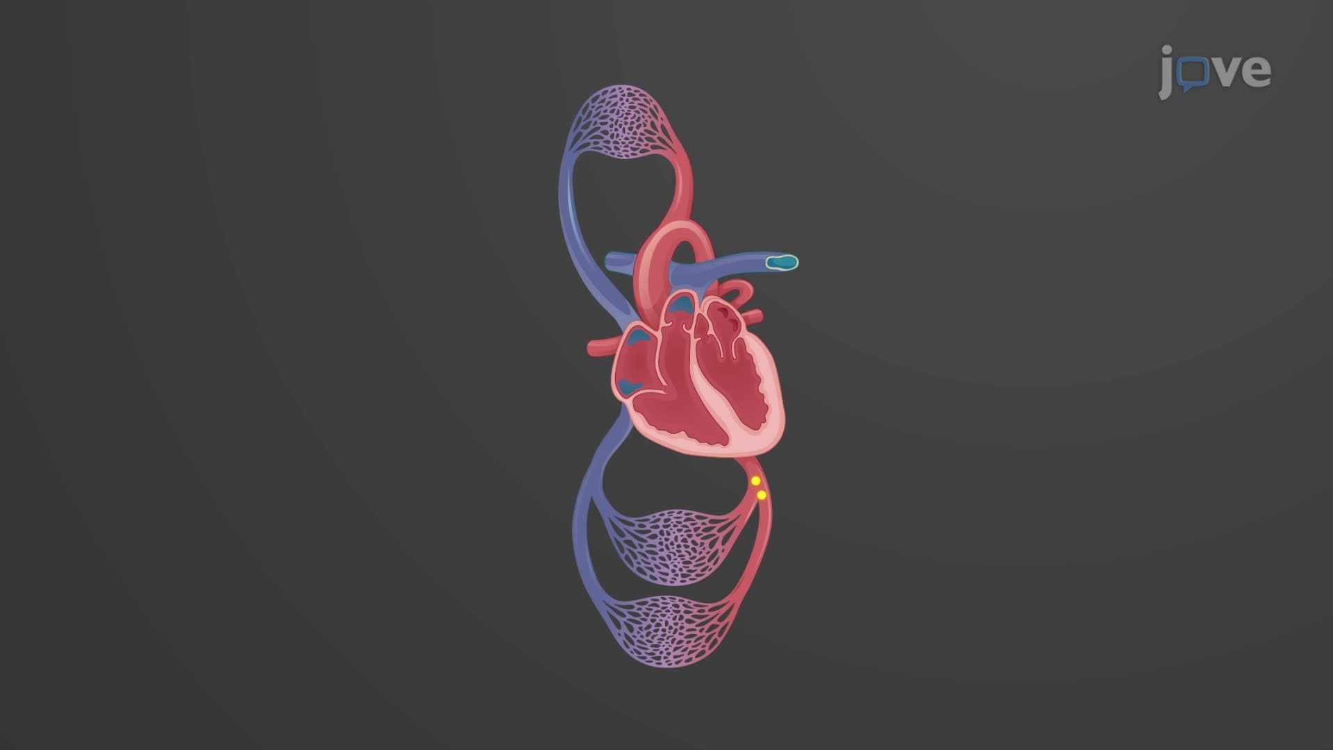
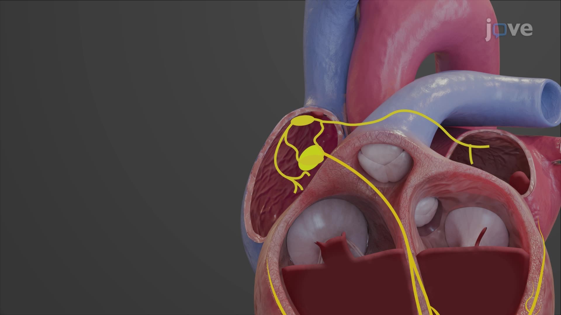
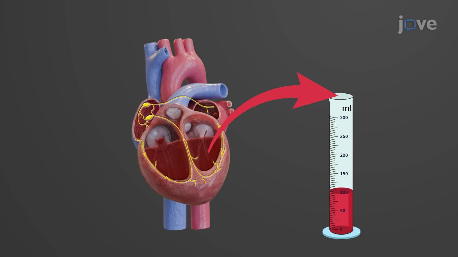
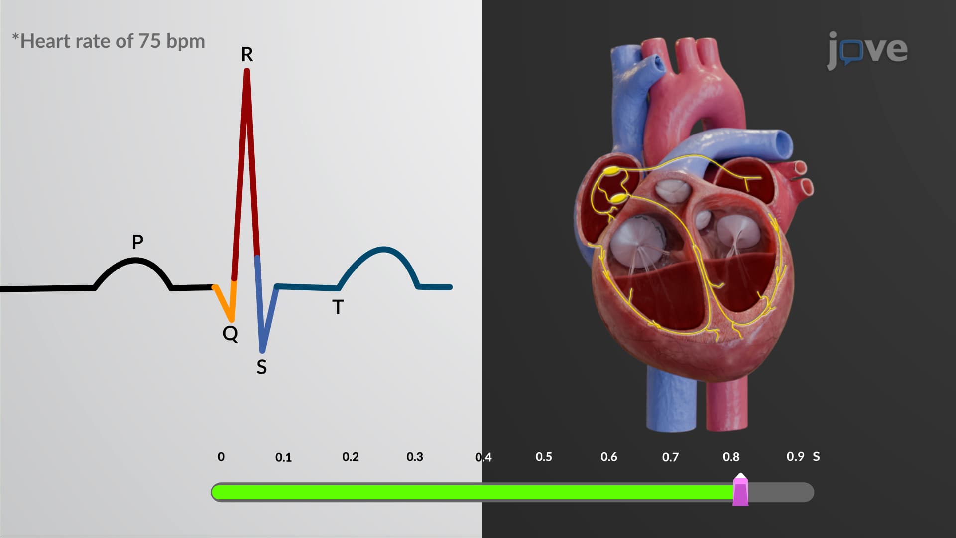
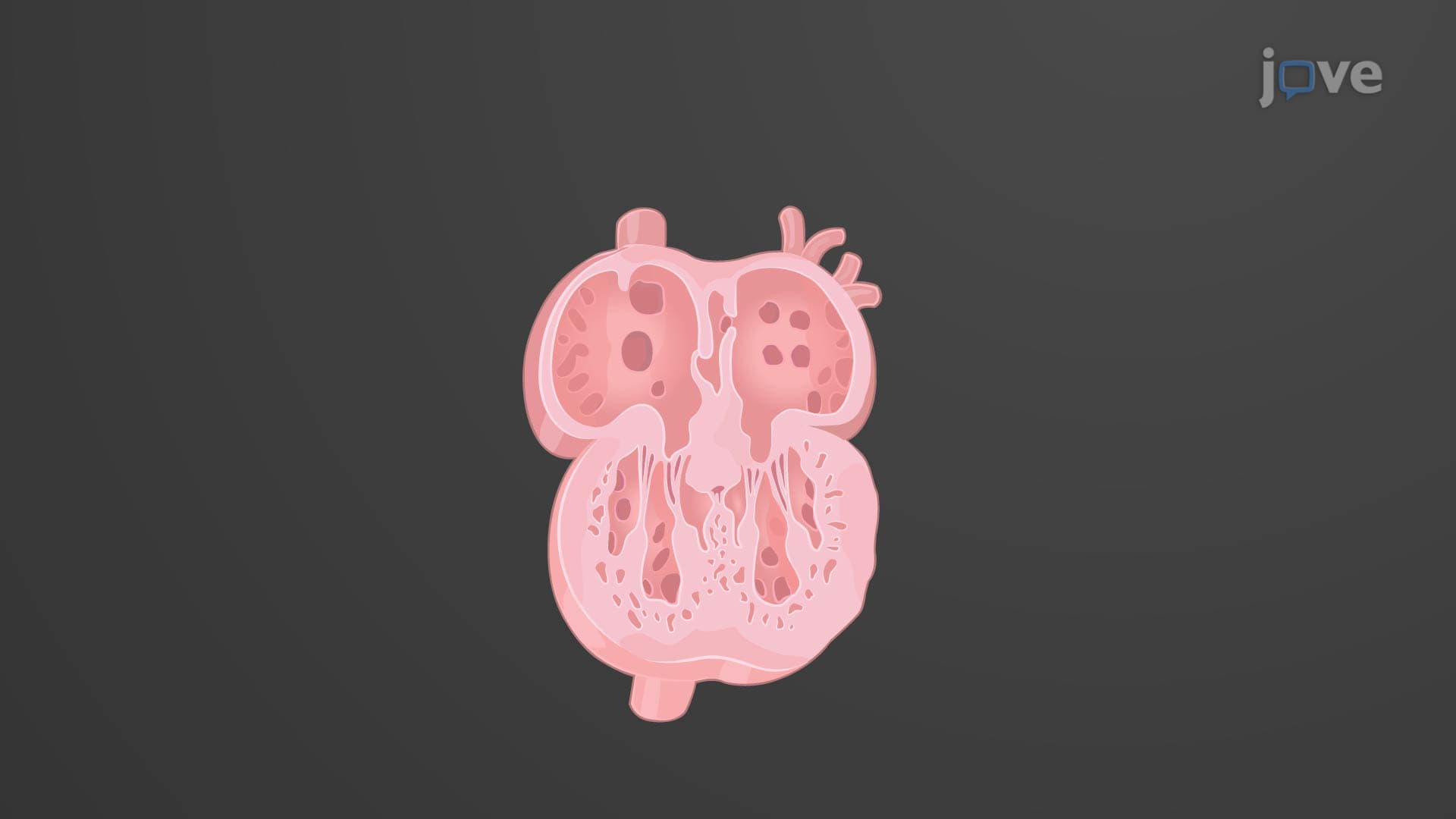
The heart four chambers create a sophisticated dual-pump system. The right side handles pulmonary circulation, while the left side manages systemic circulation. The right atrium receives deoxygenated blood from the superior and inferior vena cavae, then transfers it to the right ventricle. From there, blood travels through the pulmonary arteries to the lungs for oxygenation. The newly oxygenated blood returns via pulmonary veins to the left atrium, flows into the left ventricle, and gets pumped through the aorta to supply the entire body. This concept frequently appears on MCAT Biological Sciences sections and AP Biology exams, particularly in questions about circulatory system efficiency.
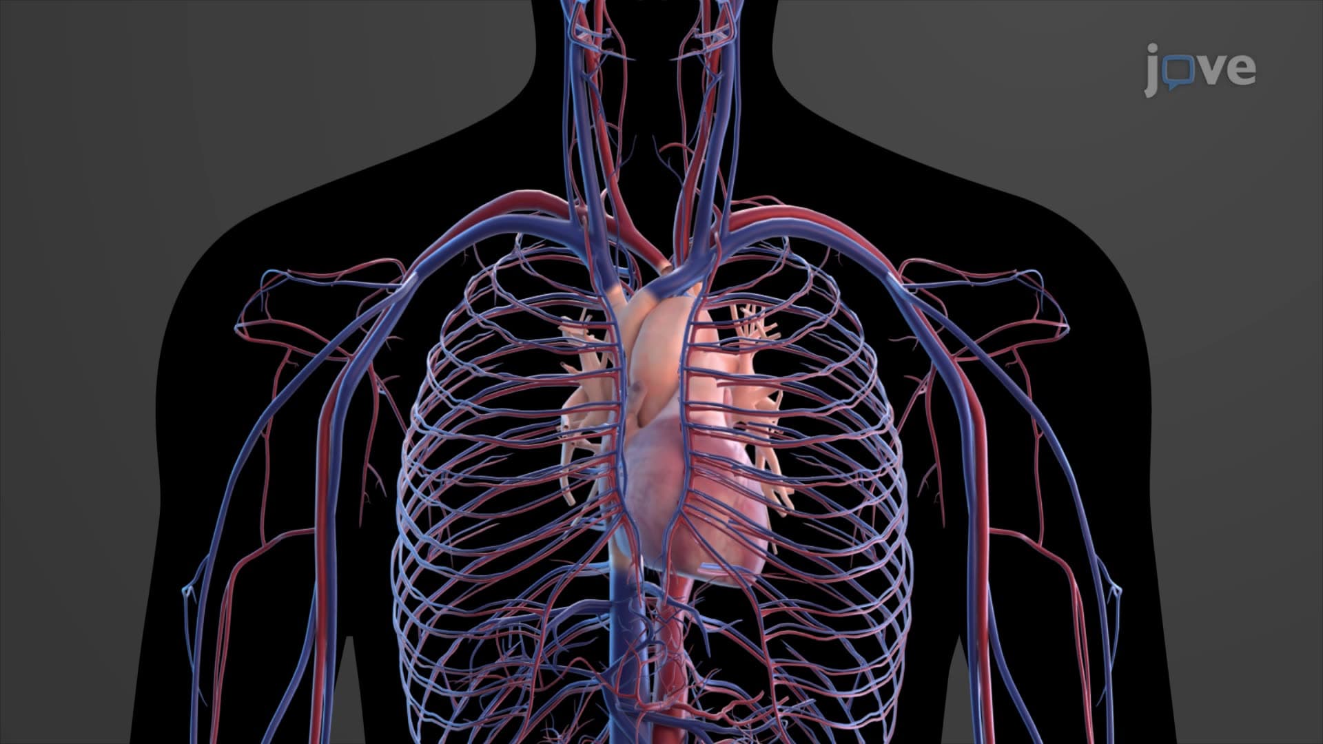
The great vessels heart connections include the aorta, pulmonary arteries, pulmonary veins, and vena cavae. These major vessels work with smaller arteries, veins, and capillaries to form an extensive network reaching every tissue. Arteries carry blood away from the heart under high pressure, while veins return blood to the heart at lower pressure. Capillaries, with their thin walls, facilitate crucial gas and nutrient exchange at the cellular level. Understanding this vascular hierarchy helps students excel in college anatomy courses and prepares pre-med students for USMLE Step 1 cardiovascular physiology questions.
Related Micro-courses