Loading subject page…


162,824 views

Video Summary: What are Cytoskeletal Accessory Proteins
Ever wonder how your muscle cells maintain their incredible strength during a marathon, or how nerve cells stretch across your entire arm without breaking? Cytoskeletal accessory proteins are the molecular engineers that make this possible, working behind the scenes to regulate and organize the cell's structural framework. These specialized proteins control everything from muscle contraction in athletes to nerve signal transmission, acting like construction managers that direct when and how cellular scaffolding should be built or torn down. Watch the full video on JoVE Coach to master this concept with expert-led visuals and step-by-step explanations.
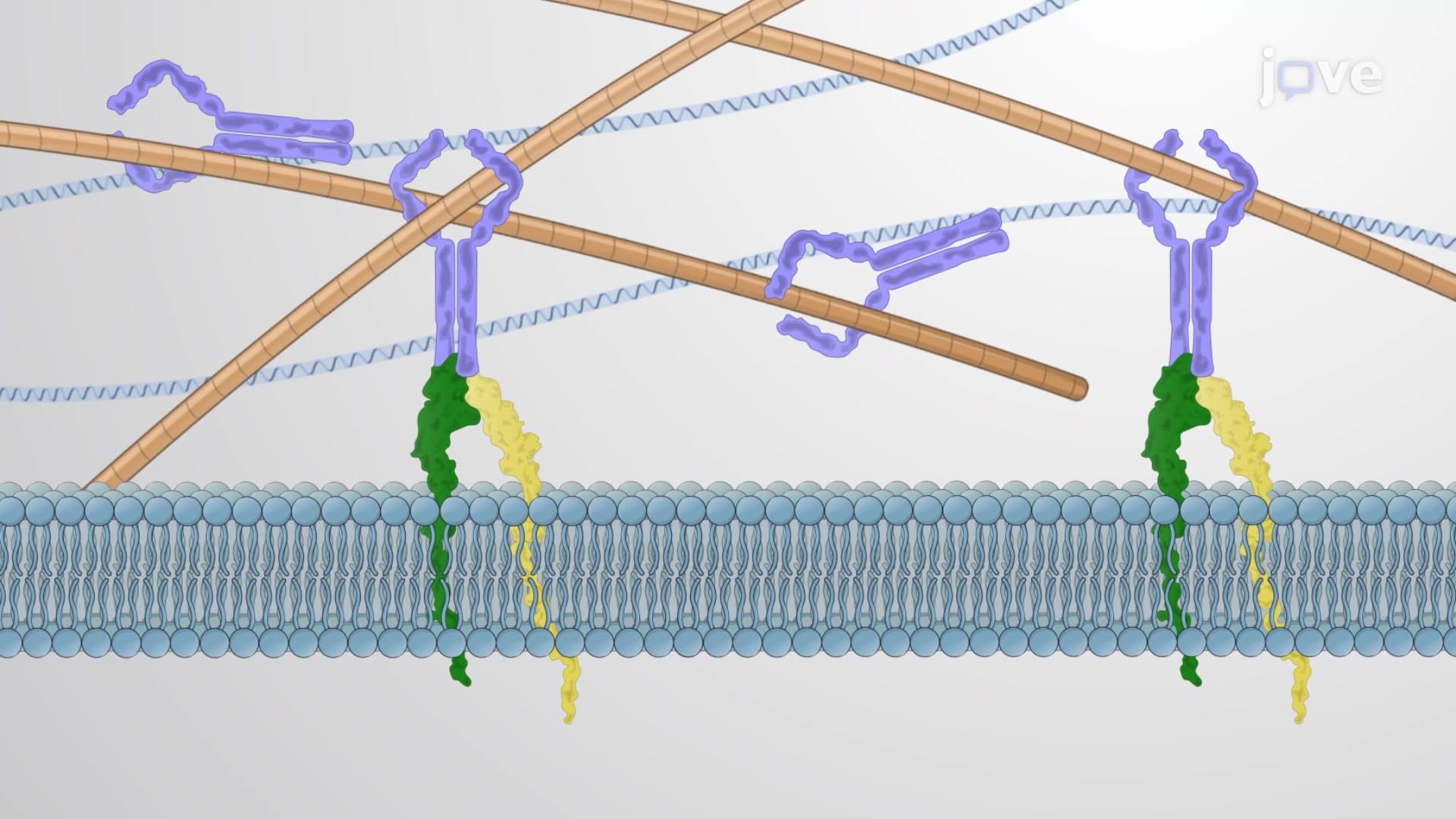
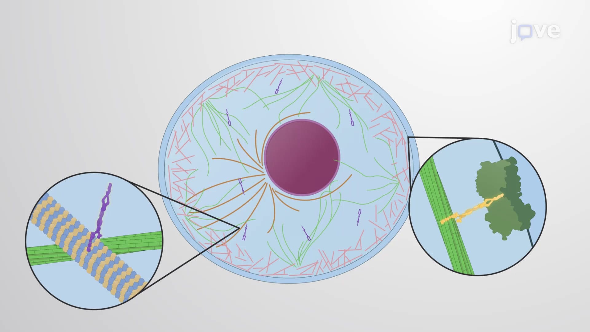
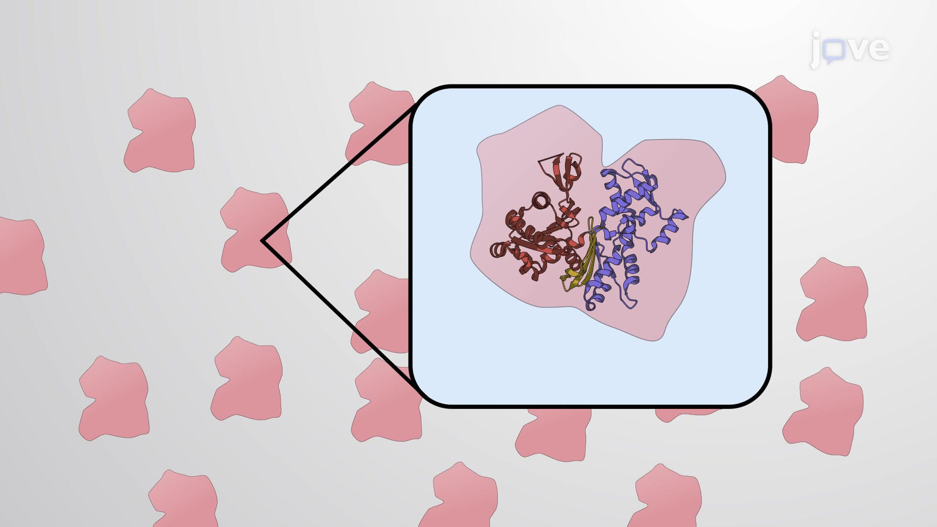
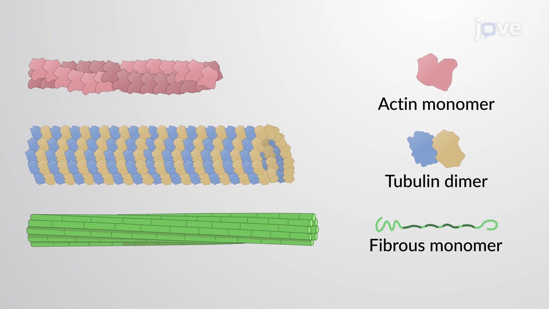
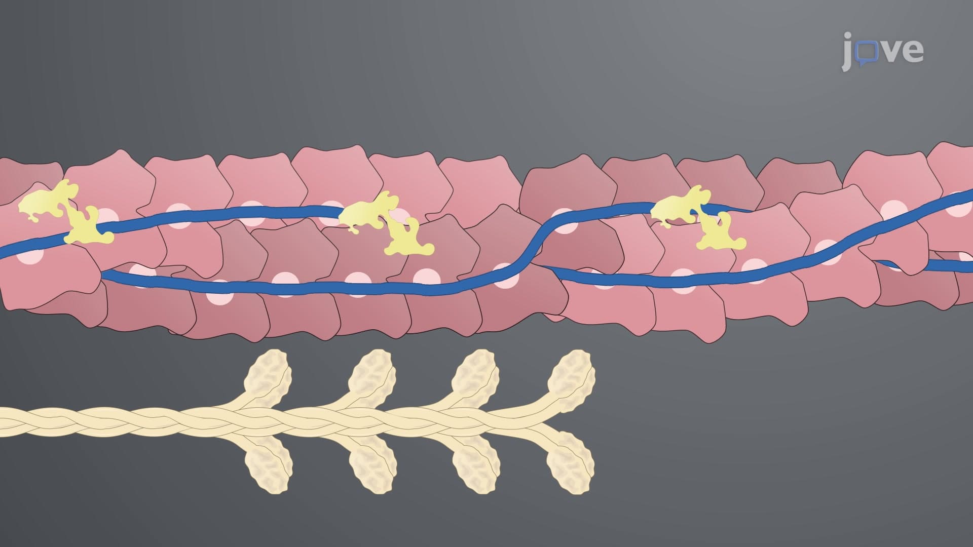
Cytoskeletal accessory proteins represent a diverse family of regulatory molecules that transform the basic cytoskeletal framework into a sophisticated, dynamic system capable of supporting complex cellular functions. Unlike the primary structural components (actin filaments, microtubules, and intermediate filaments), these accessory proteins act as molecular controllers, determining when, where, and how the cytoskeleton assembles, functions, and reorganizes.
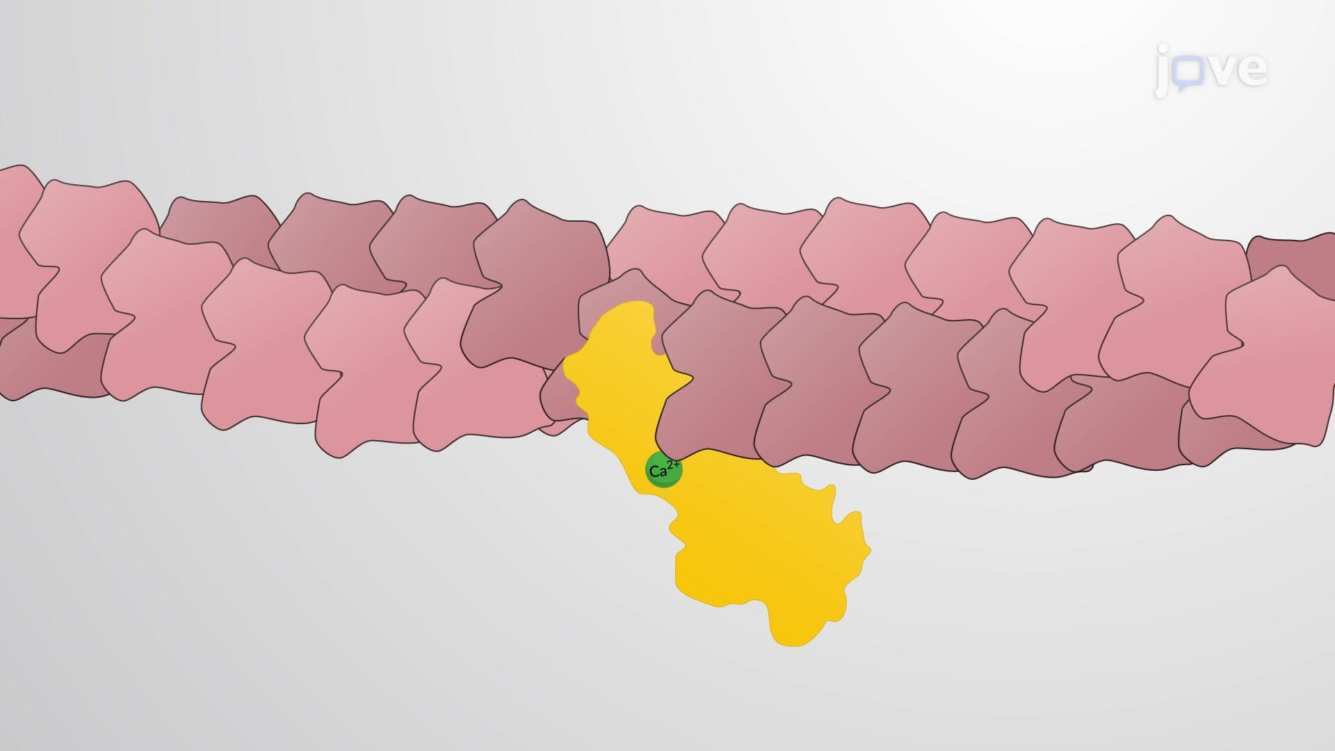
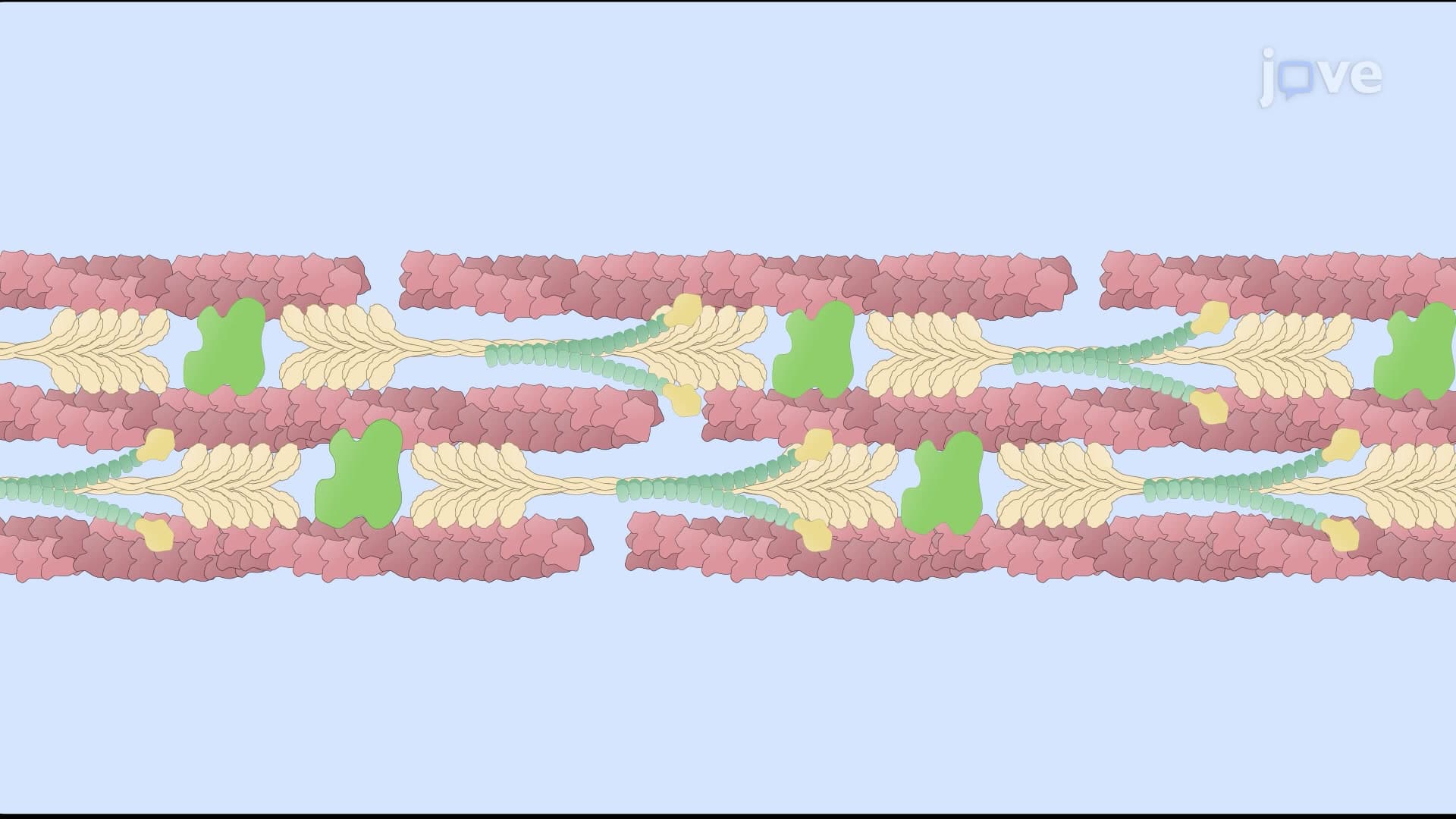
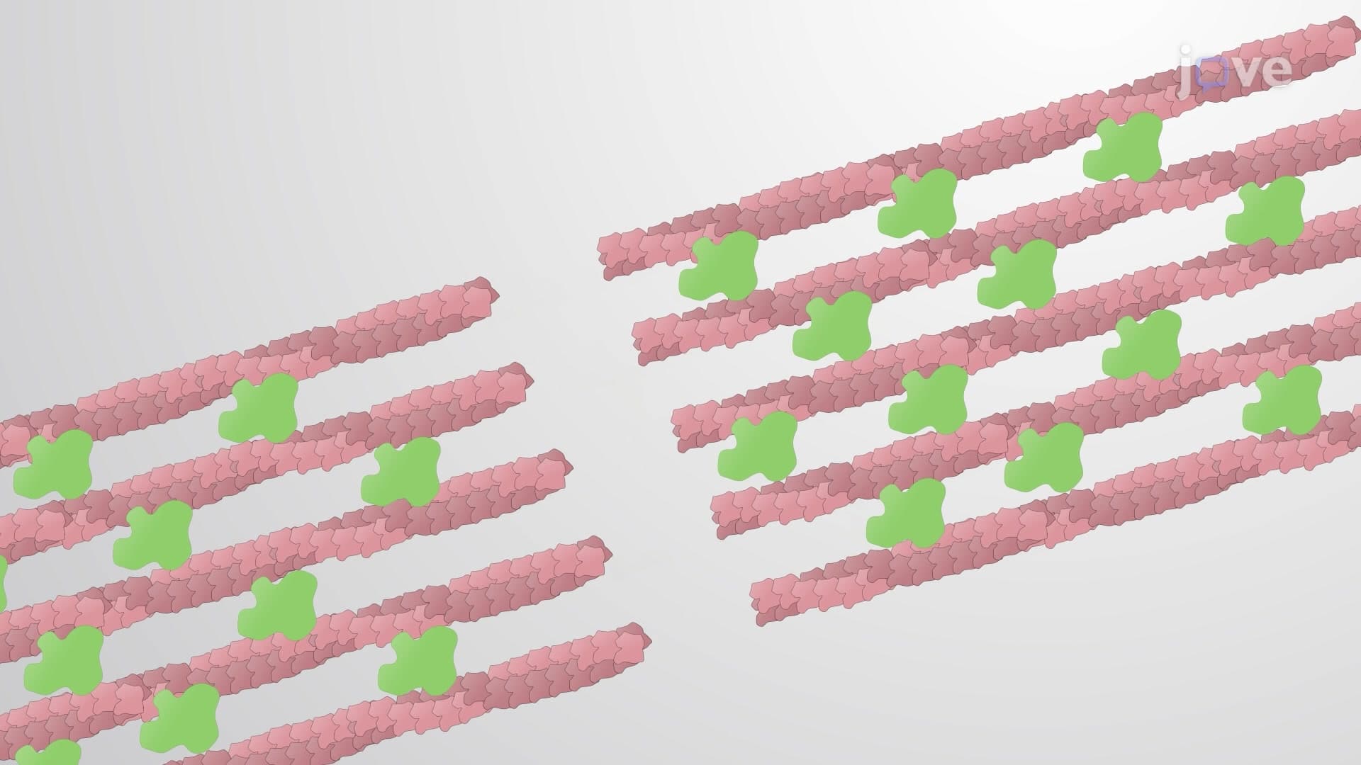
Different types of cytoskeletal accessory proteins exhibit remarkable specificity in their binding patterns. Alpha-actinin exemplifies this specificity by exclusively binding to actin filaments, where it creates loose, flexible bundles essential for muscle contraction and cell motility. This protein is particularly crucial in cardiac muscle cells, where its proper function prevents heart failure-a concept frequently tested on the MCAT and in advanced physiology courses.
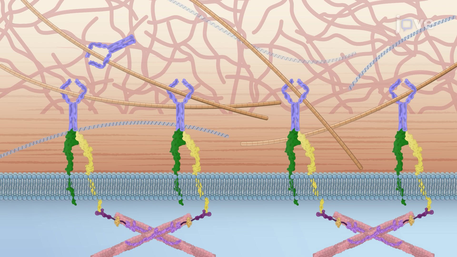
Conversely, the plakin family demonstrates remarkable versatility by cross-linking different cytoskeletal filament types. These proteins serve as molecular bridges, connecting actin filaments to intermediate filaments and anchoring the entire network to cellular junctions like desmosomes. In skin cells, plakin dysfunction leads to blistering diseases, making this a clinically relevant example for pre-med students studying for the MCAT.
The fascinating aspect of cytoskeletal accessory proteins lies in how different proteins can crosslink identical filaments to create structures with vastly different mechanical properties. Fascin creates rigid, tightly packed actin bundles that provide tremendous mechanical strength-imagine the structural integrity needed in the microvilli of intestinal cells that must withstand constant mechanical stress during digestion.
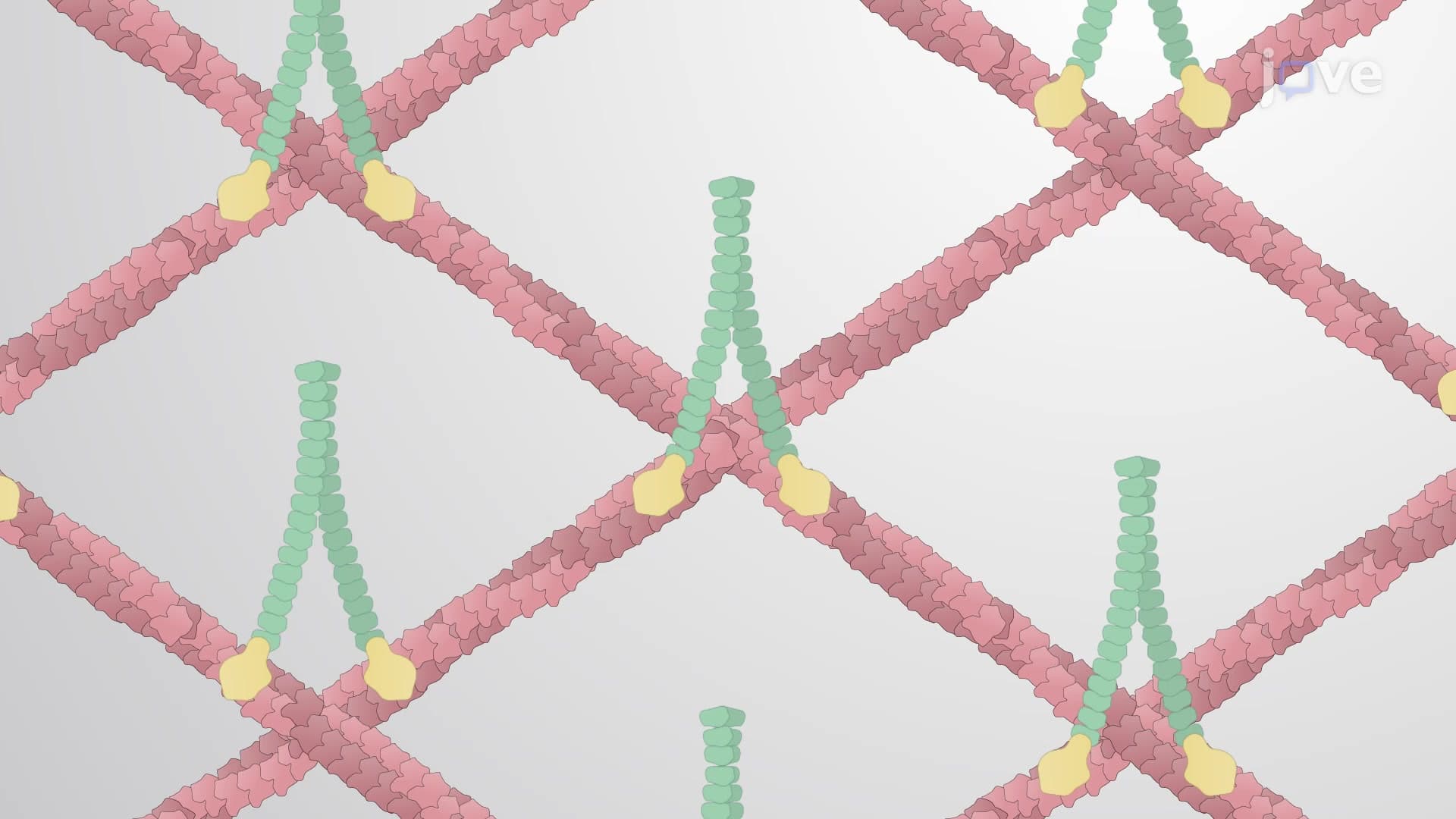
In contrast, filamin generates a flexible, gel-like network from the same actin filaments, creating a structure that can bend and stretch without breaking. This flexibility is essential in white blood cells that must squeeze through tiny blood vessel gaps during immune responses, a process called diapedesis that appears frequently on AP Biology exams.
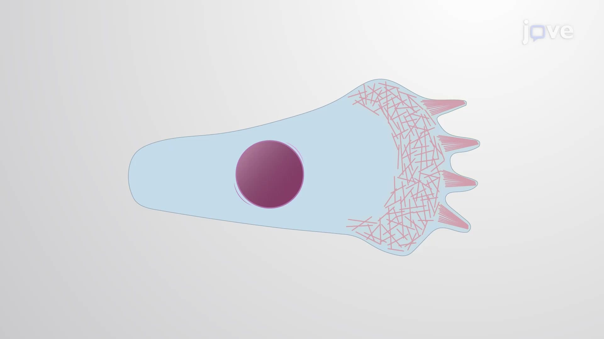
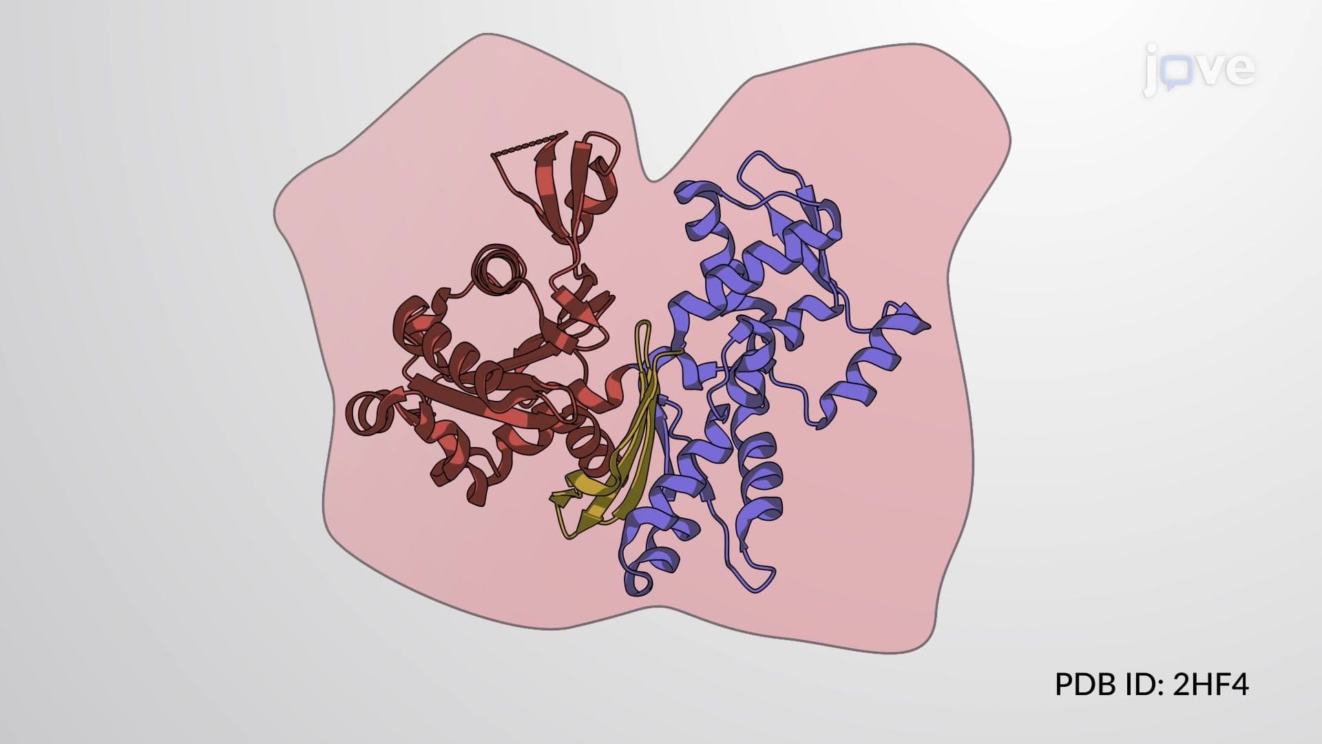
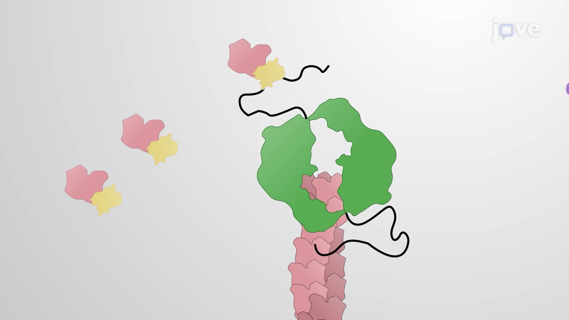
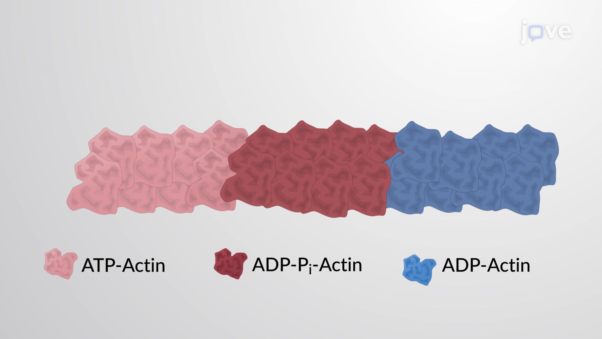
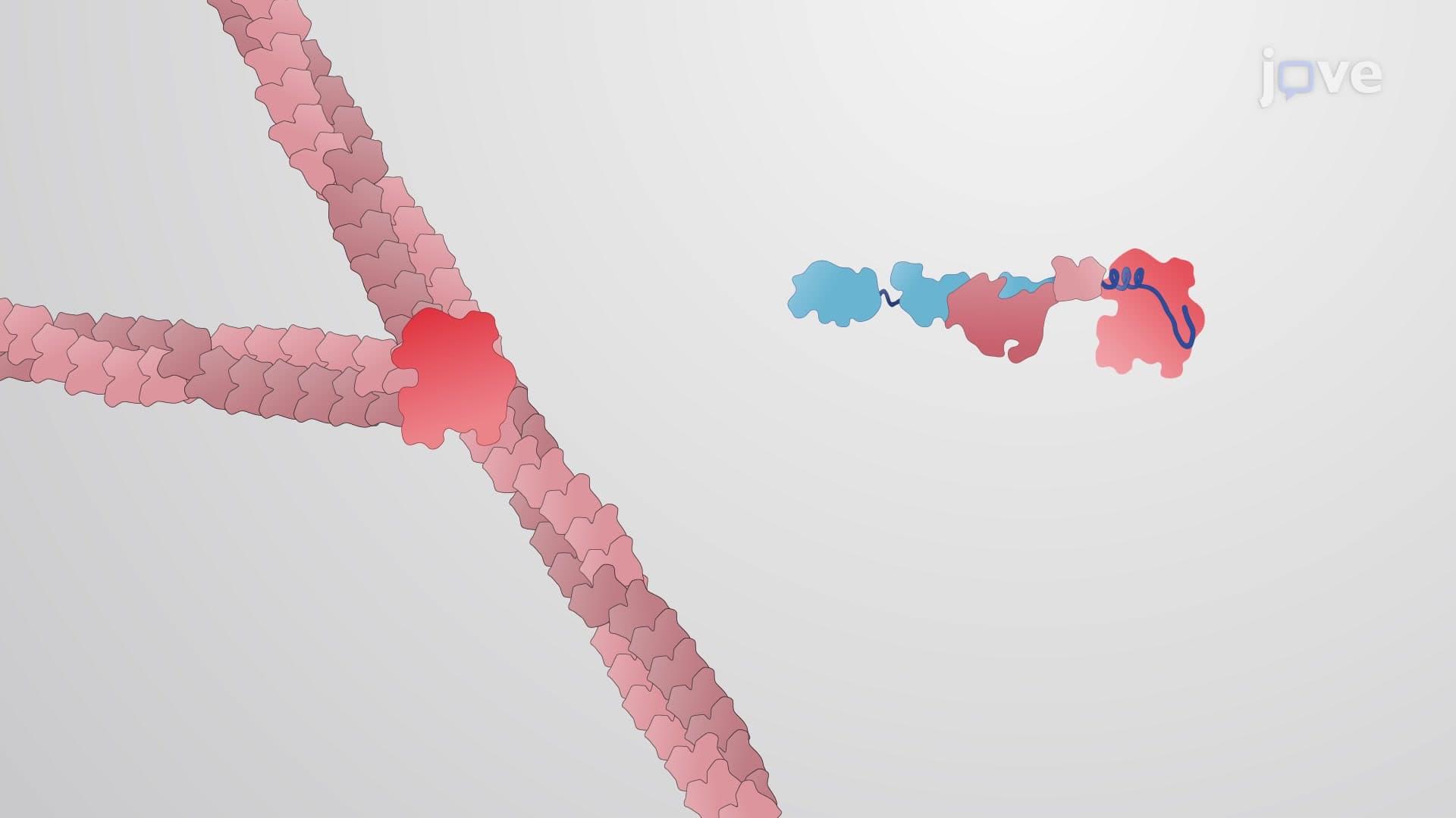
Cytoskeletal accessory proteins also function as molecular switches, controlling when filaments grow or shrink. Profilin accelerates actin polymerization by helping monomers add to growing filament ends, while cofilin acts as molecular scissors, severing actin filaments to promote disassembly. This dynamic balance is crucial during cell division, where rapid cytoskeletal reorganization must occur within minutes-a process extensively studied in college cell biology courses and featured on standardized tests like the GRE Subject Test in Biology.
Related Micro-courses