Loading video…


 18 Concepts
 162.9K views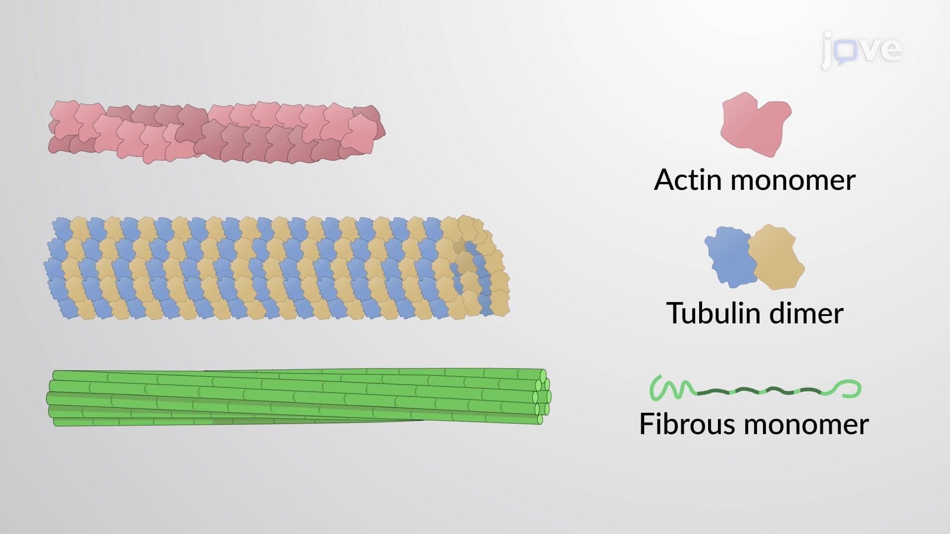

Video Summary: What Is Assembly of Cytoskeletal Filaments
Did you know that every time you flex your bicep or a neuron fires in your brain, millions of protein filaments are assembling and disassembling in perfect coordination? The assembly of cytoskeletal filaments involves three distinct protein structures-microfilaments, microtubules, and intermediate filaments-that build and rebuild themselves based on cellular needs. Just like the steel framework of the Golden Gate Bridge provides structural support, these filaments create the internal scaffold that gives cells their shape and enables movement. Watch the full video on JoVE Coach to master this concept with expert-led visuals and step-by-step explanations.
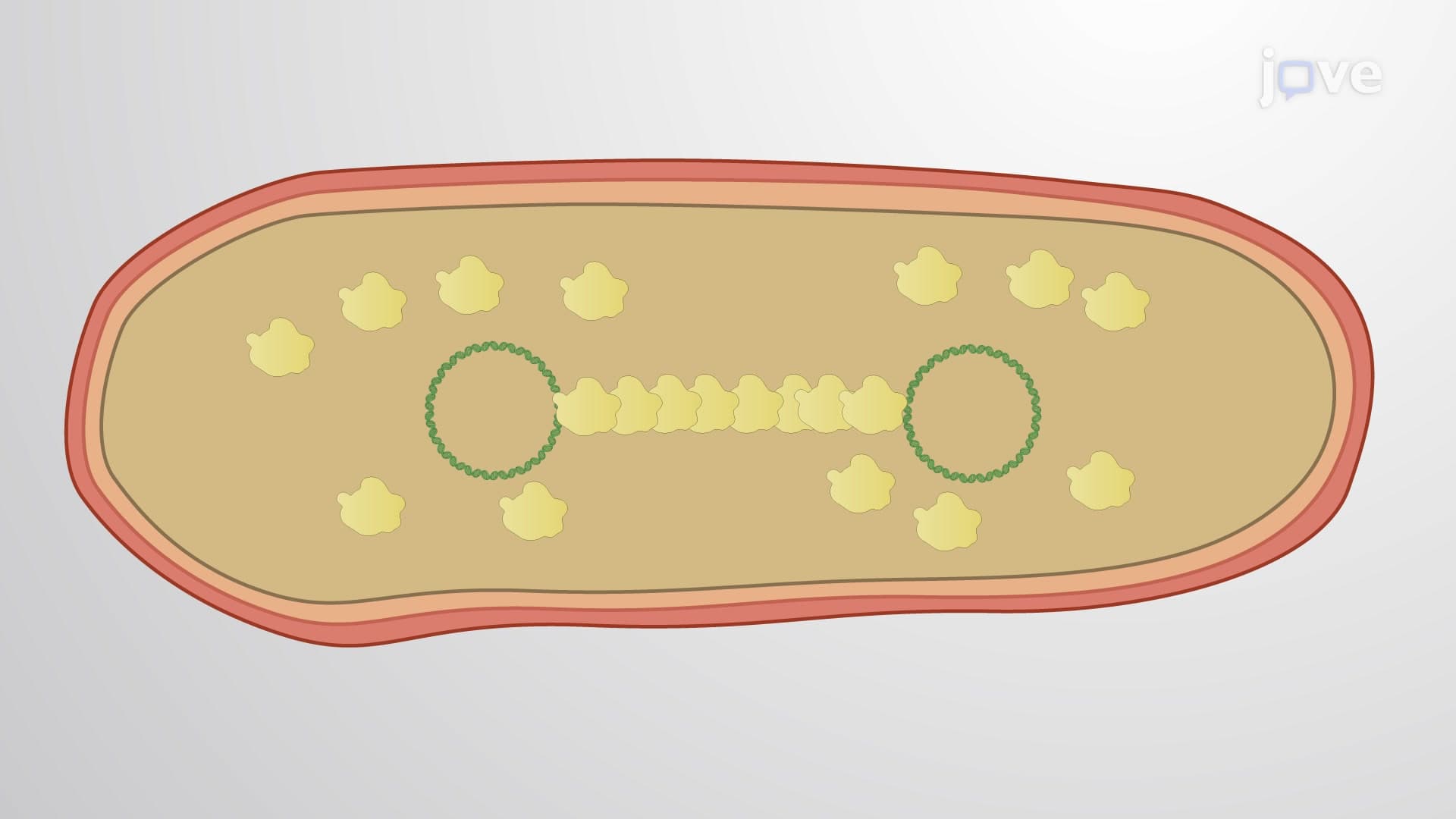
The assembly of cytoskeletal filaments represents one of biology's most elegant examples of self-organizing structures. Unlike static building materials, these protein networks continuously assemble and disassemble, responding to cellular signals with remarkable precision. This dynamic behavior enables everything from muscle contraction during a marathon to the precise division of stem cells in bone marrow.
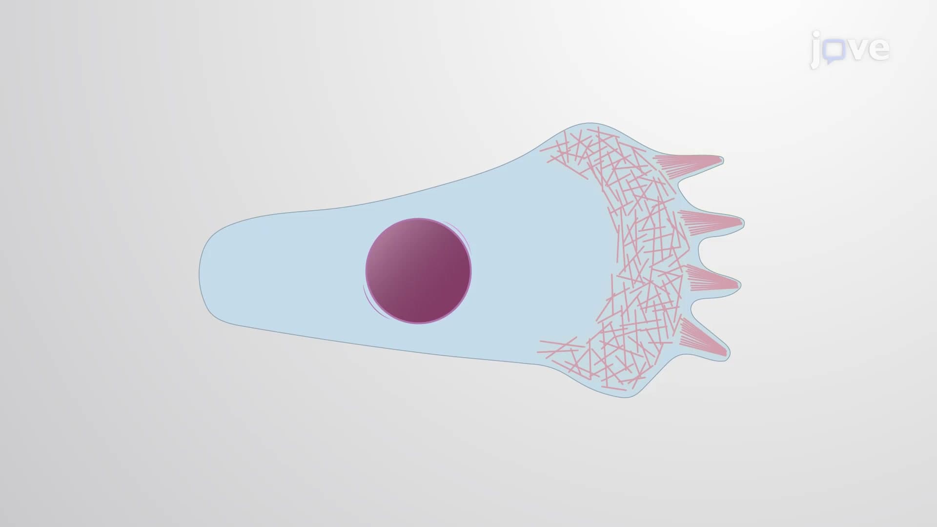
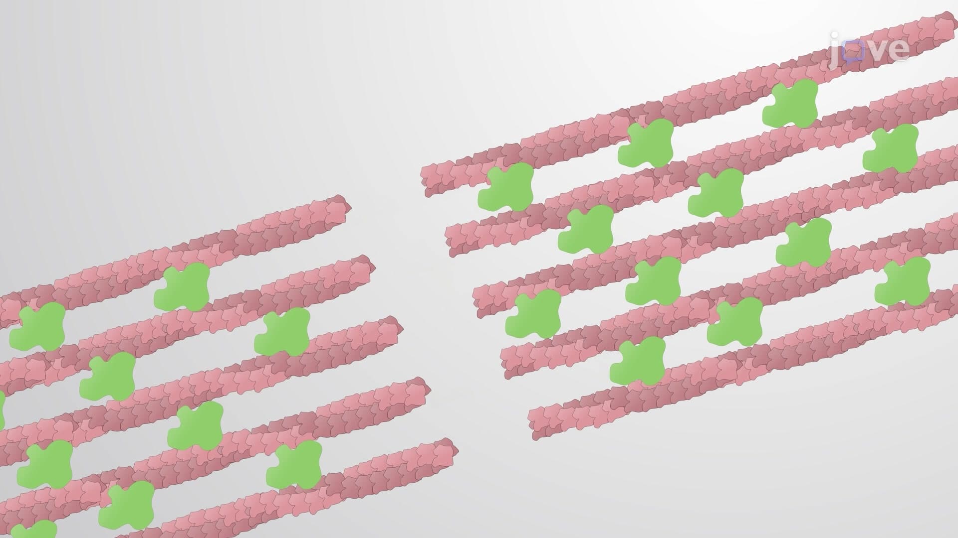
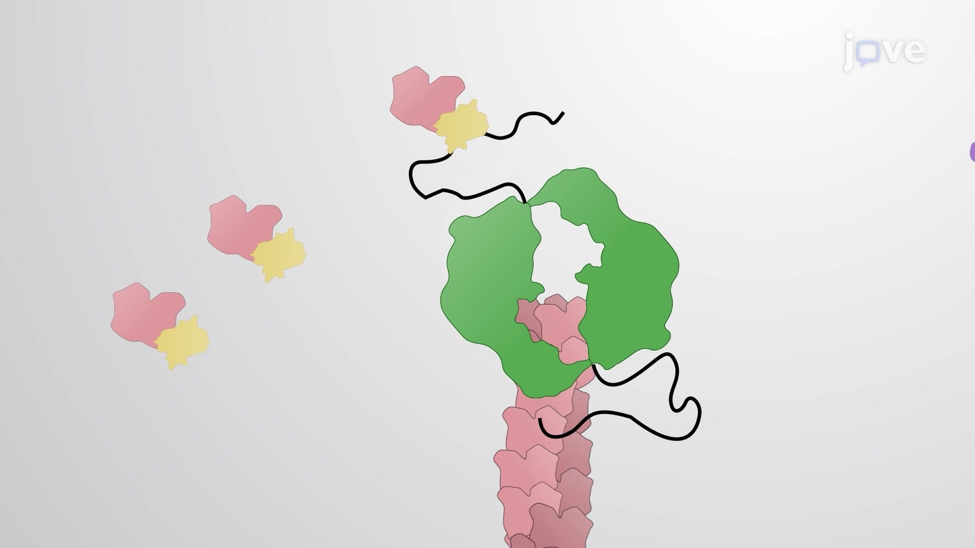
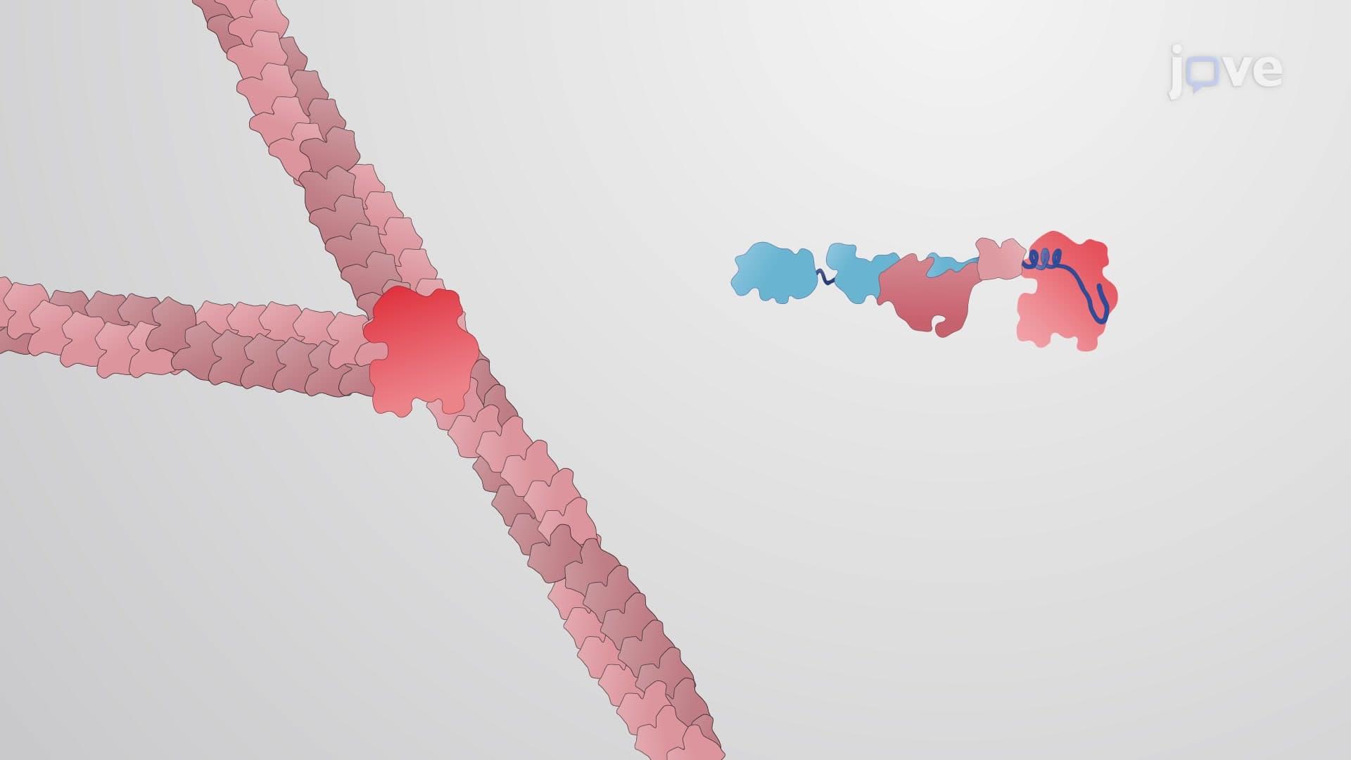
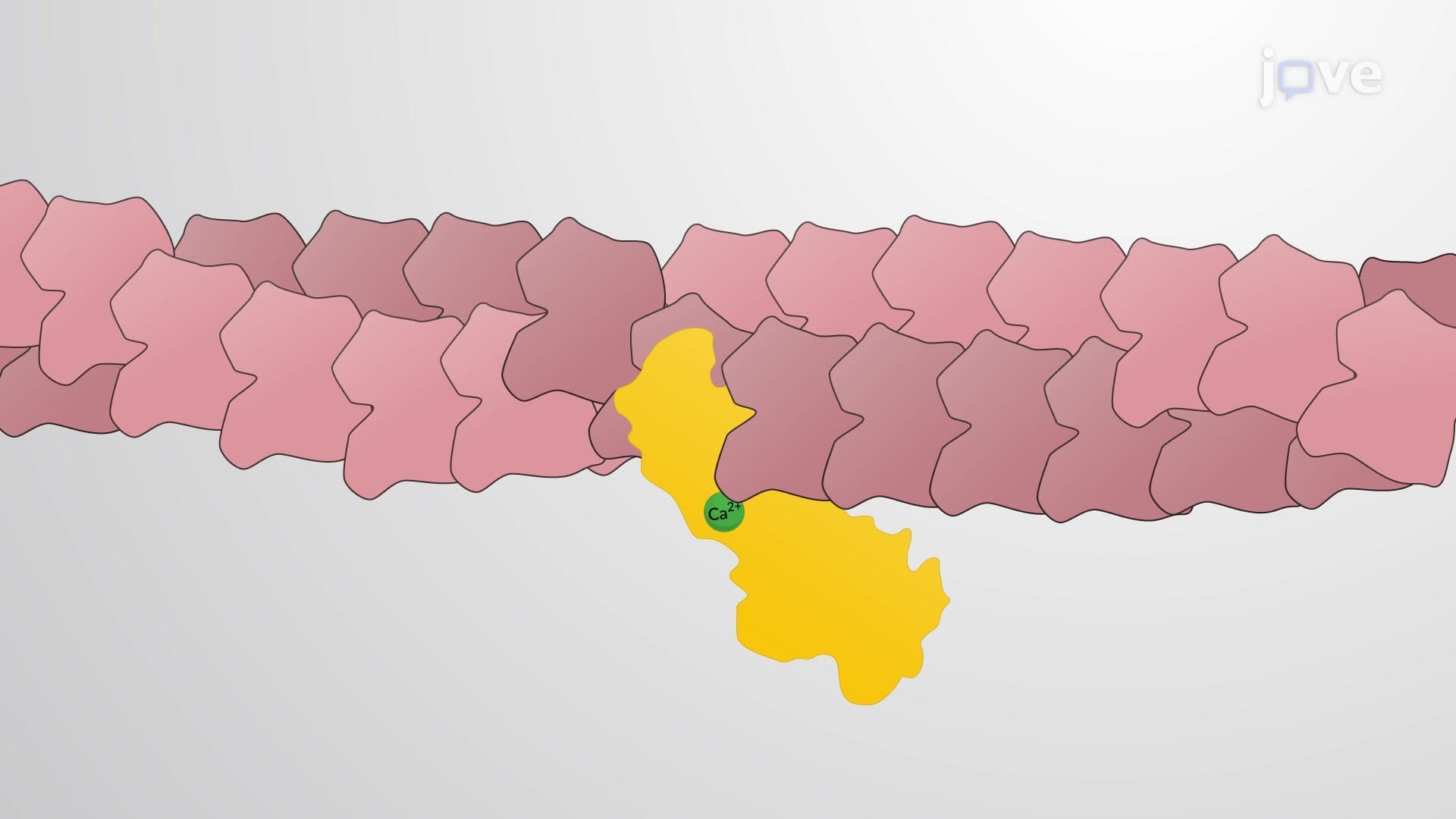
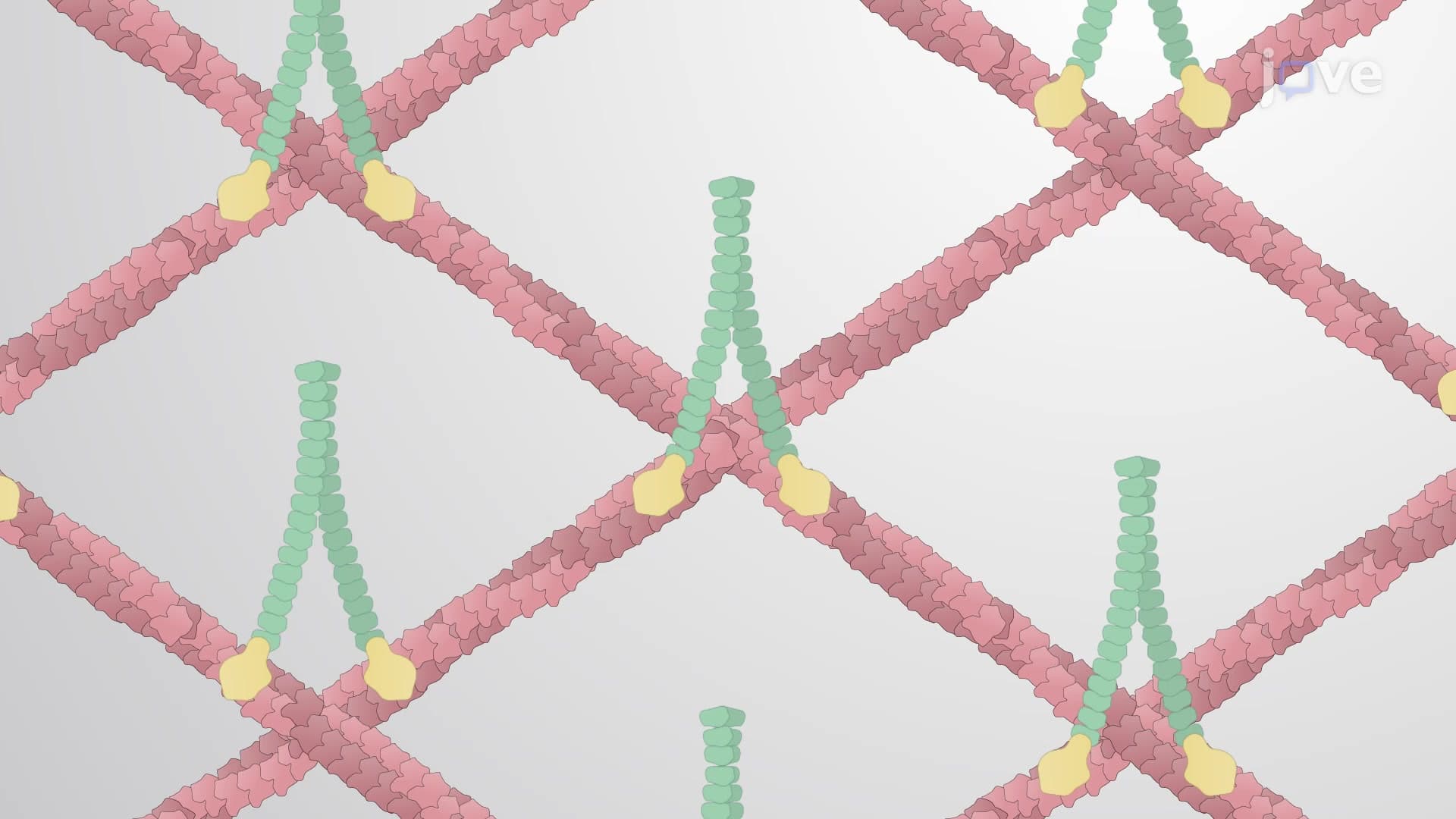
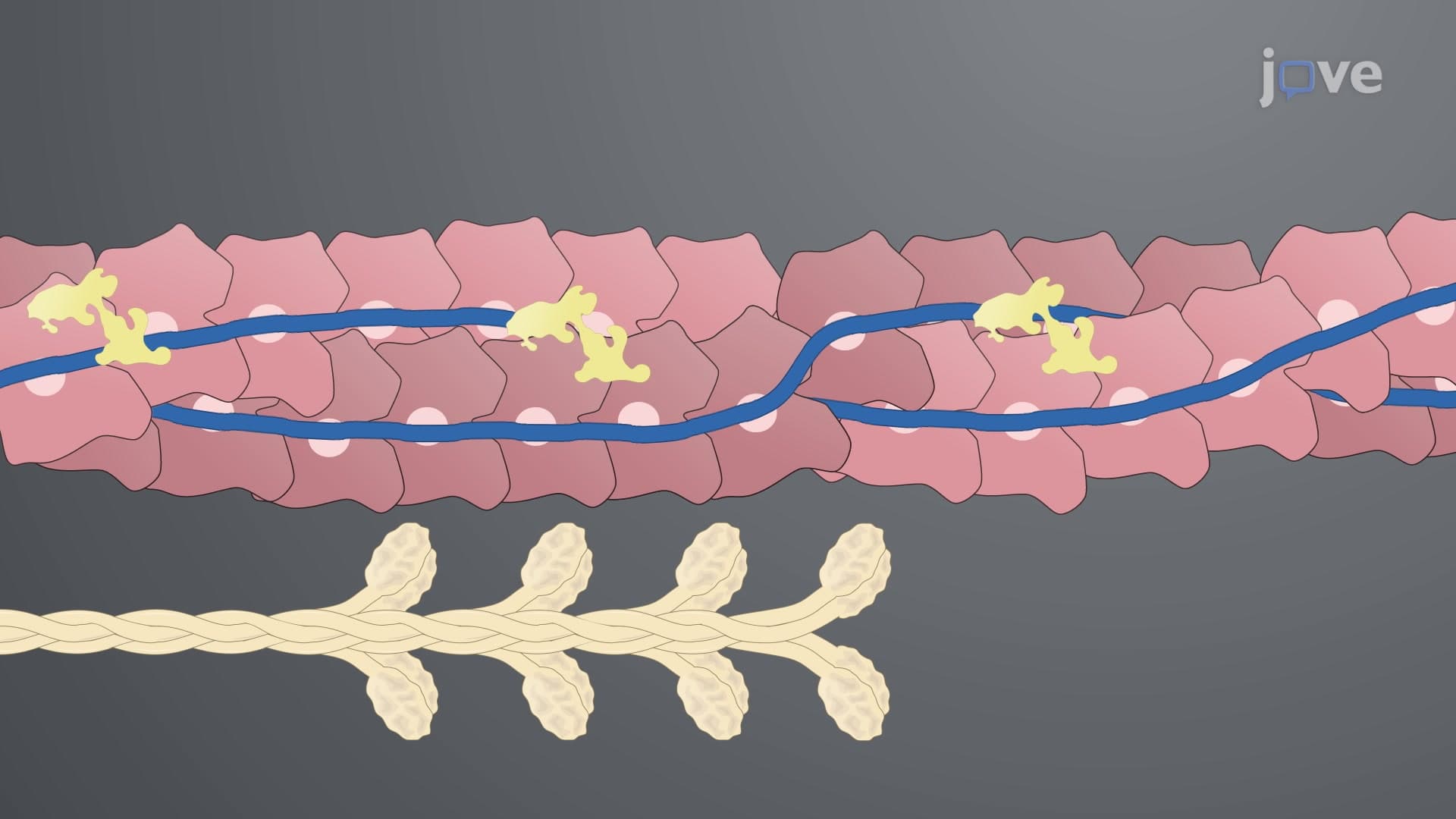
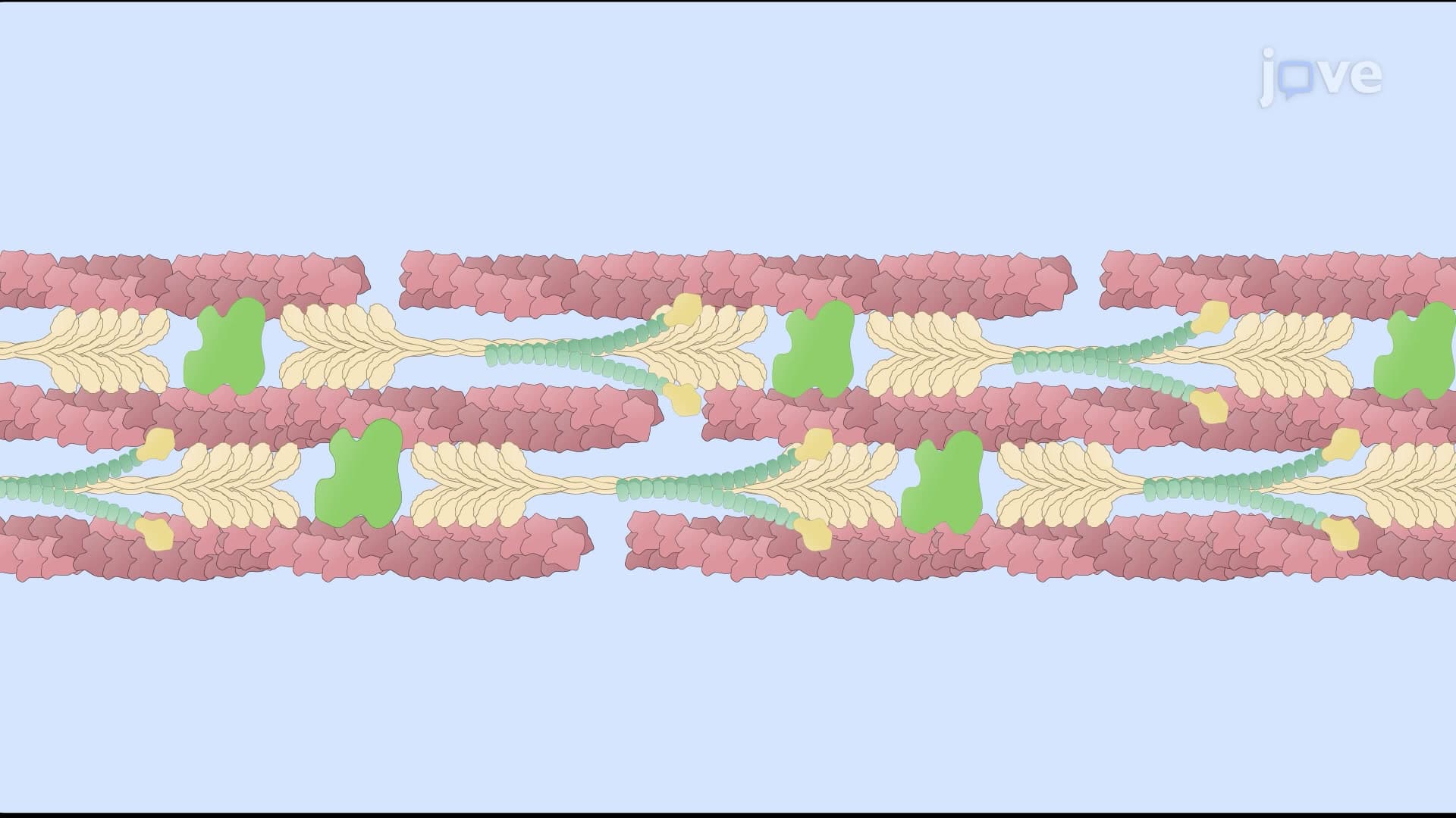
Microfilaments, composed primarily of actin proteins, demonstrate extraordinary structural versatility through their assembly patterns. These 7-nanometer-diameter filaments can organize into two distinct configurations depending on cellular needs. In muscle cells, actin-binding proteins create highly ordered parallel bundles that interact with myosin to generate contractile force-the mechanism behind every heartbeat and voluntary movement. Alternatively, in cellular regions requiring structural support like the cell cortex, these same filaments form complex three-dimensional networks that provide mechanical strength while maintaining flexibility.
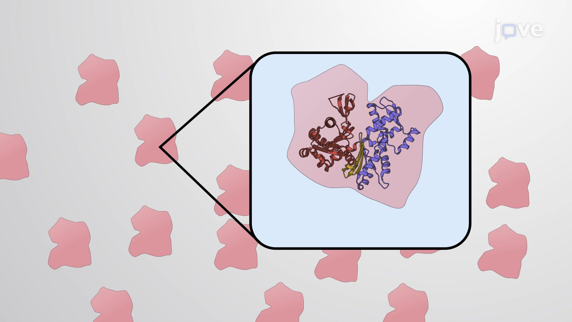
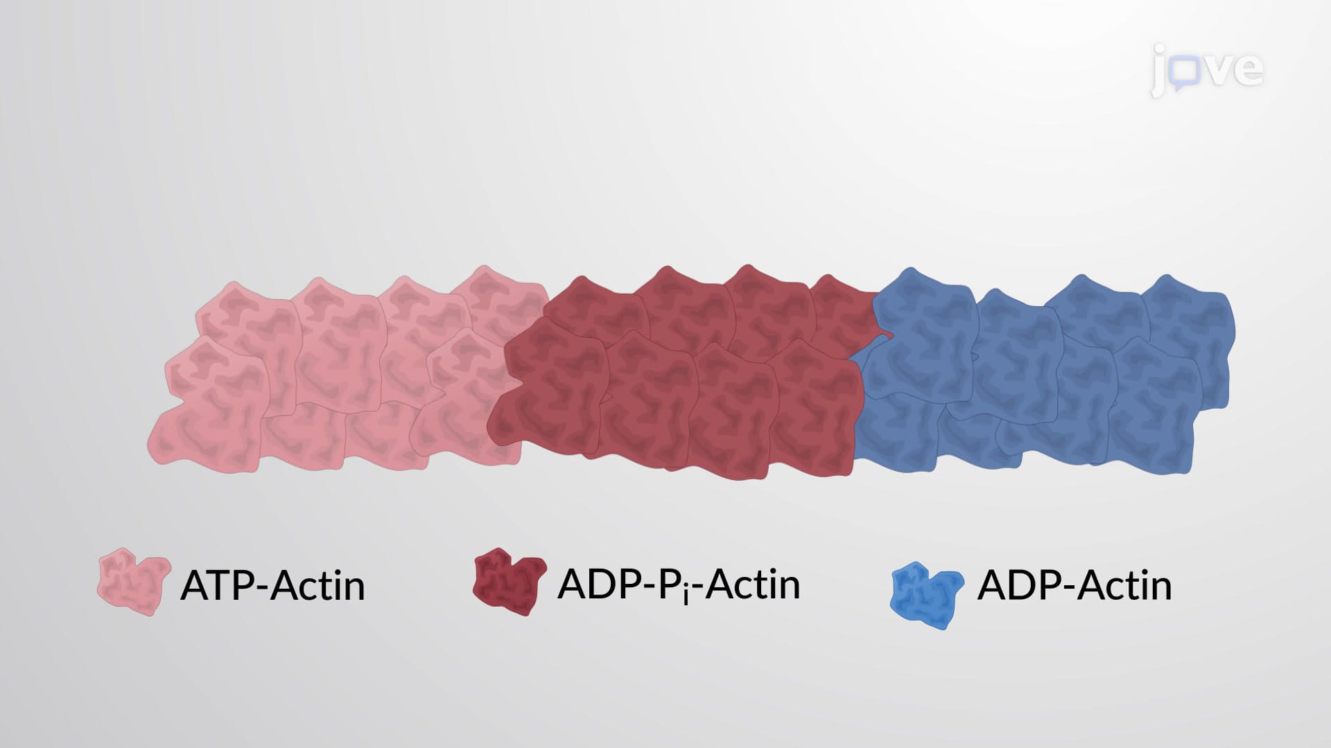
The assembly process relies on actin monomers polymerizing through end-to-end contacts, stabilized by ATP hydrolysis. This process is particularly relevant for AP Biology students studying cellular energetics, as it demonstrates how cells couple energy expenditure to structural organization. Understanding actin assembly mechanisms also appears frequently on MCAT Cell Biology sections, where students must explain how cytoskeletal dynamics enable cellular processes.
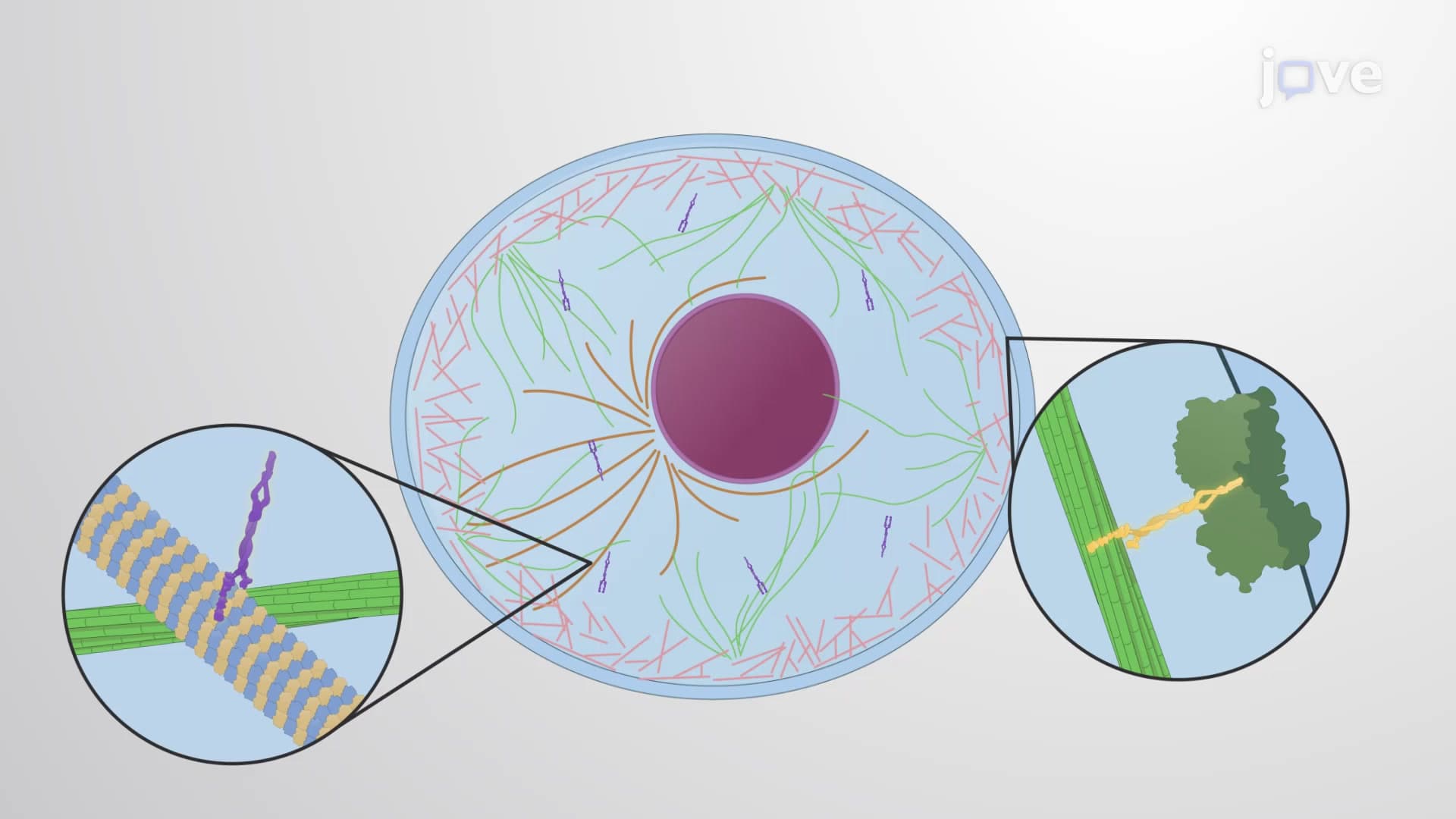
Microtubules represent the cytoskeleton's most structurally complex filaments, assembling from α and β tubulin heterodimers into hollow cylindrical structures. The thirteen-protofilament organization creates a 25-nanometer-diameter tube that serves as both structural support and transport highway. In neurons extending from the spinal cord to fingertips-distances exceeding three feet-microtubules provide the tracks along which motor proteins transport cellular cargo.
Microtubule-associated proteins (MAPs) play crucial roles in regulating assembly and stability. Tau protein, whose dysfunction contributes to Alzheimer's disease, exemplifies how MAP malfunction can have devastating consequences. College biochemistry courses frequently examine this connection, and USMLE Step 1 regularly tests understanding of cytoskeletal protein pathology.
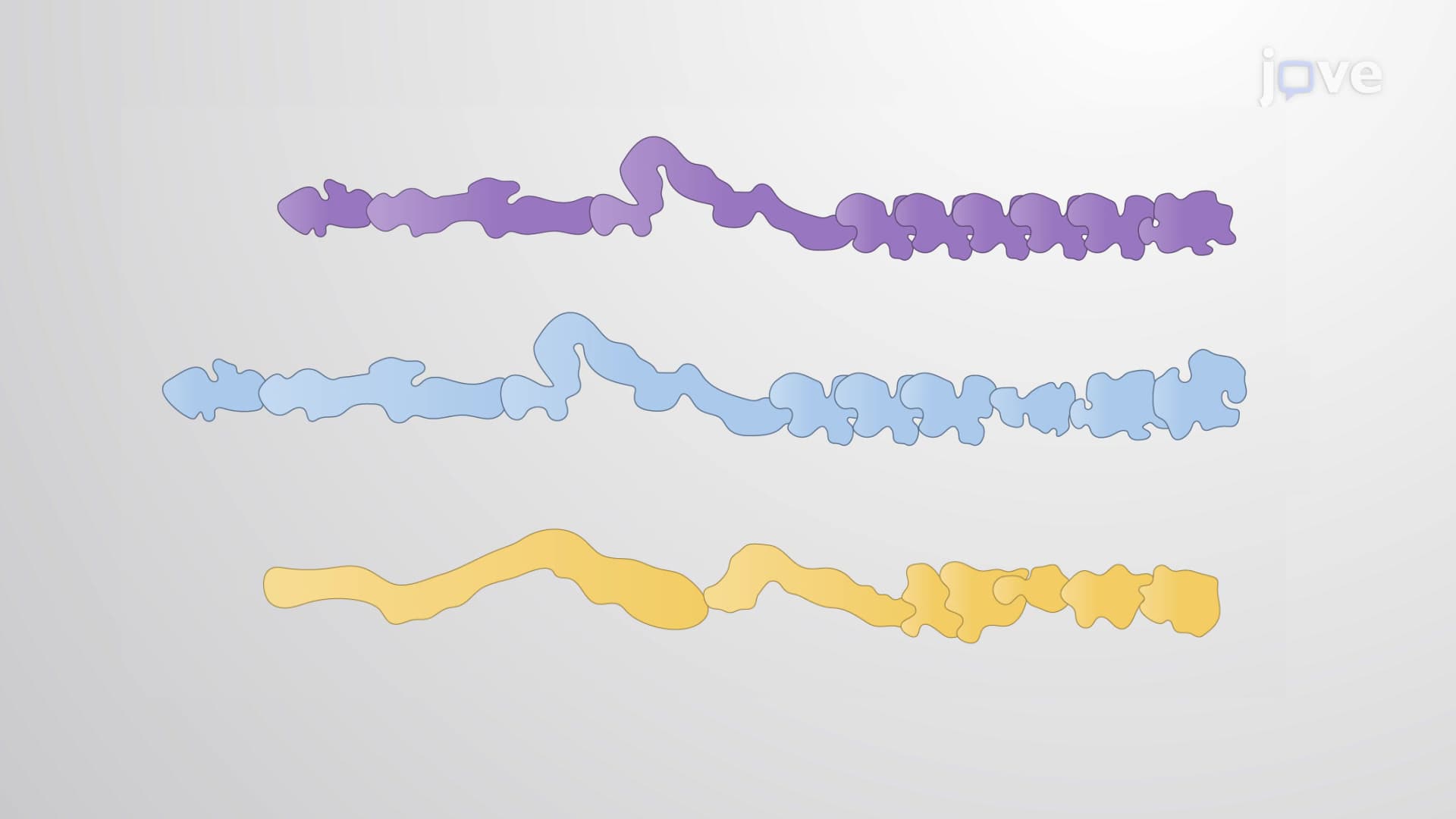
Intermediate filaments demonstrate the most cell-type-specific assembly patterns among cytoskeletal components. Keratin filaments in skin cells must withstand mechanical stress, while neurofilaments in neurons provide structural integrity across extensive axonal lengths. The multistep assembly process begins with protein dimerization, followed by tetramer formation, and finally association into the characteristic 10-nanometer-diameter rope-like structures.
This cell-type specificity makes intermediate filaments valuable diagnostic markers in clinical pathology. Pathologists routinely use keratin staining to identify epithelial tumors, while neurofilament markers help diagnose neurological conditions-applications that nursing students encounter in NCLEX-RN preparation and medical students see on clinical rotations.
Related Micro-courses