Loading subject page…


162,854 views

Video Summary: What Is Cytoskeletal Proteins in Bacteria
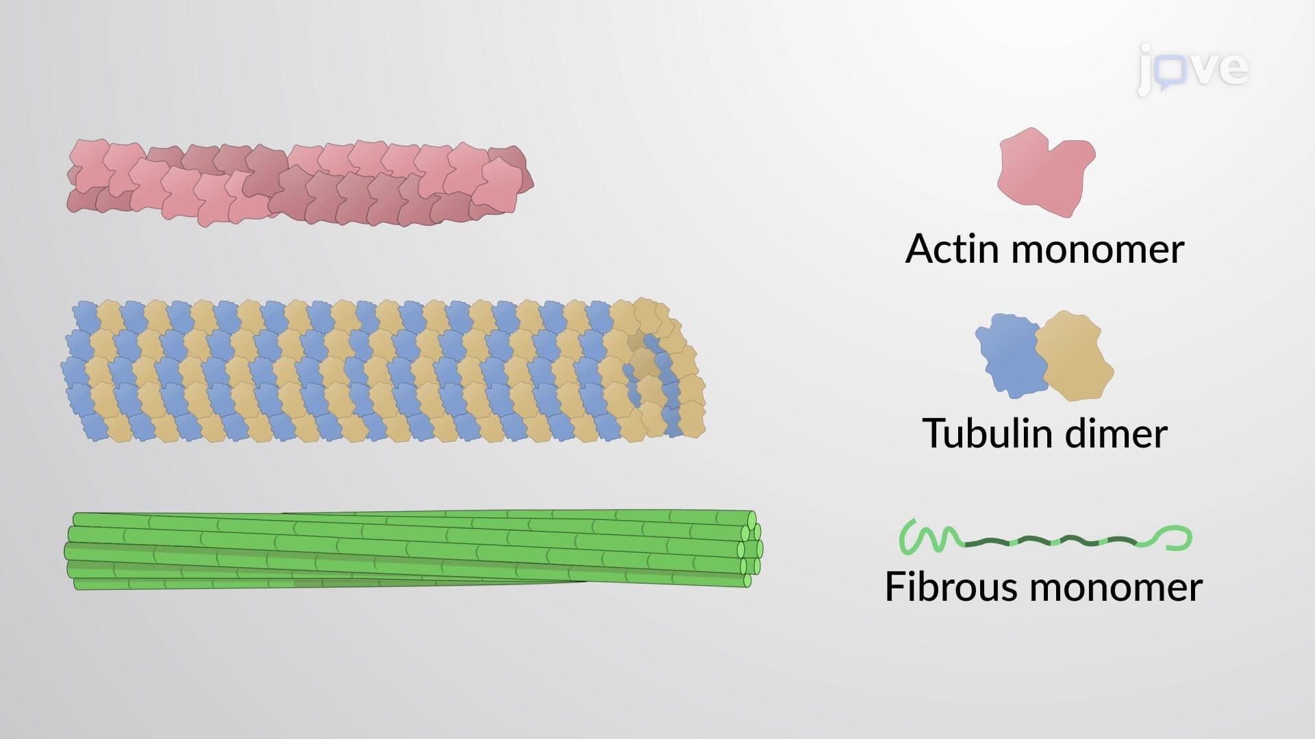
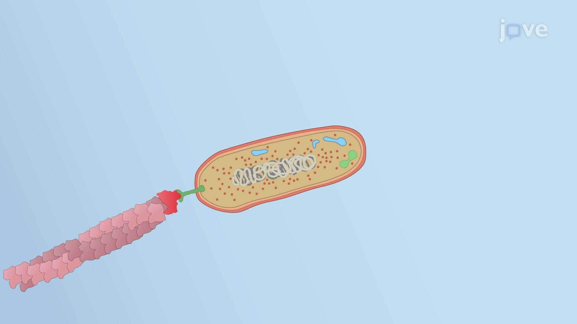
Did you know that bacterial cells, despite lacking a nucleus, possess sophisticated protein scaffolds that rival those in human cells? Cytoskeletal proteins in bacteria include familiar homologs of actin and tubulin, plus unique bacterial proteins that orchestrate cell division and shape. These molecular frameworks enable E. coli and other bacteria to maintain their distinctive rod shapes and successfully reproduce in laboratory cultures across American universities. Understanding what is cytoskeletal proteins in bacteria reveals how these microscopic organisms achieve remarkable structural organization without membrane-bound organelles. Watch the full video on JoVE Coach to master this concept with expert-led visuals and step-by-step explanations.
Cytoskeletal proteins in bacteria represent a fascinating convergence of evolutionary biology and cellular mechanics that challenges traditional views of prokaryotic simplicity. Unlike the static perception of bacterial cells taught in introductory biology courses, these organisms possess dynamic protein networks that rival eukaryotic cytoskeletons in complexity and function.
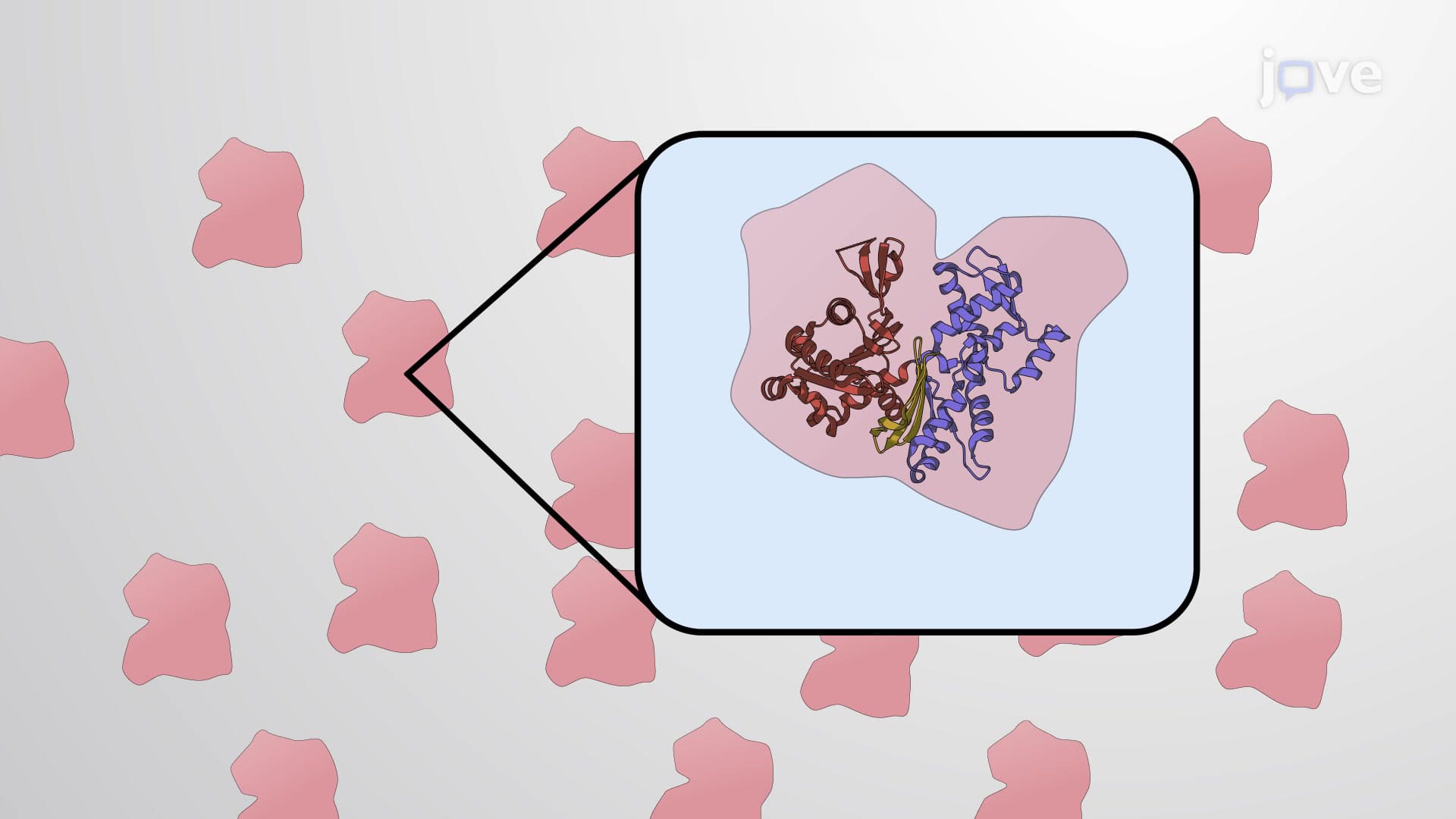
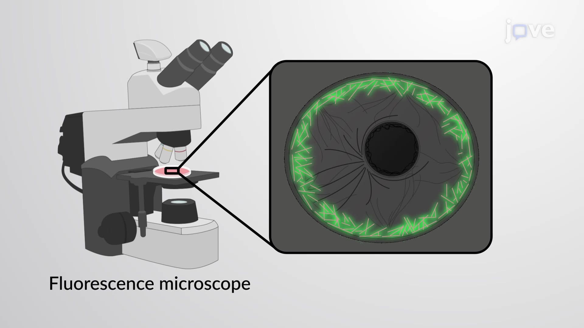
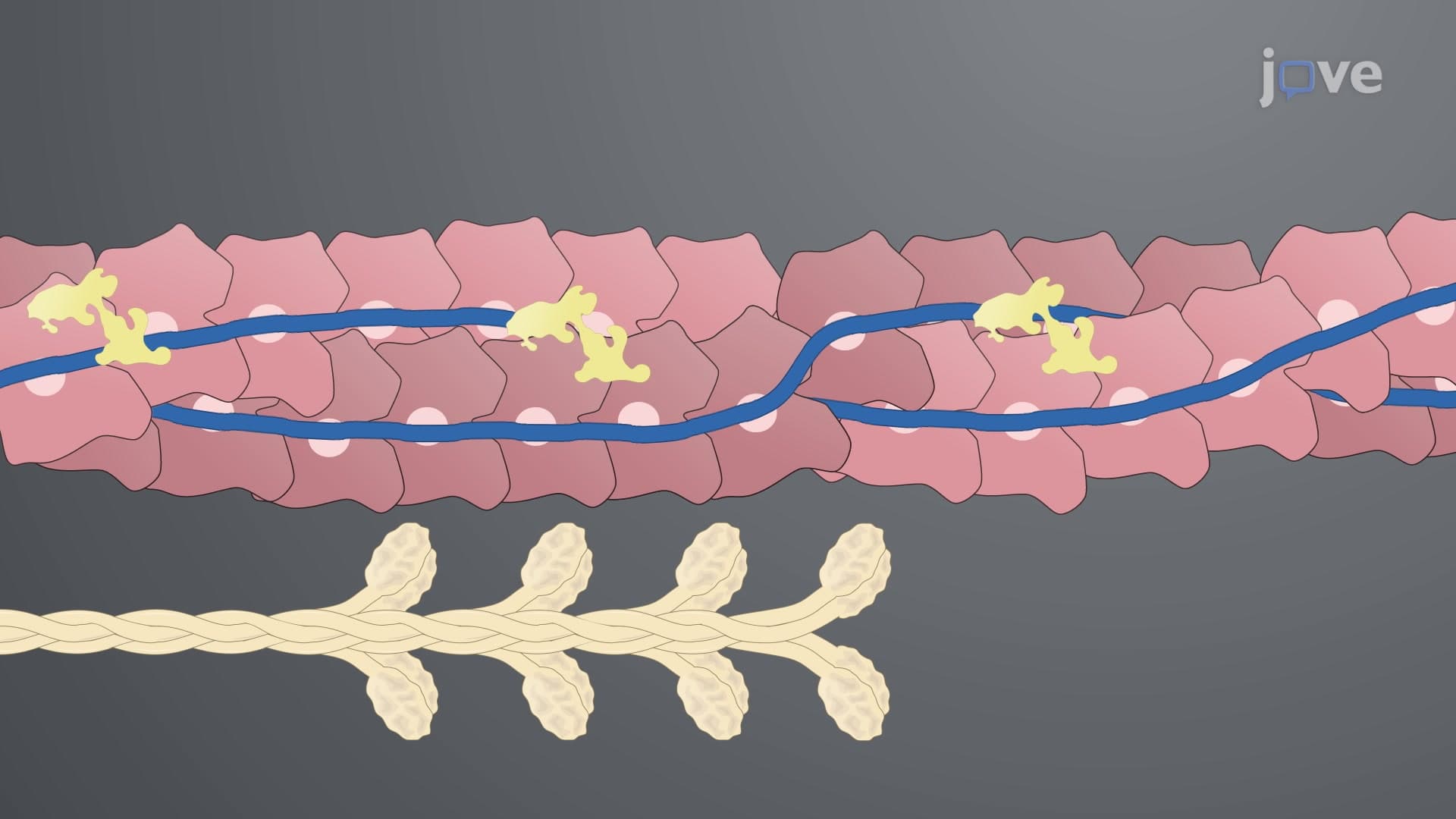
The MreB and Mbl proteins serve as bacterial actin homologs, forming helical scaffolds that spiral around the cell's interior. These proteins don't merely provide structural support-they actively guide the synthesis of peptidoglycan, the crucial polymer that gives bacteria their distinctive shapes. In laboratory studies at institutions like MIT and Stanford, researchers have demonstrated how MreB depletion causes rod-shaped bacteria like E. coli to become spherical, highlighting these proteins' critical role in morphogenesis.
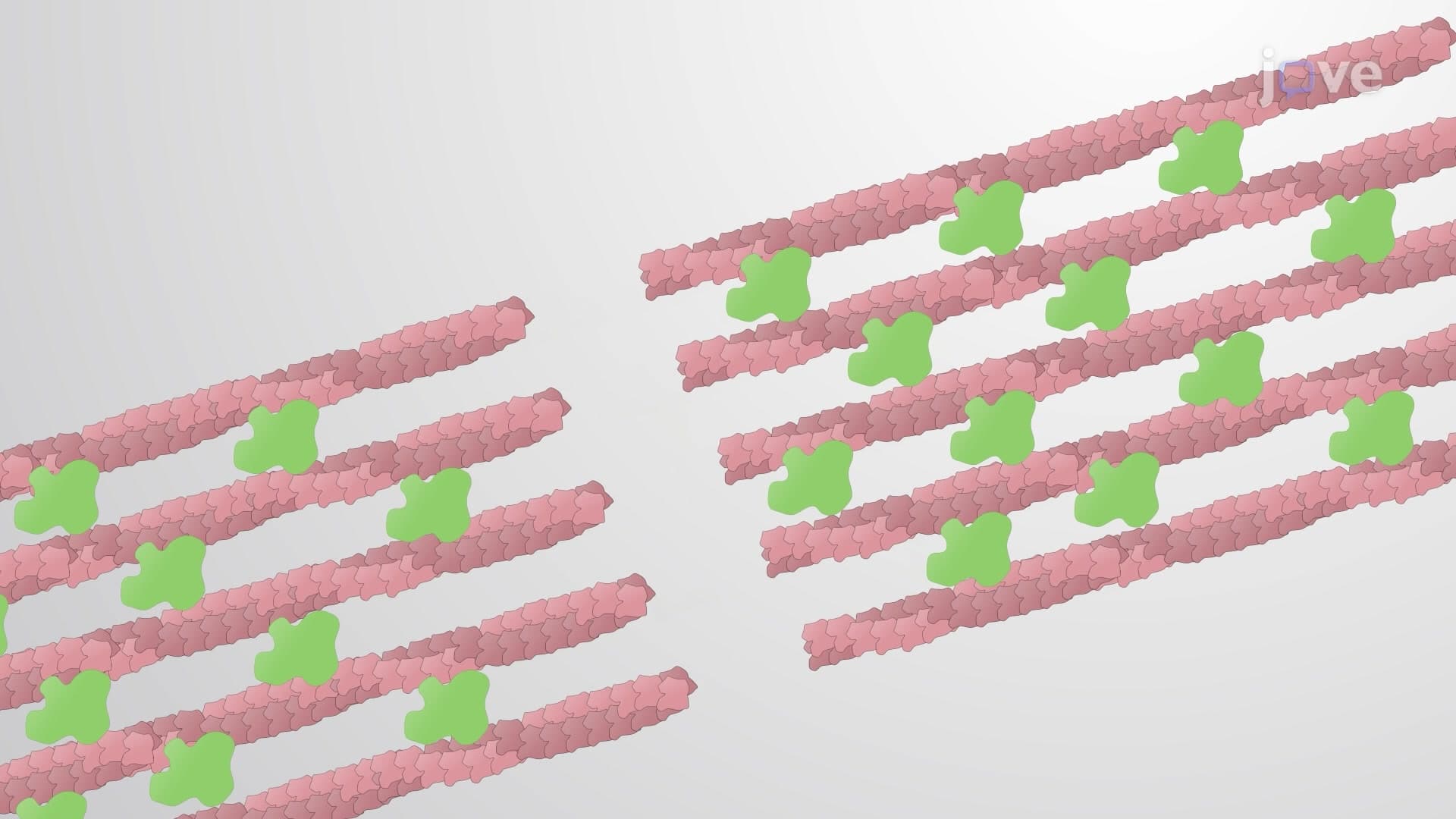
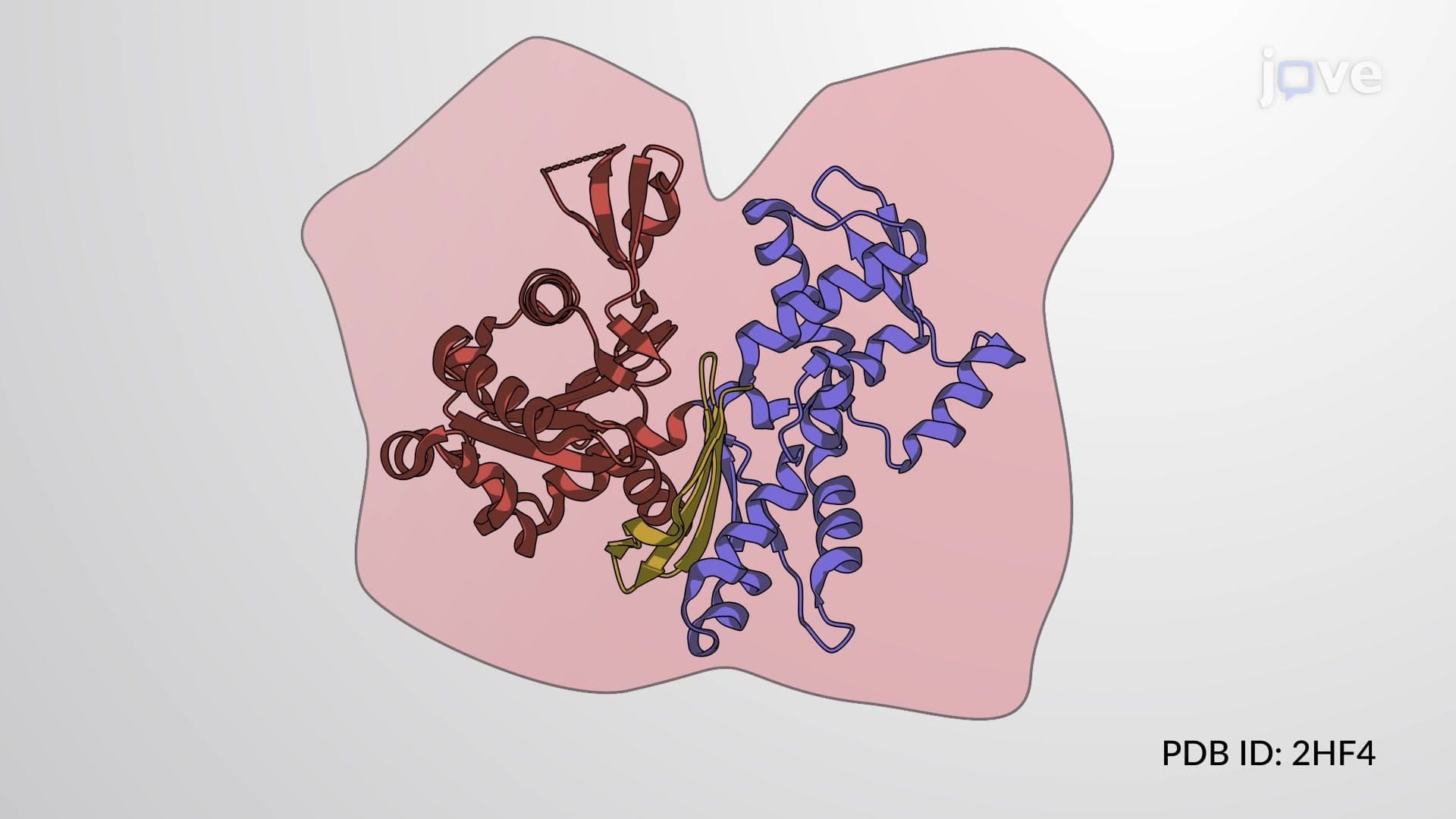
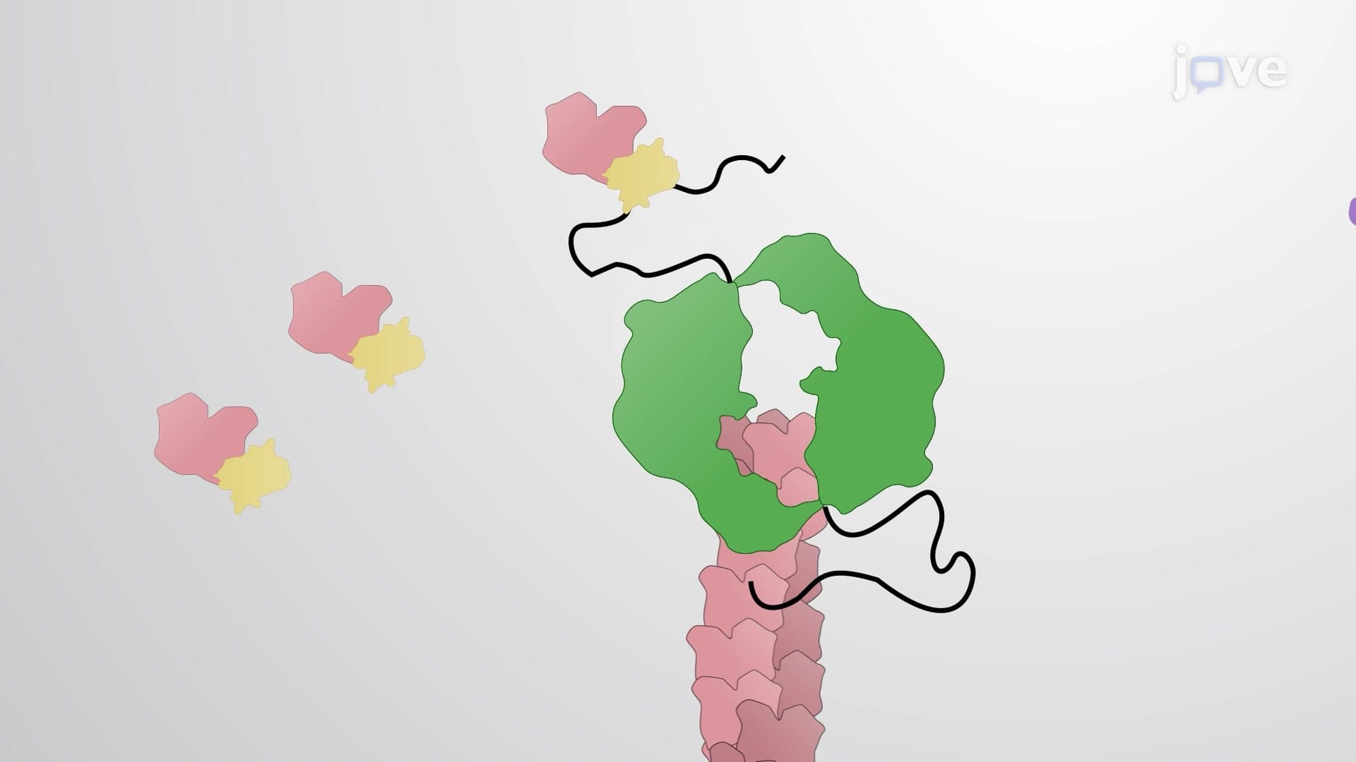
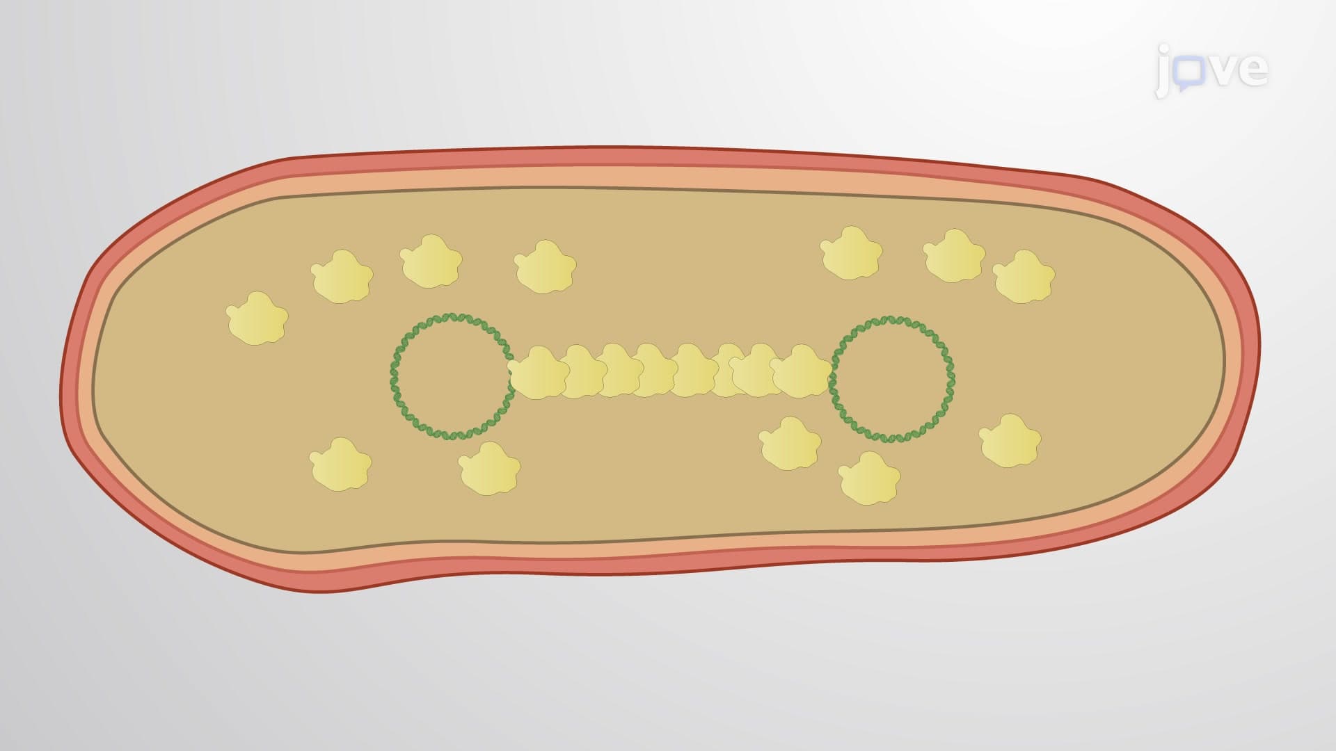
ParM represents another actin homolog with unique properties. Found on antibiotic-resistance plasmids, ParM filaments exhibit dynamic instability similar to eukaryotic actin. During bacterial replication, ParM polymerization physically pushes plasmid copies to opposite cell poles-a mechanism that ensures antibiotic resistance genes are inherited by both daughter cells, a phenomenon particularly relevant in clinical settings across American hospitals.
FtsZ, the bacterial tubulin homolog, forms the foundation of the divisome-the molecular machine responsible for bacterial cell division. This protein assembles into a contractile ring at the cell's midpoint, creating the Z-ring structure. Students preparing for the MCAT or AP Biology exams should understand that FtsZ polymerization and constriction coordinate with peptidoglycan synthesis to achieve cytokinesis. Recent research from University of California laboratories has shown how FtsZ inhibitors could serve as novel antibiotics, making this protein a target of significant clinical interest.
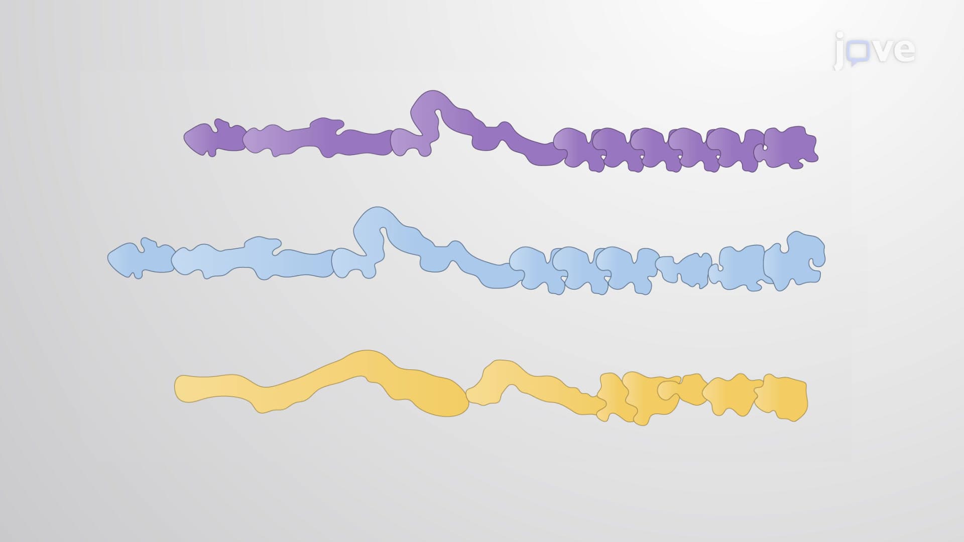
The MinD-ParA protein family represents bacterial innovation in cytoskeletal organization. These ATPases oscillate within cells to establish division sites and coordinate chromosome segregation. The Min system's oscillatory behavior, first characterized in studies at Princeton University, demonstrates sophisticated spatial regulation that ensures symmetric cell division.
Crescentin proteins create the distinctive curved morphology of Caulobacter crescentus, serving as bacterial intermediate filament analogs. When crescentin genes are deleted, these naturally crescent-shaped bacteria become rod-shaped, proving the direct relationship between cytoskeletal proteins and bacterial morphology-a concept frequently tested in college-level microbiology courses.
Related Micro-courses