Loading subject page…


162,819 views

Video Summary: What Is Studying the Cytoskeleton
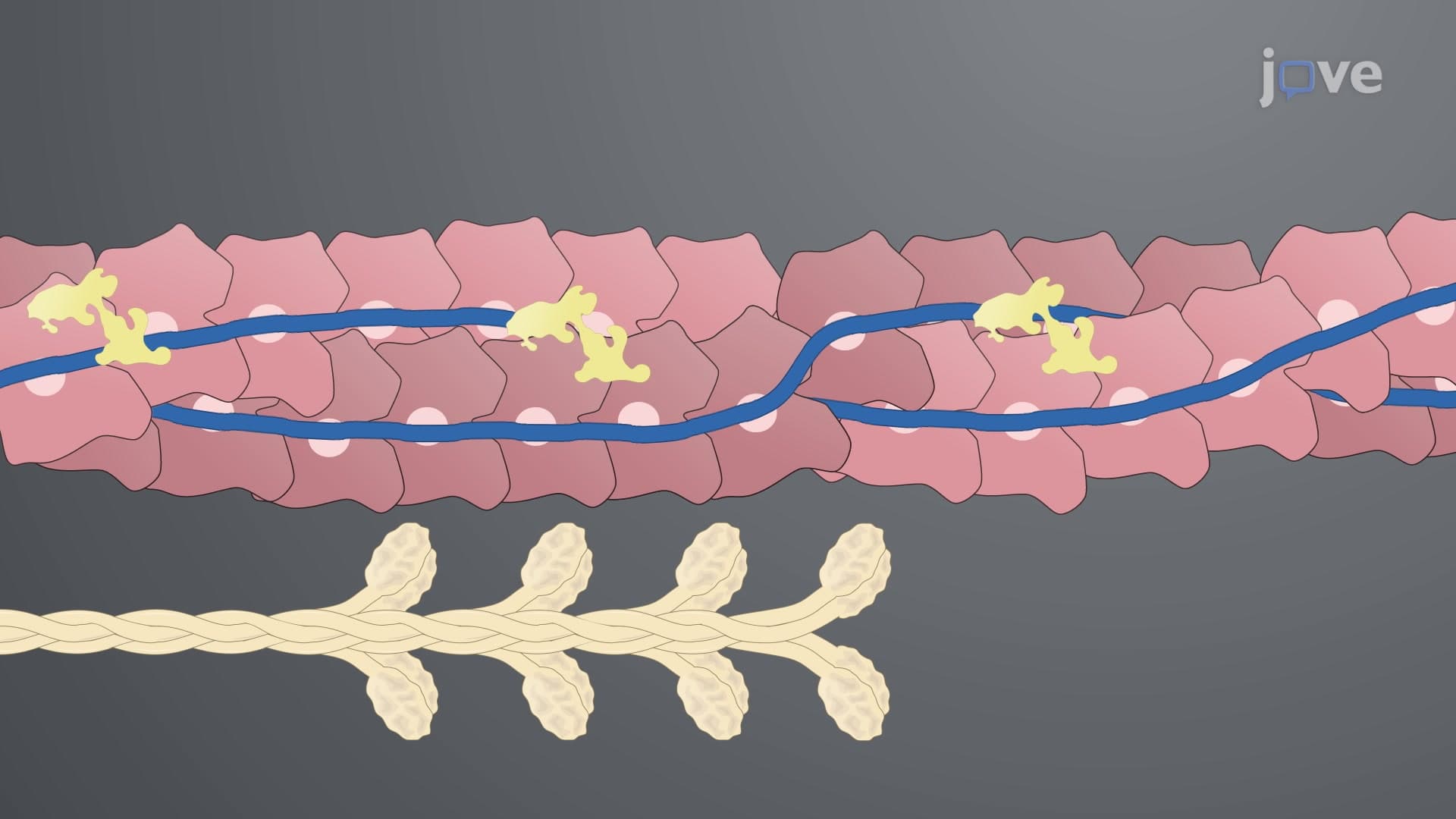
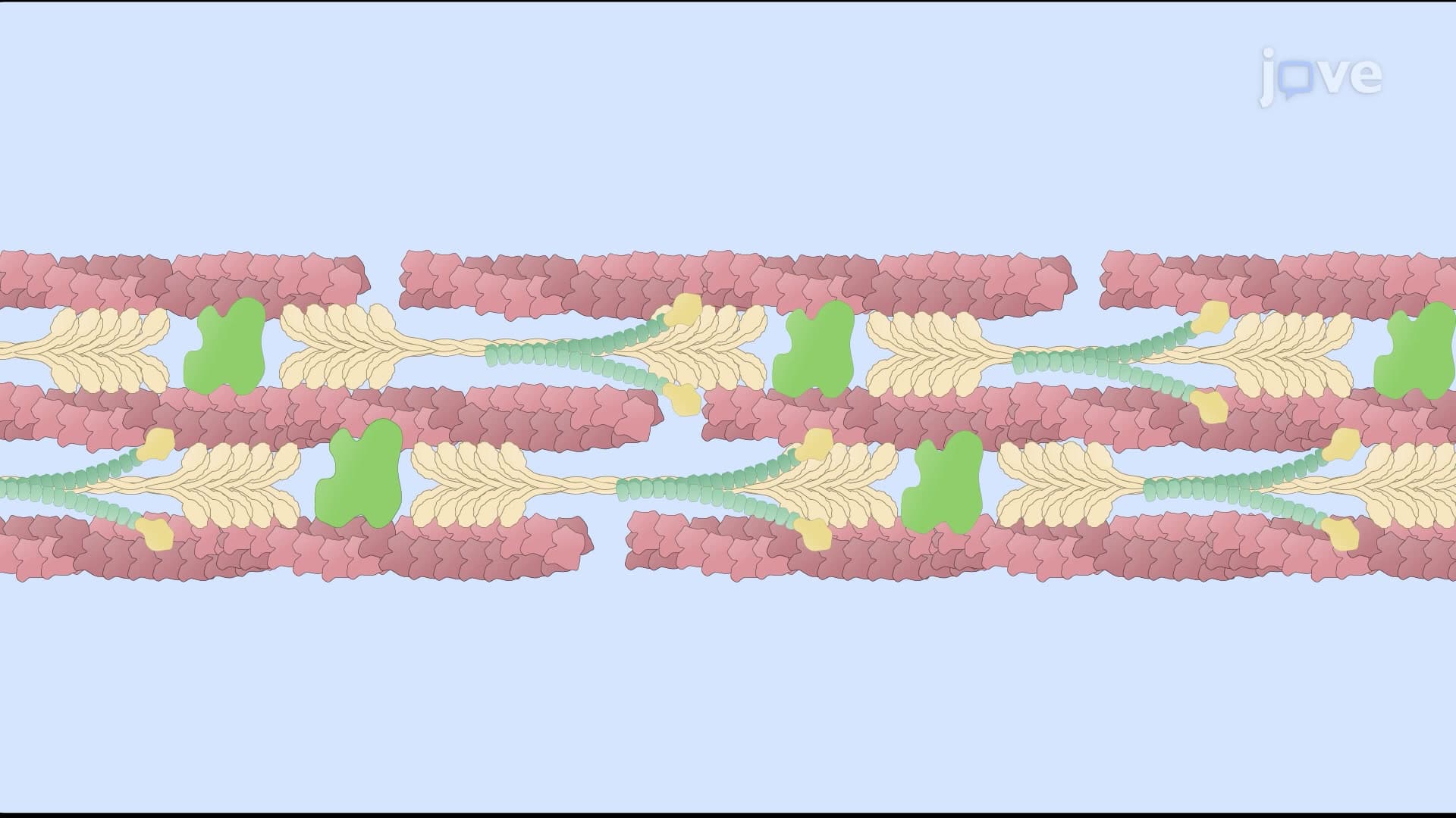
Ever wonder how researchers at Stanford University discovered that cancer cells can literally squeeze through spaces 10 times smaller than themselves? Studying the cytoskeleton reveals the microscopic machinery that makes this cellular flexibility possible. Scientists use advanced techniques like atomic force microscopy and fluorescence imaging to understand how these protein networks stretch, contract, and reorganize within living cells. From determining the tensile strength of intermediate filaments to visualizing motor proteins walking along microtubules, what is studying the cytoskeleton opens up the hidden world of cellular mechanics that governs everything from muscle contraction to cell division. Watch the full video on JoVE Coach to master this concept with expert-led visuals and step-by-step explanations.
The cytoskeleton represents one of cell biology's most dynamic and mechanically sophisticated systems, and studying the cytoskeleton requires a multidisciplinary approach combining advanced microscopy, biochemistry, and biophysics. Unlike static cellular components, the cytoskeleton constantly remodels itself, making its investigation both challenging and fascinating for researchers worldwide.
Atomic force microscopy (AFM) revolutionized our understanding of cytoskeletal mechanics by enabling direct measurement of individual filament properties. This technique employs nanoscale tips to mechanically manipulate cytoskeletal components, essentially performing "molecular stress tests" on proteins smaller than a virus. Researchers at institutions like MIT and Harvard Medical School use AFM to determine tensile strength, elasticity, and breaking points of different cytoskeletal elements.
The most striking discovery involves intermediate filaments, which demonstrate remarkable extensibility compared to actin filaments and microtubules. These rope-like structures can stretch up to 3.5 times their resting length before failure, explaining why cells can withstand significant mechanical stress. This property proves crucial for understanding diseases like muscular dystrophy, where intermediate filament defects compromise cellular integrity.
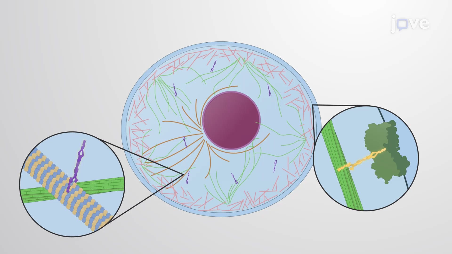
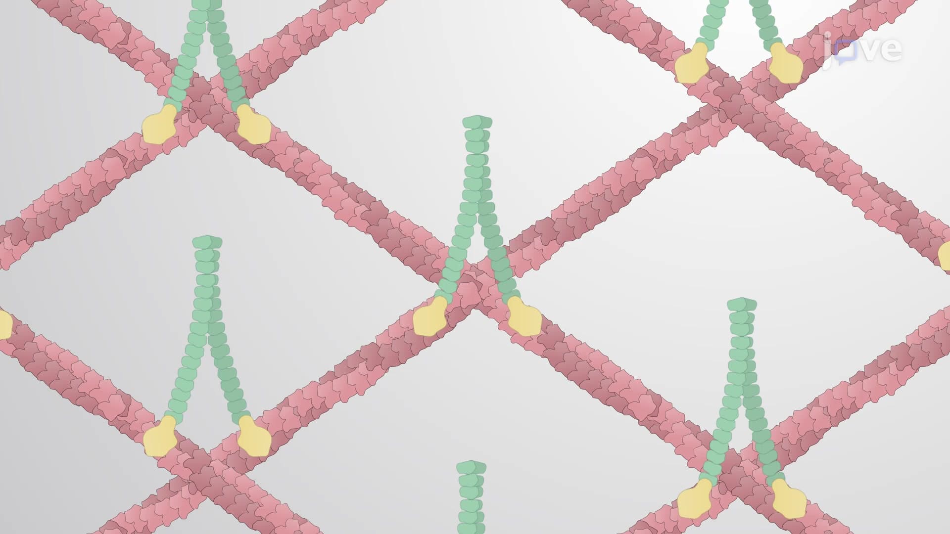
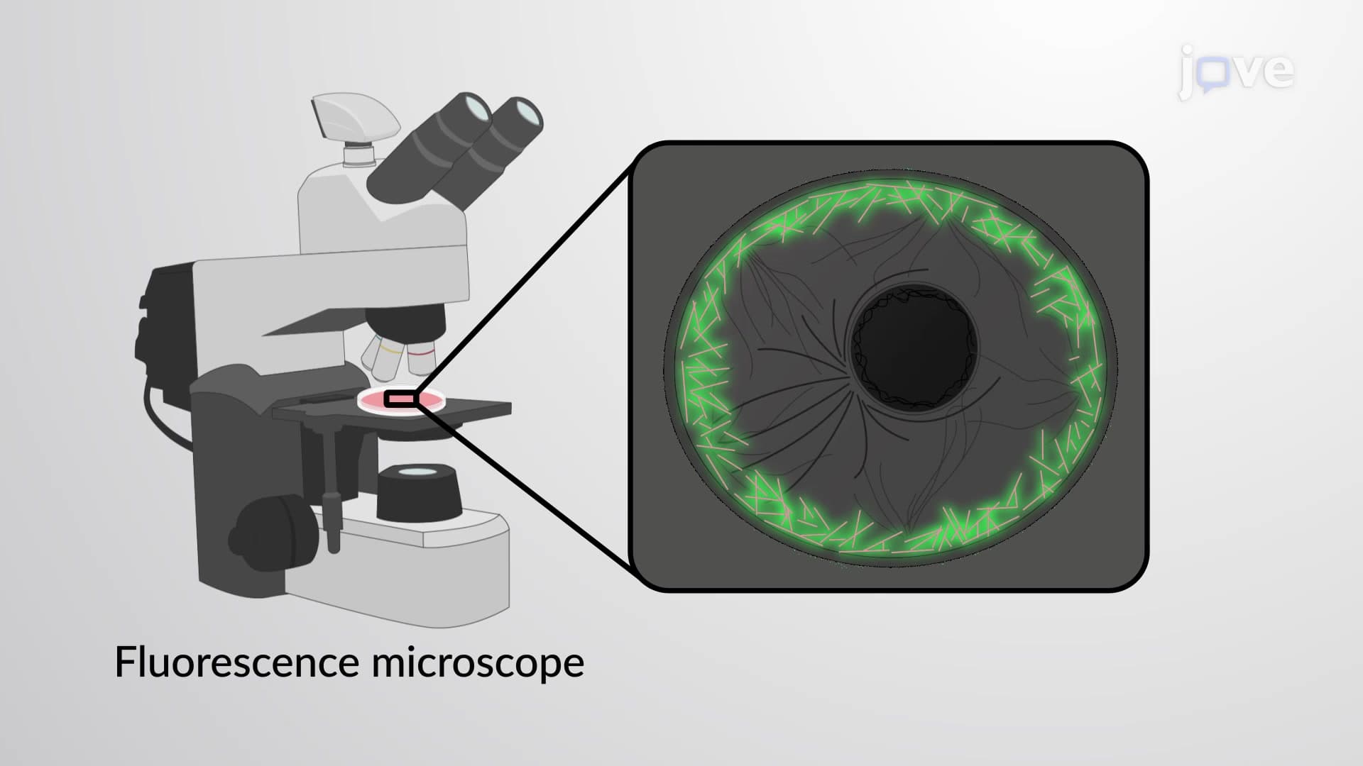
Modern cell biology relies heavily on fluorescence-based techniques to observe living cytoskeletal processes. Scientists employ green fluorescent protein (GFP) labeling and synthetic fluorescent dyes to create "molecular movies" of cytoskeletal behavior. For instance, researchers can watch kinesin motor proteins literally walk along microtubule highways, carrying cellular cargo with remarkable precision and efficiency.
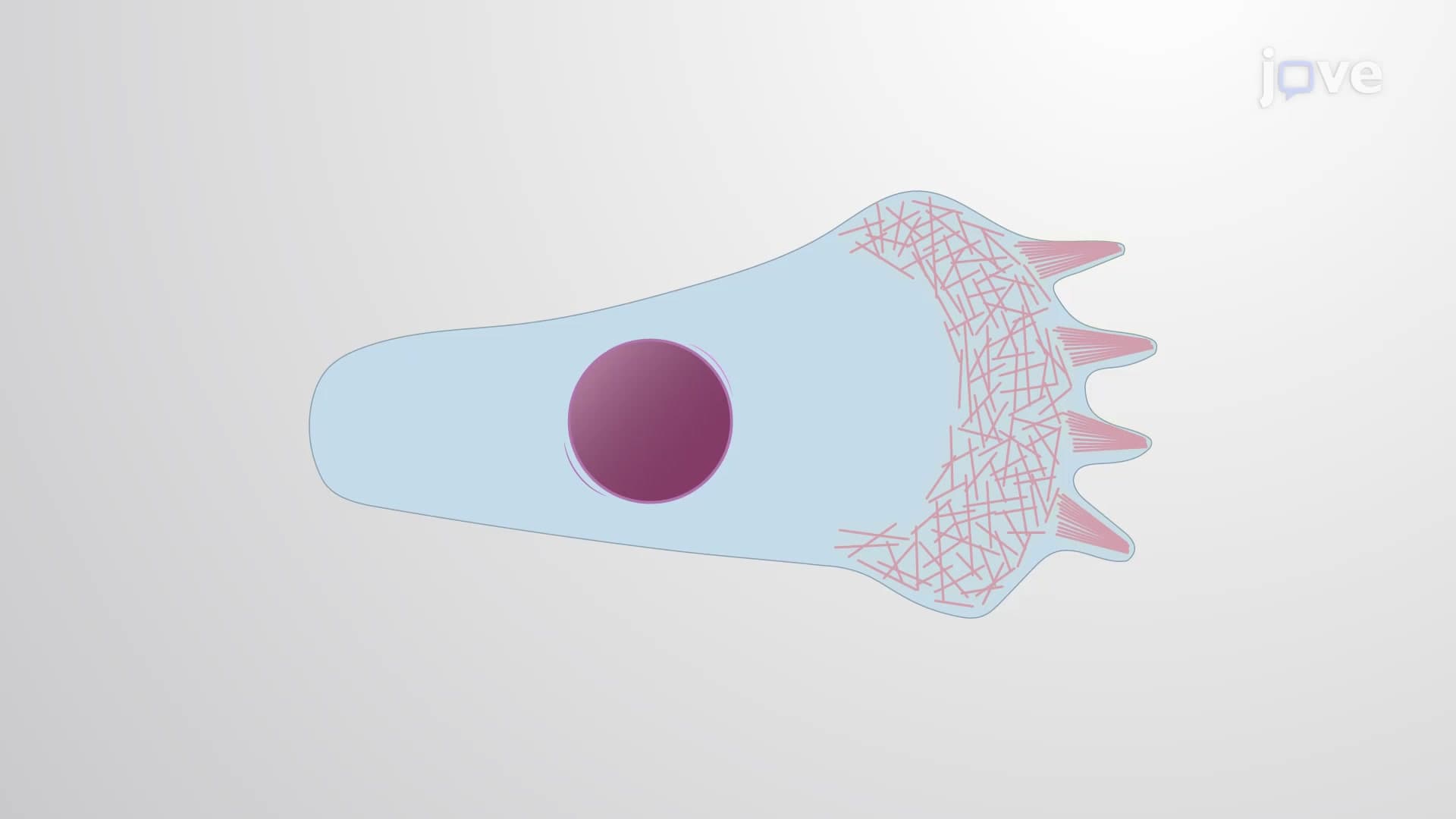
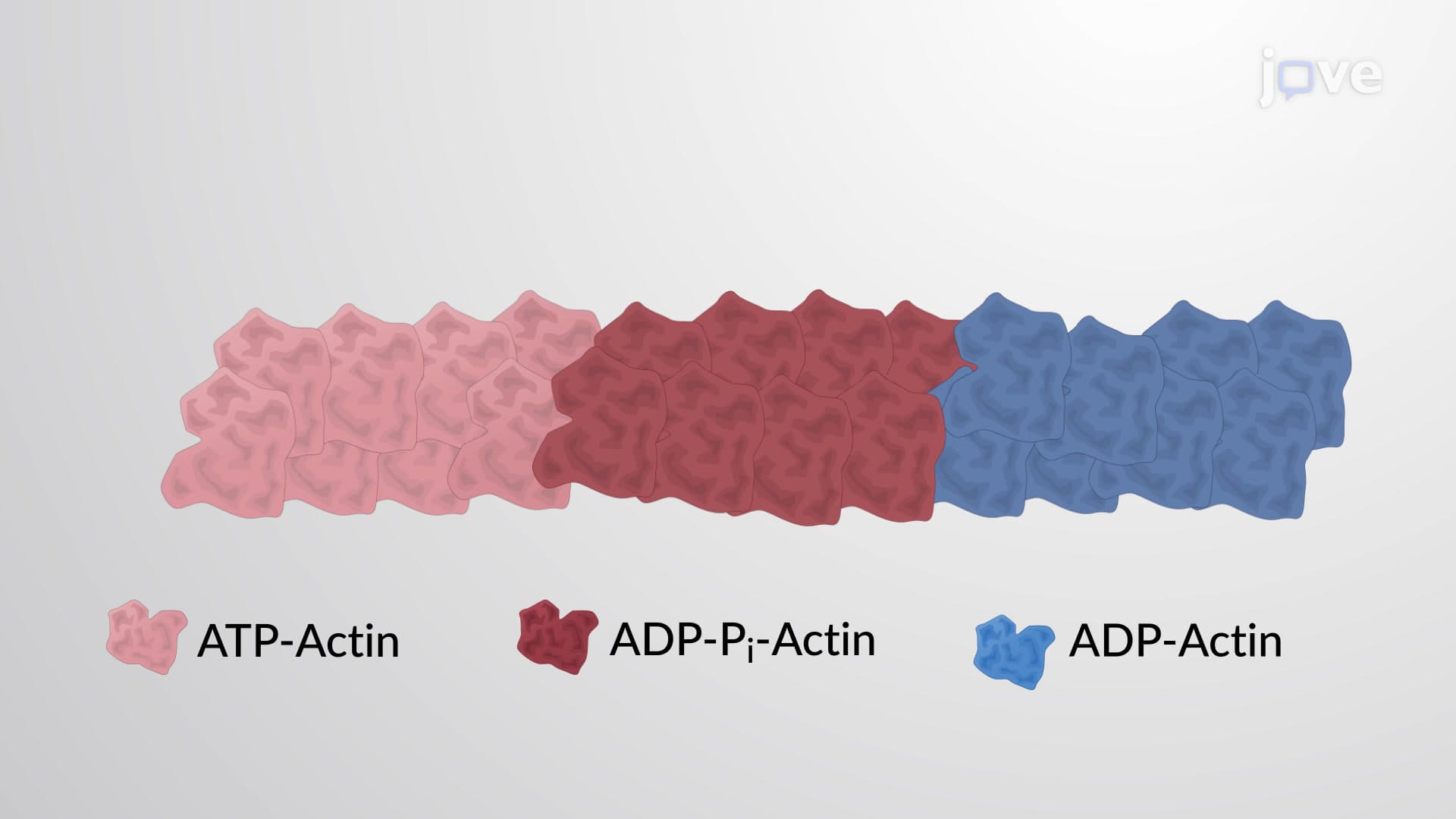
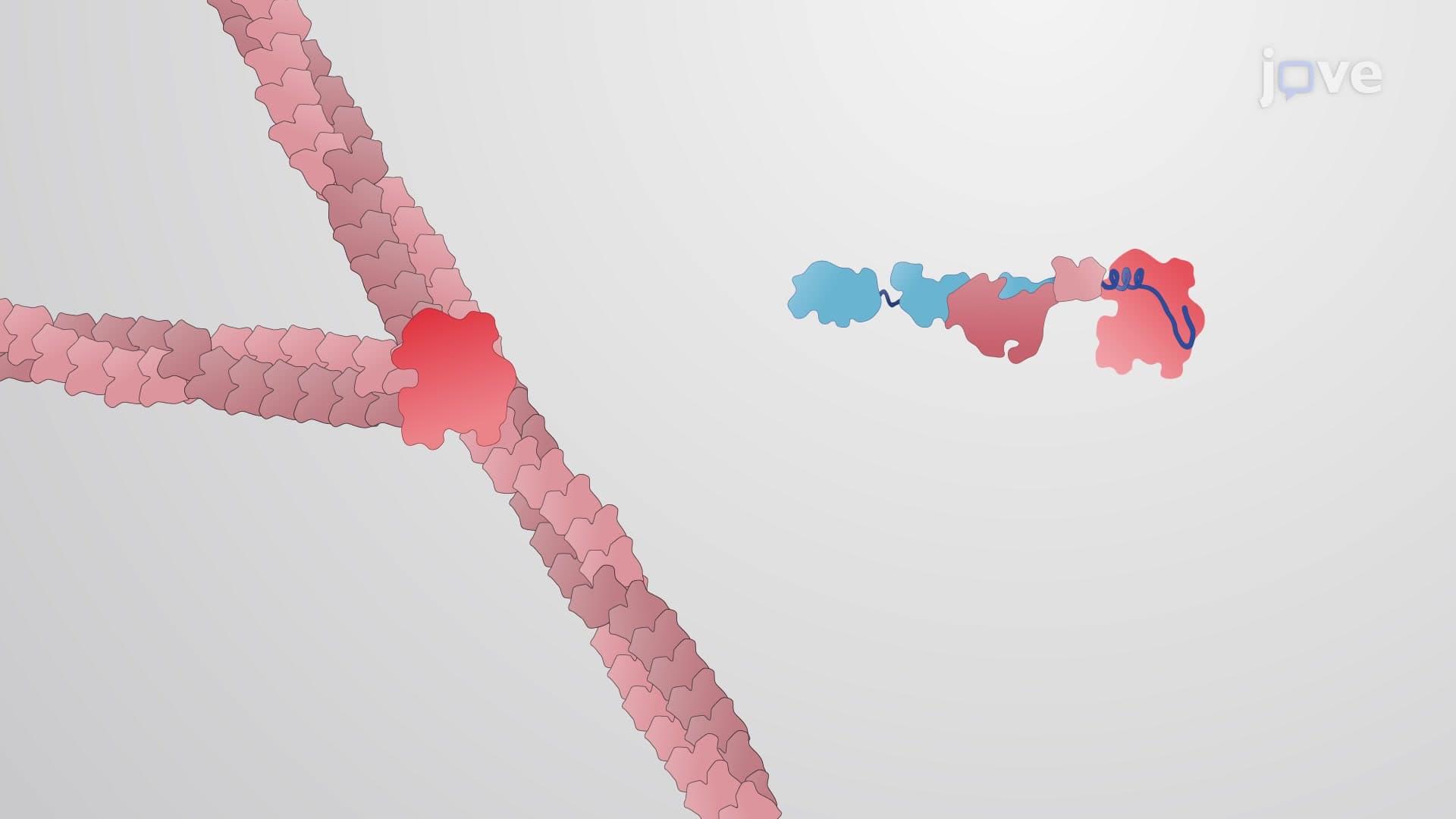
The visualization of F-actin polymerization represents another breakthrough application. By microinjecting fluorescently labeled G-actin monomers into living cells, scientists observe real-time filament assembly and disassembly. This technique proved instrumental in understanding how cells migrate, divide, and respond to mechanical stimuli-concepts frequently tested on the MCAT and AP Biology exams.
Understanding cytoskeletal dynamics has profound implications for medical research and drug development. Pharmaceutical companies like Genentech and Pfizer invest heavily in cytoskeleton-targeting therapies for cancer treatment, since many chemotherapy drugs work by disrupting microtubule function during cell division. Similarly, research into cytoskeletal diseases helps explain conditions ranging from Alzheimer's disease to cardiomyopathy, making this knowledge essential for pre-med students preparing for the MCAT.
Related Micro-courses