Loading subject page…


162,819 views

Video Summary: What Is the Cytoskeleton
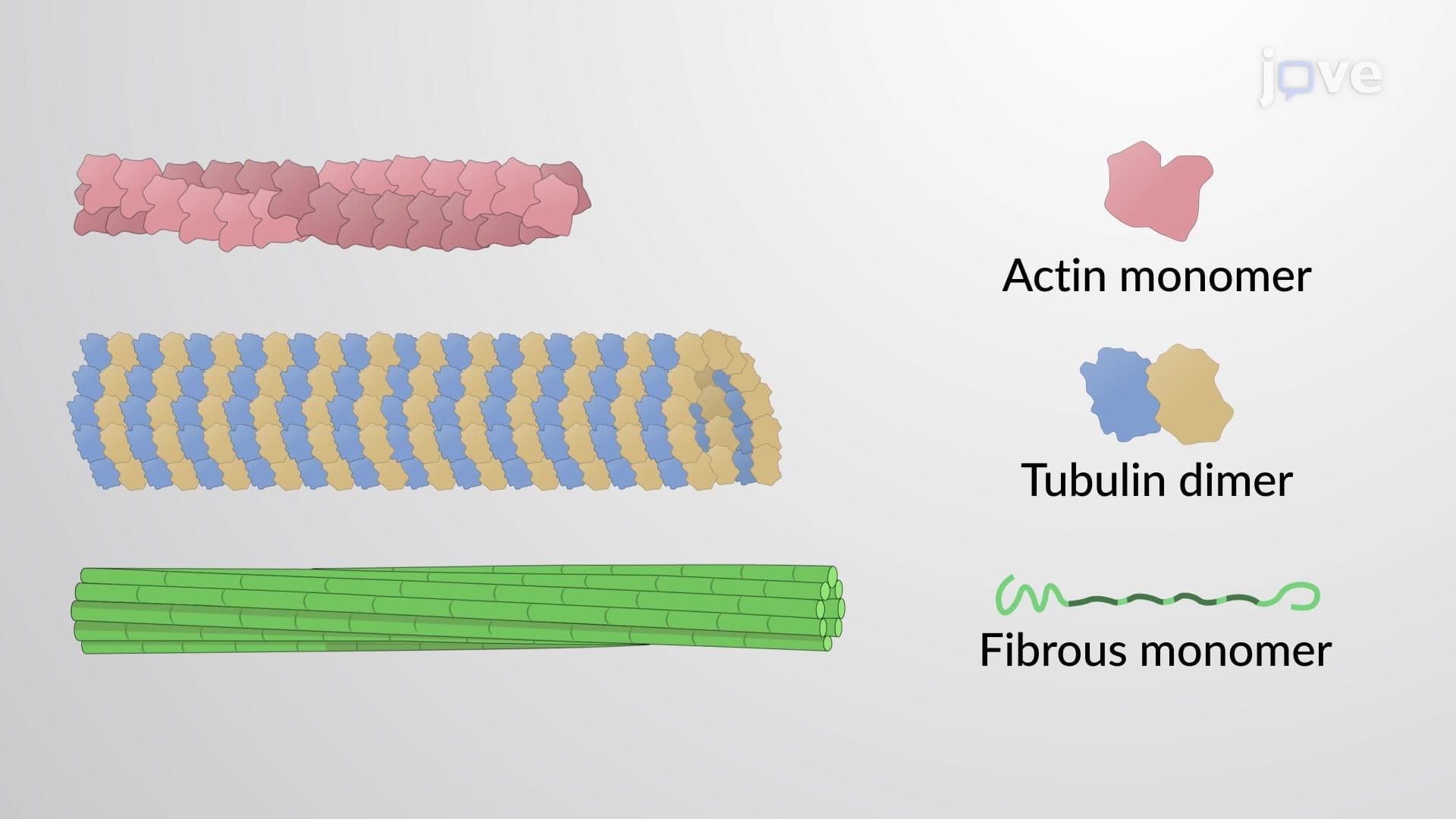
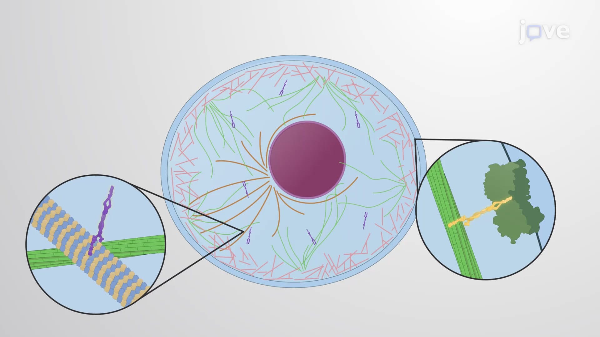
Did you know that every time you move your arm or blink your eye, billions of microscopic protein highways inside your cells are coordinating that motion? The cytoskeleton is the dynamic network of protein filaments that gives cells their shape, internal organization, and ability to move-much like how the steel framework supports a skyscraper in New York City. This cellular scaffolding consists of three distinct types of filaments that work together to maintain cell structure and enable critical functions like cell division and migration. Watch the full video on JoVE Coach to master this concept with expert-led visuals and step-by-step explanations.
The cytoskeleton represents one of the most sophisticated structural systems in biology, functioning as the cellular equivalent of a city's infrastructure. Unlike static building frameworks, the cytoskeleton definition encompasses a highly dynamic network that can rapidly reorganize within minutes or maintain stability for hours, depending on cellular needs. This adaptability makes it essential for processes ranging from basic cell shape maintenance to complex functions like wound healing and embryonic development.
For students preparing for AP Biology or college-level cell biology courses, understanding the cytoskeleton concept provides crucial insight into how cells maintain their three-dimensional architecture while remaining functionally flexible. The system's complexity becomes apparent when considering that a single cell contains thousands of these protein filaments working in concert.
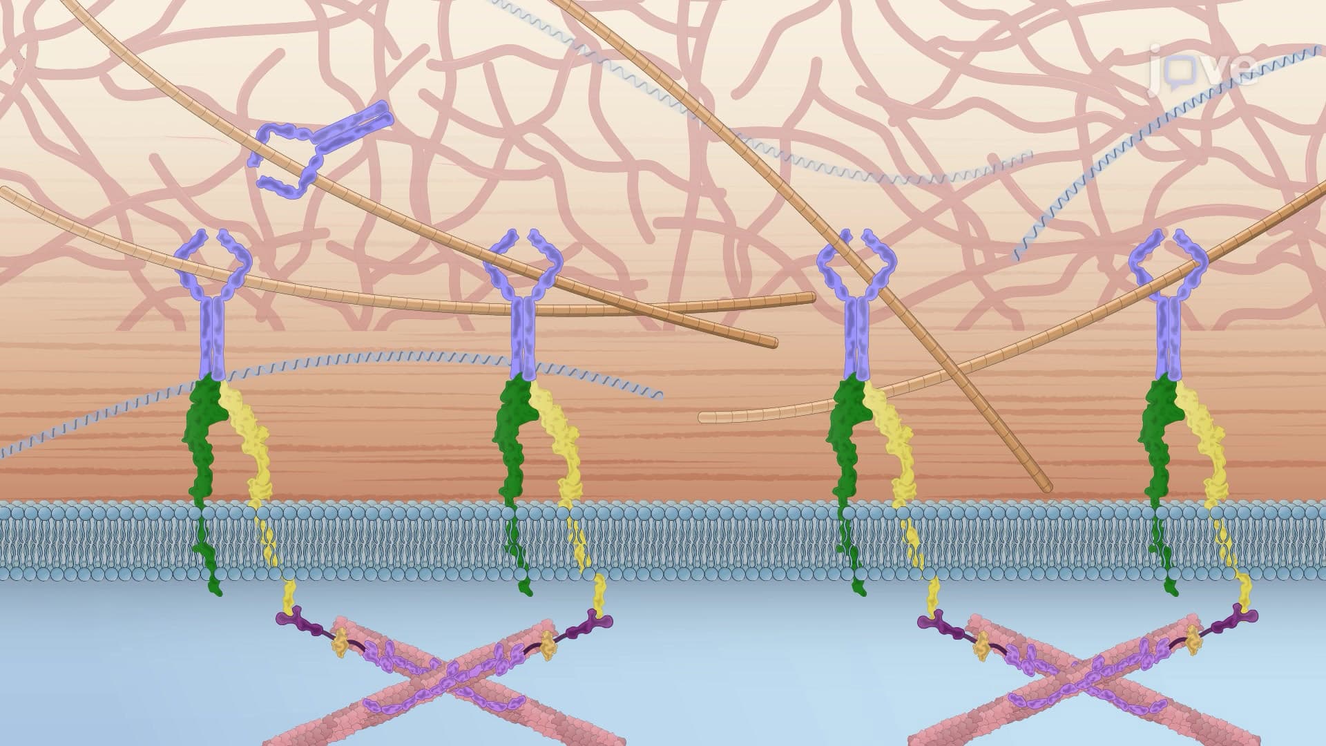
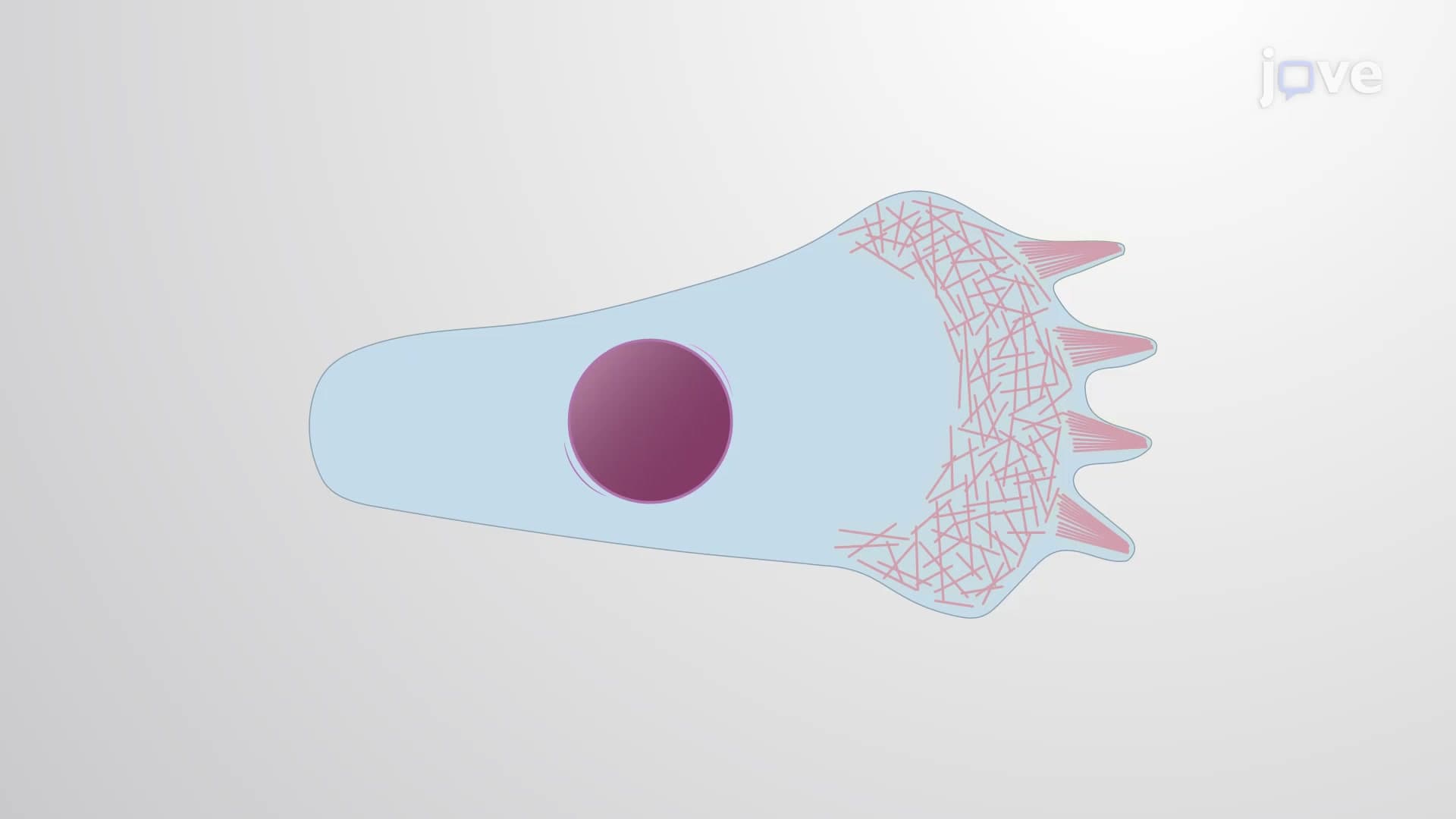
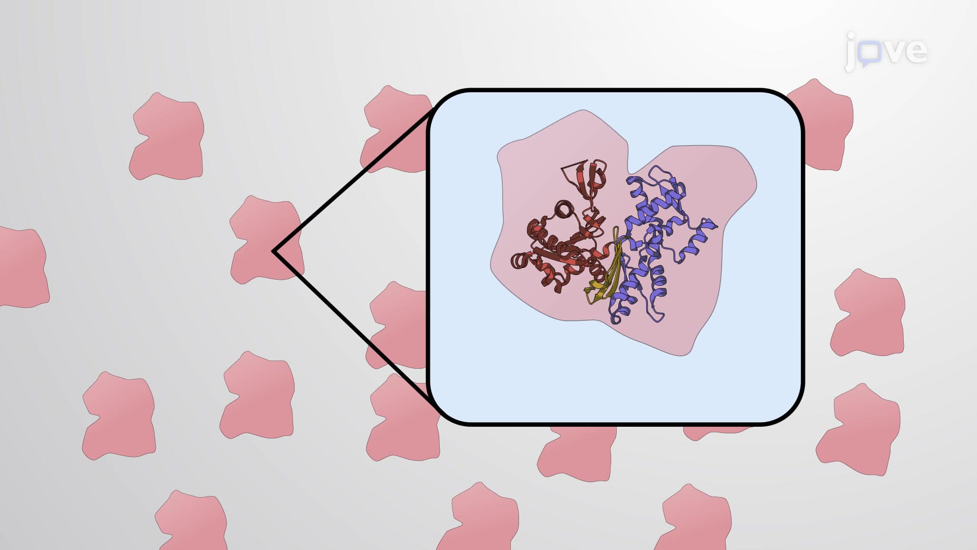
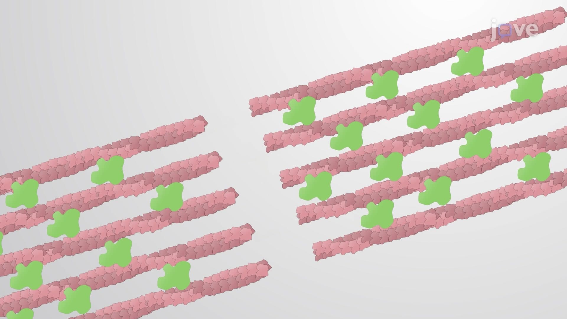
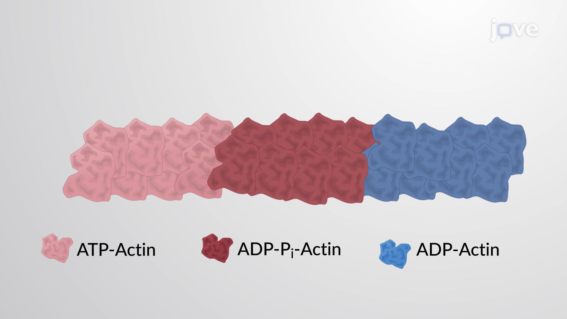
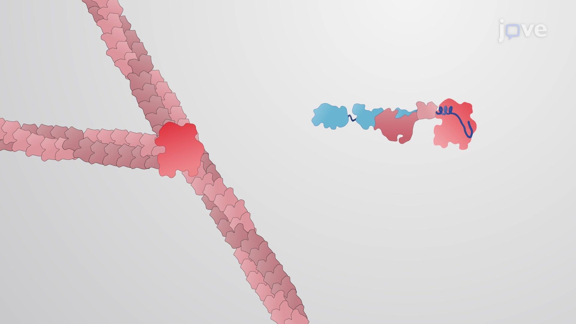
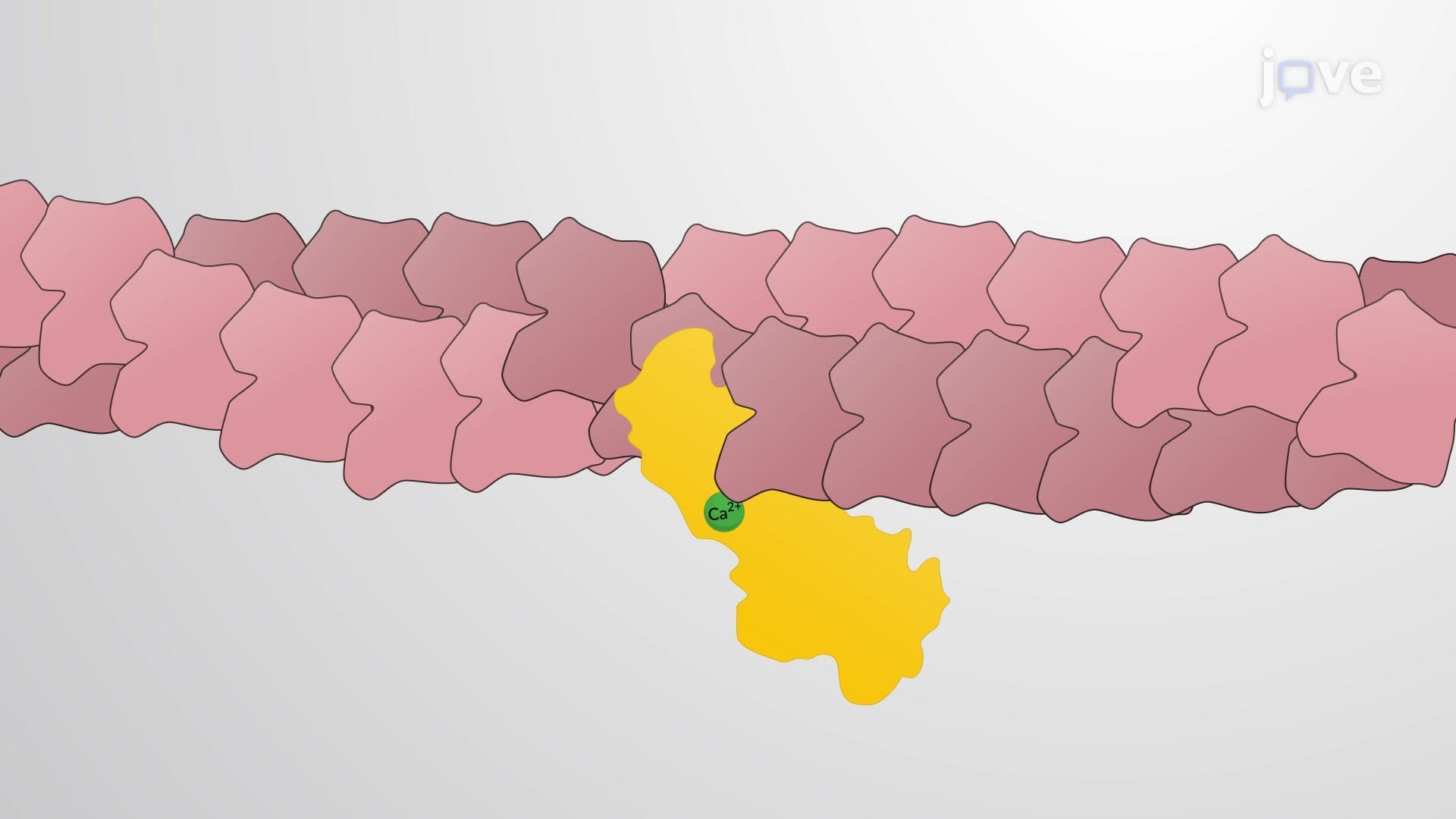
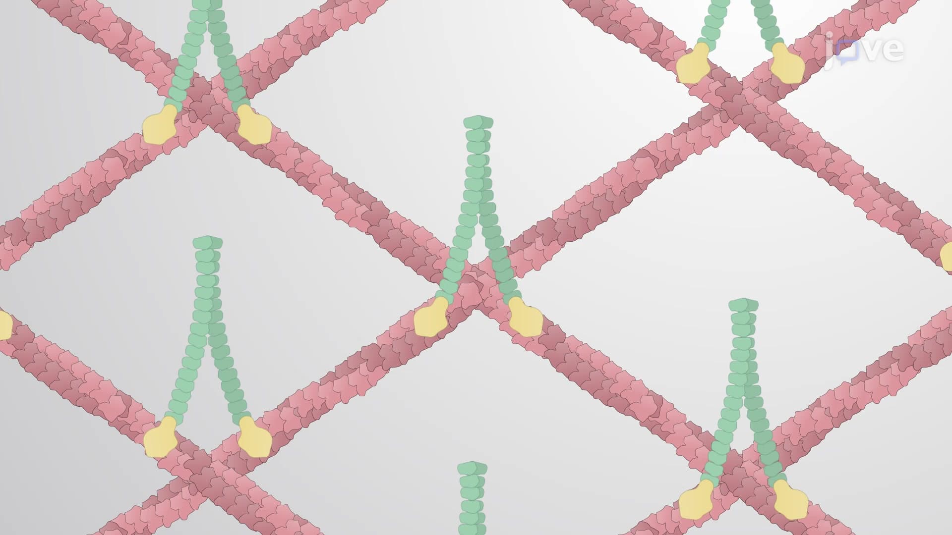
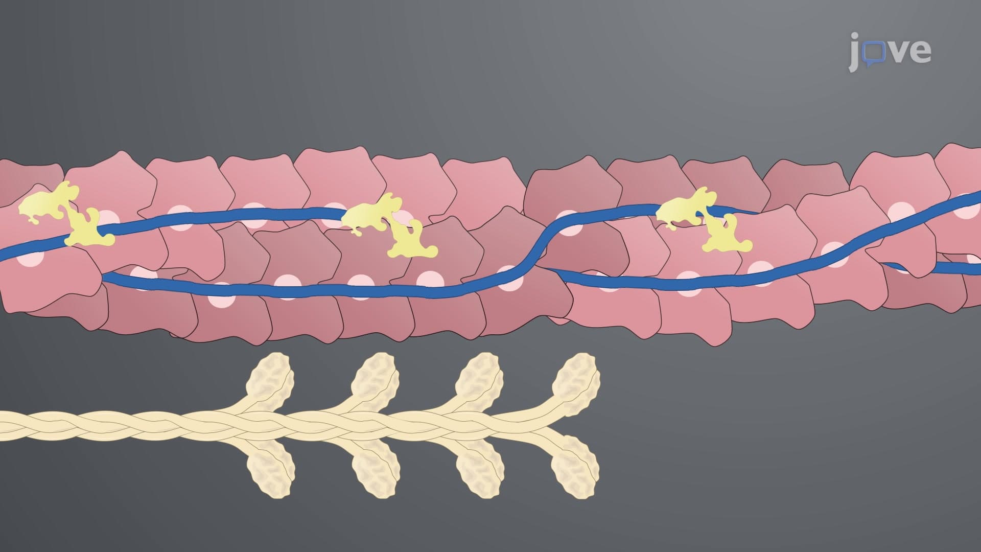
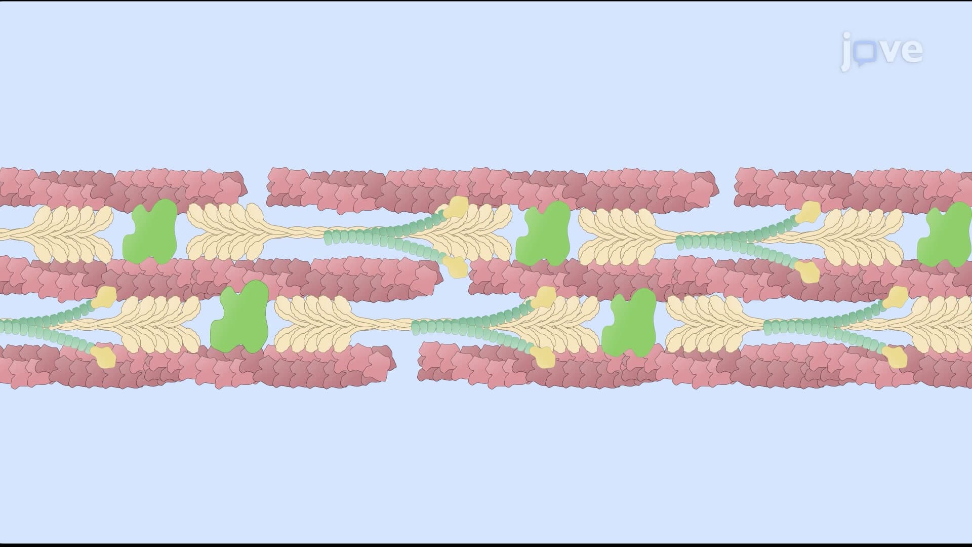
Microfilaments, composed of actin protein polymers, form the thinnest component of the cytoskeletal network. These structures are particularly abundant in muscle cells, where they interact with myosin proteins to generate the contractile force responsible for muscle movement. In non-muscle cells, microfilaments concentrate near the cell membrane, creating a cortical network that maintains cell shape and enables processes like cell crawling during immune responses.
Microtubules serve as the "highways" of the cell, formed by tubulin protein subunits arranged in hollow tubes. These structures extend from organizing centers called centrosomes and provide tracks for molecular motors that transport cellular cargo. During cell division, microtubules reorganize to form the mitotic spindle, a critical structure that ensures accurate chromosome distribution to daughter cells-a process frequently tested on MCAT and advanced placement exams.
Intermediate filaments provide mechanical strength and act as cellular "shock absorbers." Their composition varies dramatically between cell types, making them valuable diagnostic markers in medical practice. For instance, keratin intermediate filaments in skin cells provide the durability needed to protect against physical damage, while neurofilaments in neurons maintain axon structure across distances that can exceed three feet in humans.
The cytoskeleton study guide applications extend far beyond academic learning. In cancer research, understanding cytoskeletal dynamics helps explain how tumor cells acquire the ability to invade surrounding tissues and metastasize. Pharmaceutical companies develop drugs that target cytoskeletal components, such as taxanes used in chemotherapy that stabilize microtubules and prevent cancer cell division.
For pre-medical students, recognizing cytoskeletal pathologies becomes crucial for future clinical practice. Conditions like Alzheimer's disease involve cytoskeletal protein abnormalities, while genetic disorders affecting intermediate filaments can cause skin fragility diseases studied extensively at institutions like the National Institutes of Health.
Related Micro-courses