Loading subject page…


27,706 views

Video Summary: What Is Activation of Integrins
Did you know that your immune cells constantly patrol your bloodstream, ready to squeeze through blood vessel walls to fight infection? The activation of integrins makes this cellular migration possible by allowing cells to rapidly form and break attachments with their surroundings. When a white blood cell encounters inflammation signals at a wound site in your body, integrins switch between active and inactive states to enable precise movement through tissue barriers. This molecular on-off switch is essential for immune responses, wound healing, and tissue repair throughout the human body. Watch the full video on JoVE Coach to master this concept with expert-led visuals and step-by-step explanations.
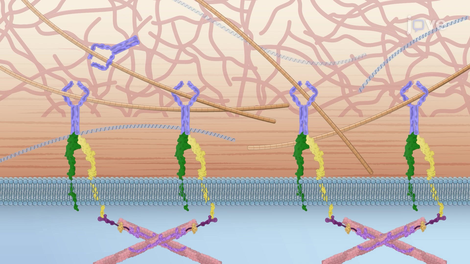
Activation of integrins represents one of biology's most elegant molecular switches, controlling when and where cells can attach to their surrounding environment. Integrins are transmembrane proteins that span the cell membrane, connecting the cell's internal cytoskeleton to the external extracellular matrix (ECM). This connection is crucial for processes ranging from embryonic development to immune system function.
The concept of integrin activation is fundamental to understanding cellular biology and appears frequently in advanced placement biology courses and pre-medical education. Students preparing for the MCAT will encounter questions about integrin function in the context of cell signaling and tissue organization.
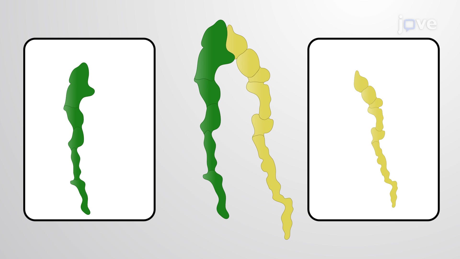
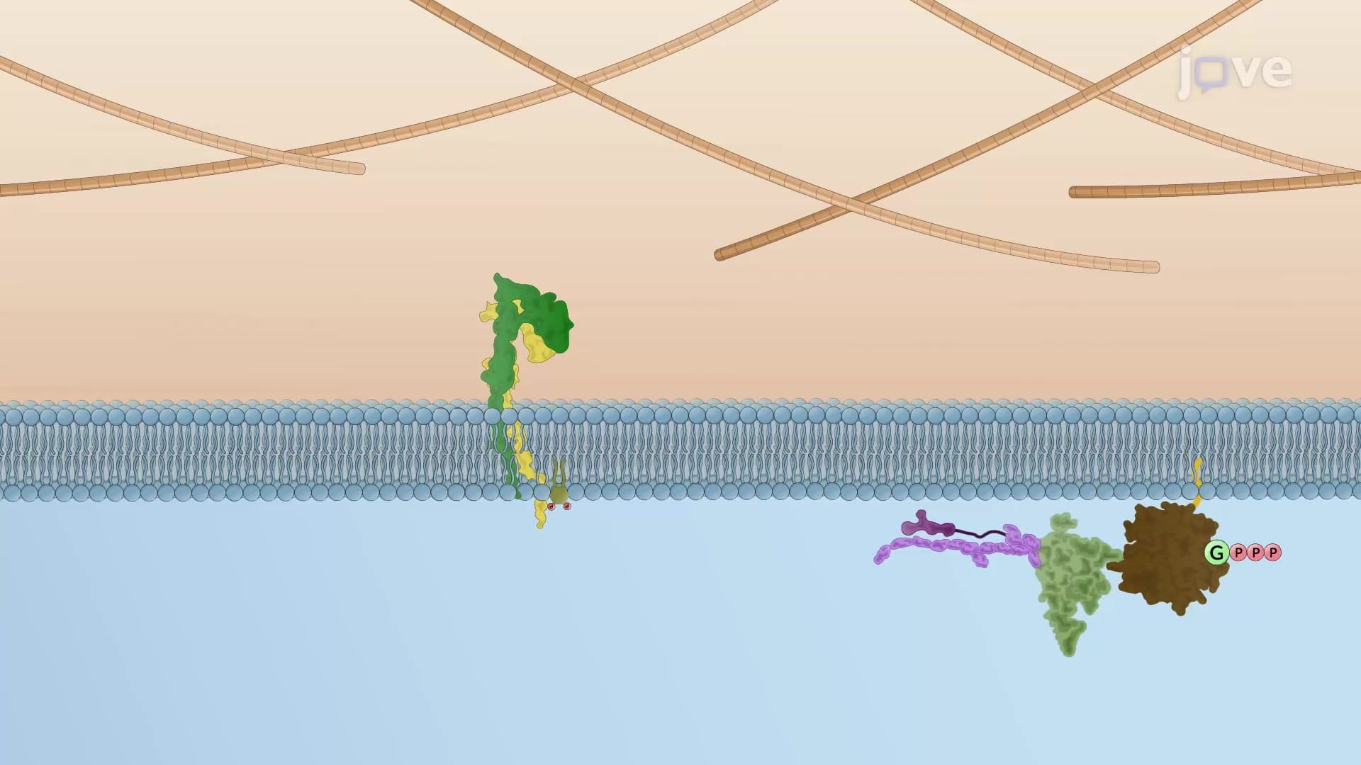
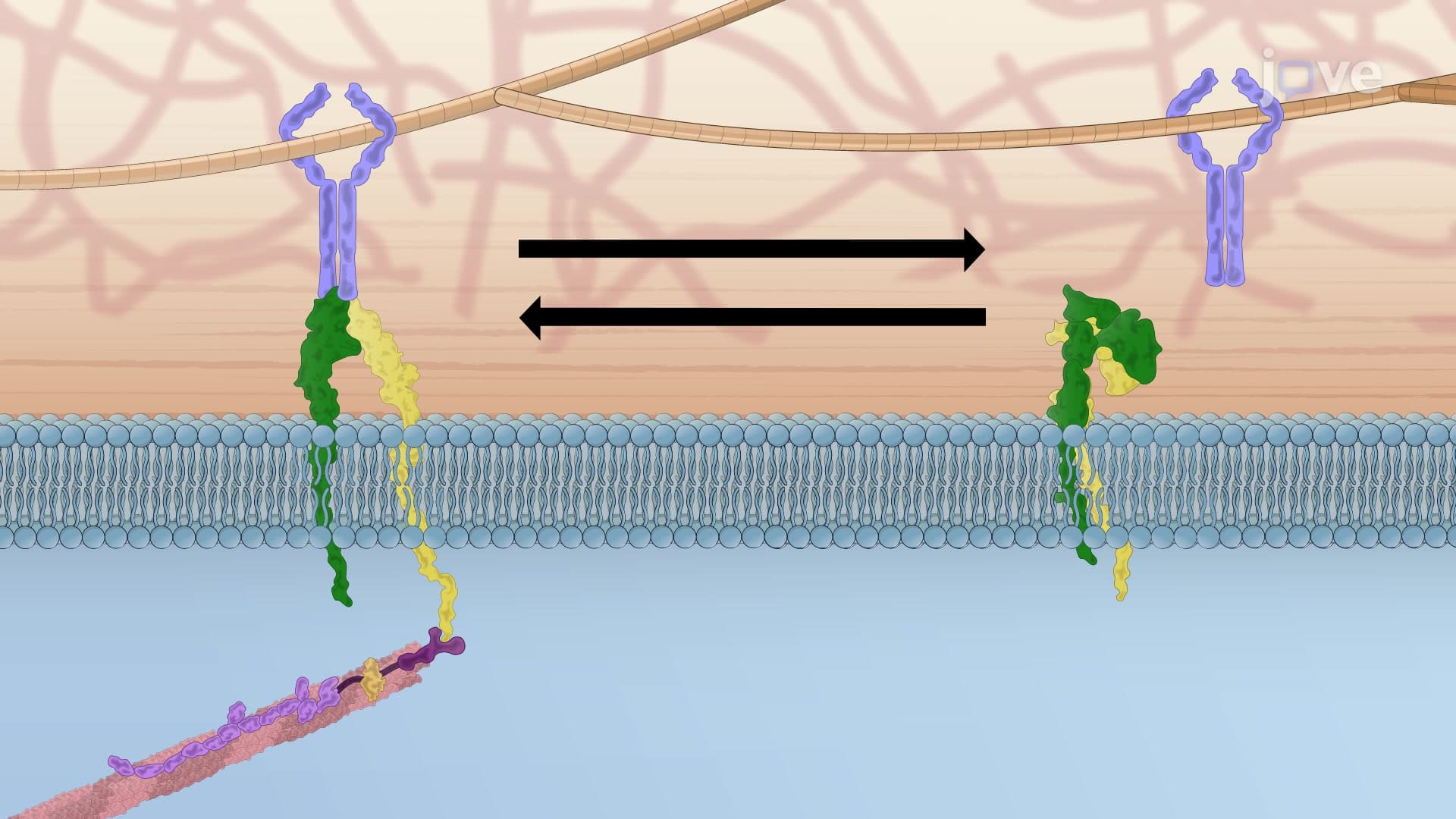
In their inactive state, integrins adopt a compact, bent conformation where the alpha and beta subunit tails remain tightly associated. This locked configuration prevents the integrin from binding to ECM proteins or connecting to internal cytoskeletal elements. Think of inactive integrins as molecular "safety locks" that prevent unwanted cellular adhesions.
Upon activation, integrins undergo dramatic conformational changes, extending outward from the cell surface and exposing binding sites previously hidden in the inactive state. This structural transformation is comparable to a spring-loaded mechanism being released, allowing the integrin to engage with its molecular partners both inside and outside the cell.
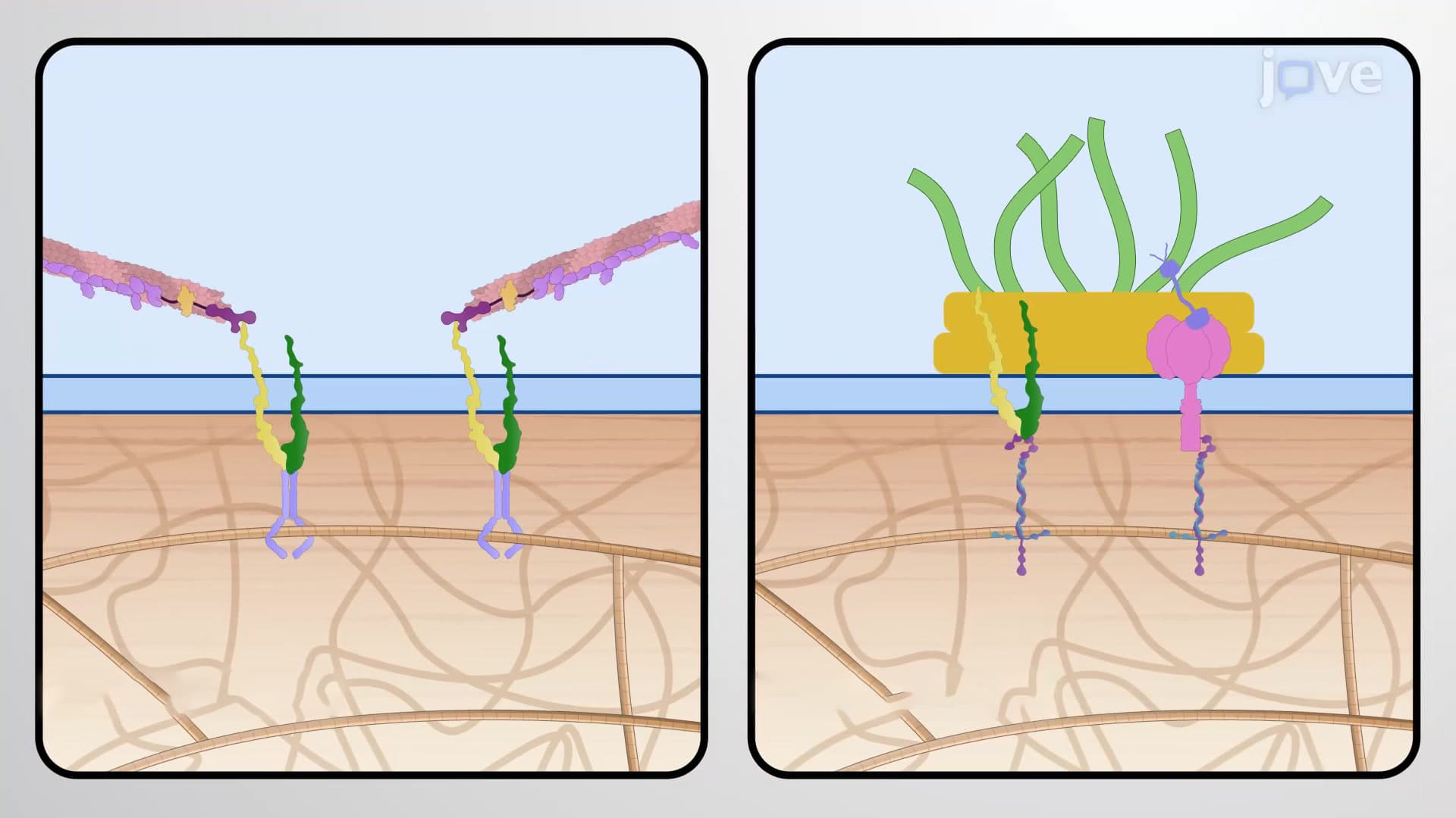
The outside-in pathway begins when ECM proteins such as fibronectin, collagen, or laminin bind to the integrin's extracellular domain. This binding event triggers a cascade of conformational changes that propagate through the entire integrin structure. As the extracellular domains engage their targets, the transmembrane regions shift position, causing the cytoplasmic tails to separate and expose binding sites for intracellular adaptor proteins.
This mechanism is particularly important in wound healing, where platelets use integrin activation to form blood clots. When platelets encounter damaged blood vessel walls, fibrinogen in the blood plasma binds to platelet integrins, triggering their activation and enabling clot formation.
Conversely, the inside-out pathway is initiated by intracellular signaling molecules. Adaptor proteins like talin bind to the beta integrin subunit's cytoplasmic tail, physically separating the alpha and beta chains. This separation forces the integrin into its extended, active conformation, ready to engage ECM proteins.
Inside-out activation is crucial during immune responses. When T-cells recognize foreign antigens, internal signaling cascades activate integrins on the cell surface, enabling these immune cells to adhere to blood vessel walls and migrate into infected tissues. Understanding this process is essential for students studying immunology and preparing for medical school entrance exams.
Related Micro-courses