Loading subject page…


15 Concepts
12 Concepts
22 Concepts
12 Concepts
10 Concepts
12 Concepts
10 Concepts
17 Concepts
18 Concepts
11 Concepts
14 Concepts
16 Concepts
14 Concepts
18 Concepts
20 Concepts
18 Concepts
21 Concepts
17 Concepts
12 Concepts
7 Concepts
12 Concepts
10 Concepts
6 Concepts
6 Concepts
18 Concepts
19 Concepts
11 Concepts
6 Concepts
12 Concepts
11 Concepts
7 Concepts
20 Concepts
17 Concepts
11 Concepts
17 Concepts
6 Concepts
8 Concepts
21 Concepts
13 Concepts
9 Concepts
8 Concepts
8 Concepts
8 Concepts
 10 Concepts
 76.5K Views
 Popular topics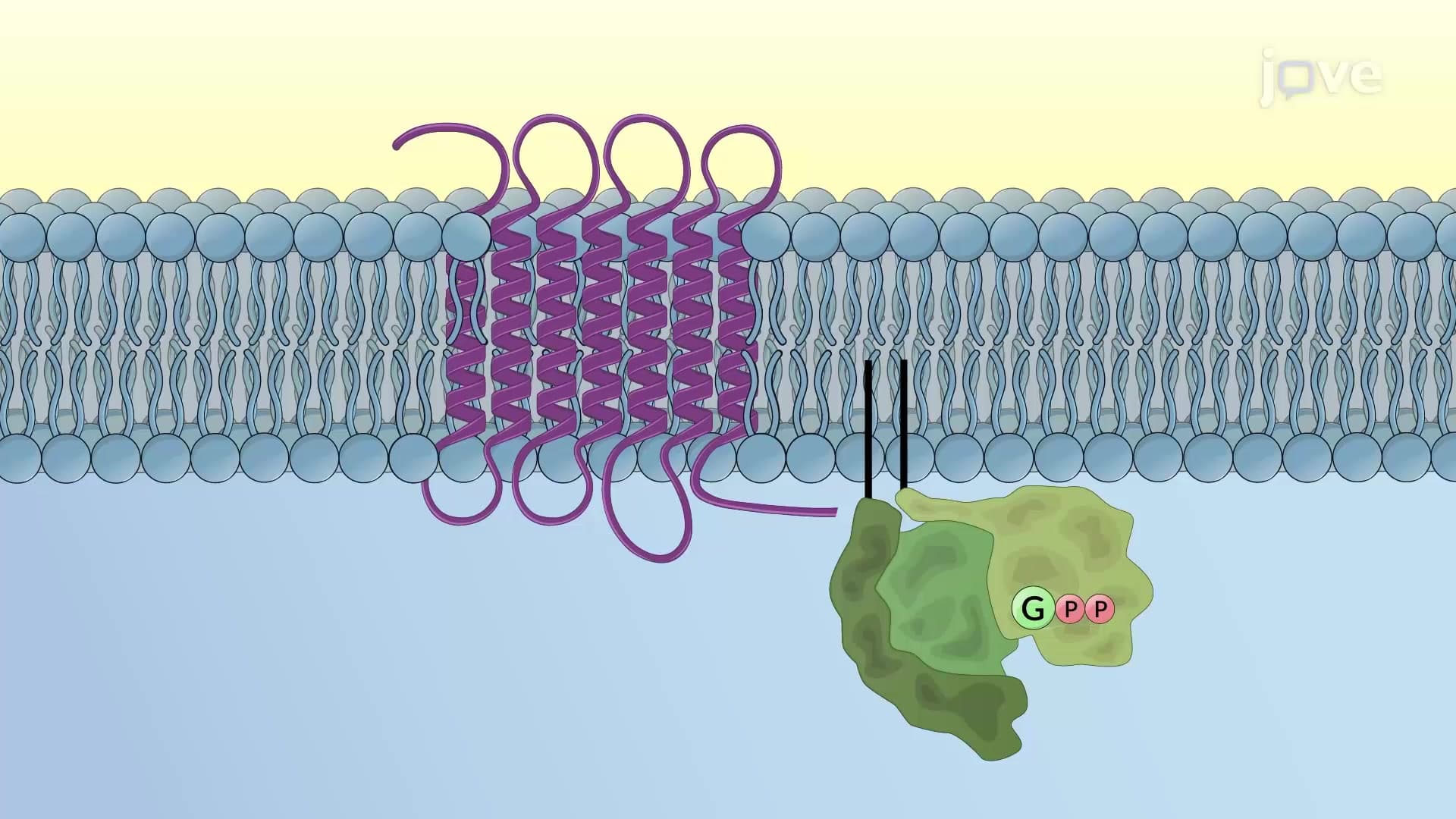


GPCR signaling forms the foundation of cellular communication in the human body, enabling responses to hormones, neurotransmitters, and sensory stimuli. This comprehensive course explores how G protein coupled receptors transmit signals through adenylyl cyclase, phospholipase C pathway, and ion channel regulation. Students will master the molecular mechanisms behind heart rate control, blood sugar regulation, and muscle contraction-essential knowledge for MCAT preparation and advanced biology coursework. JoVE Coach provides detailed animations of each signaling cascade.
1. GPCR Structure and Ligand Binding G protein coupled receptors contain seven transmembrane alpha-helices with extracellular ligand-binding domains and intracellular G protein-binding regions. When hormones like glucagon or neurotransmitters like acetylcholine bind these receptors, conformational changes expose the G protein binding site. This structural transformation initiates signal transduction cascades that regulate processes from blood glucose homeostasis in diabetic patients to heart rate control during exercise. Understanding GPCR structure helps explain how medications like beta-blockers work by competing with natural ligands at receptor binding sites.
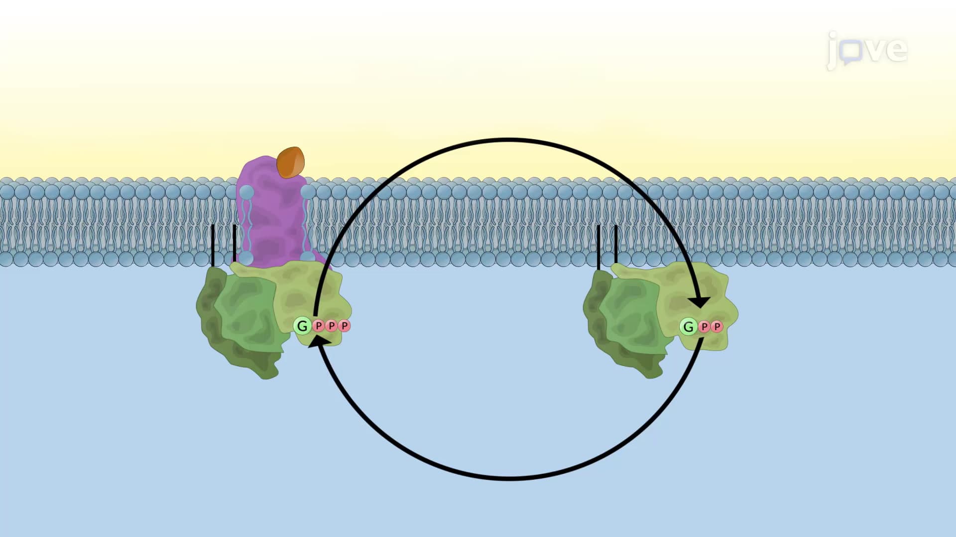
2. G Protein Activation and GTP/GDP Exchange Heterotrimeric G proteins cycle between inactive GDP-bound and active GTP-bound states to relay cellular signals. The alpha subunit exchanges GDP for GTP upon GPCR activation, causing dissociation from beta-gamma subunits and enabling interaction with downstream effectors like adenylyl cyclase. This molecular switch mechanism amplifies weak extracellular signals into robust intracellular responses. GTPase activity eventually hydrolyzes GTP to GDP, terminating the signal and allowing G protein reassembly. This cycling process explains how stimulant medications can have temporary effects before cellular responses diminish.
3. GPCR Desensitization and Receptor Regulation Prolonged hormone exposure triggers protective desensitization mechanisms involving GPCR kinases and beta-arrestin proteins. Phosphorylated receptors bind beta-arrestin, blocking additional G protein interactions and facilitating receptor internalization through clathrin-mediated endocytosis. This process prevents cellular overstimulation during chronic stress responses or drug treatments. Internalized receptors can be recycled to the plasma membrane or degraded in lysosomes, regulating cellular sensitivity to hormones like insulin or adrenaline. Understanding desensitization mechanisms helps explain drug tolerance development and the need for medication dosage adjustments.
4. Ion Channel Regulation and Membrane Potential GPCRs directly control ion channel activity to modulate cellular excitability and membrane potential changes. When acetylcholine binds heart muscle GPCRs, activated Gi proteins open potassium channels, causing hyperpolarization and slowing heart rate during rest periods. Simultaneously, Gi inhibits adenylyl cyclase, reducing cAMP levels and closing calcium channels. This dual mechanism explains how parasympathetic nervous system activation promotes cardiovascular relaxation after exercise. These pathways are targeted by cardiac medications and demonstrate how single receptor activation can coordinate multiple cellular responses.
5. Adenylyl Cyclase and cAMP Signaling Pathways Stimulatory G proteins activate adenylyl cyclase to convert ATP into the second messenger cyclic AMP, which amplifies hormonal signals throughout the cell. cAMP activates protein kinase A, leading to phosphorylation of metabolic enzymes and transcription factors like CREB. This cascade regulates glucose metabolism during fasting states and stress responses. PKA phosphorylates glycogen phosphorylase kinase to promote glucose release and inhibits glycogen synthase to prevent glucose storage. Understanding cAMP signaling explains how hormones like glucagon and epinephrine coordinate the body's response to low blood sugar emergencies.
6. Phospholipase C, IP3, and DAG Signaling Gq-coupled receptors activate phospholipase C-beta to cleave PIP2 into two second messengers: membrane-bound DAG and diffusible IP3. IP3 triggers calcium release from endoplasmic reticulum stores, while DAG activates protein kinase C at the plasma membrane. This dual-messenger system coordinates cellular responses requiring both calcium mobilization and protein phosphorylation. The pathway mediates platelet aggregation during blood clotting, smooth muscle contraction in blood vessels, and hormone secretion from endocrine glands. Dysregulation of this signaling system contributes to cardiovascular disease and metabolic disorders commonly studied in medical curricula.
7. Calcium Regulation and Cellular Oscillations Intracellular calcium levels are precisely controlled through positive and negative feedback mechanisms that generate oscillatory patterns. IP3-gated calcium channels on the ER membrane create calcium waves that propagate through the cytoplasm, triggering cellular responses like muscle contraction and neurotransmitter release. When calcium levels become excessive, channels close and plasma membrane pumps restore resting concentrations. These oscillations enable repetitive cellular functions like heartbeat rhythms and breathing patterns. Understanding calcium signaling explains how medications affect muscle function and why calcium channel blockers are effective treatments for hypertension and cardiac arrhythmias.
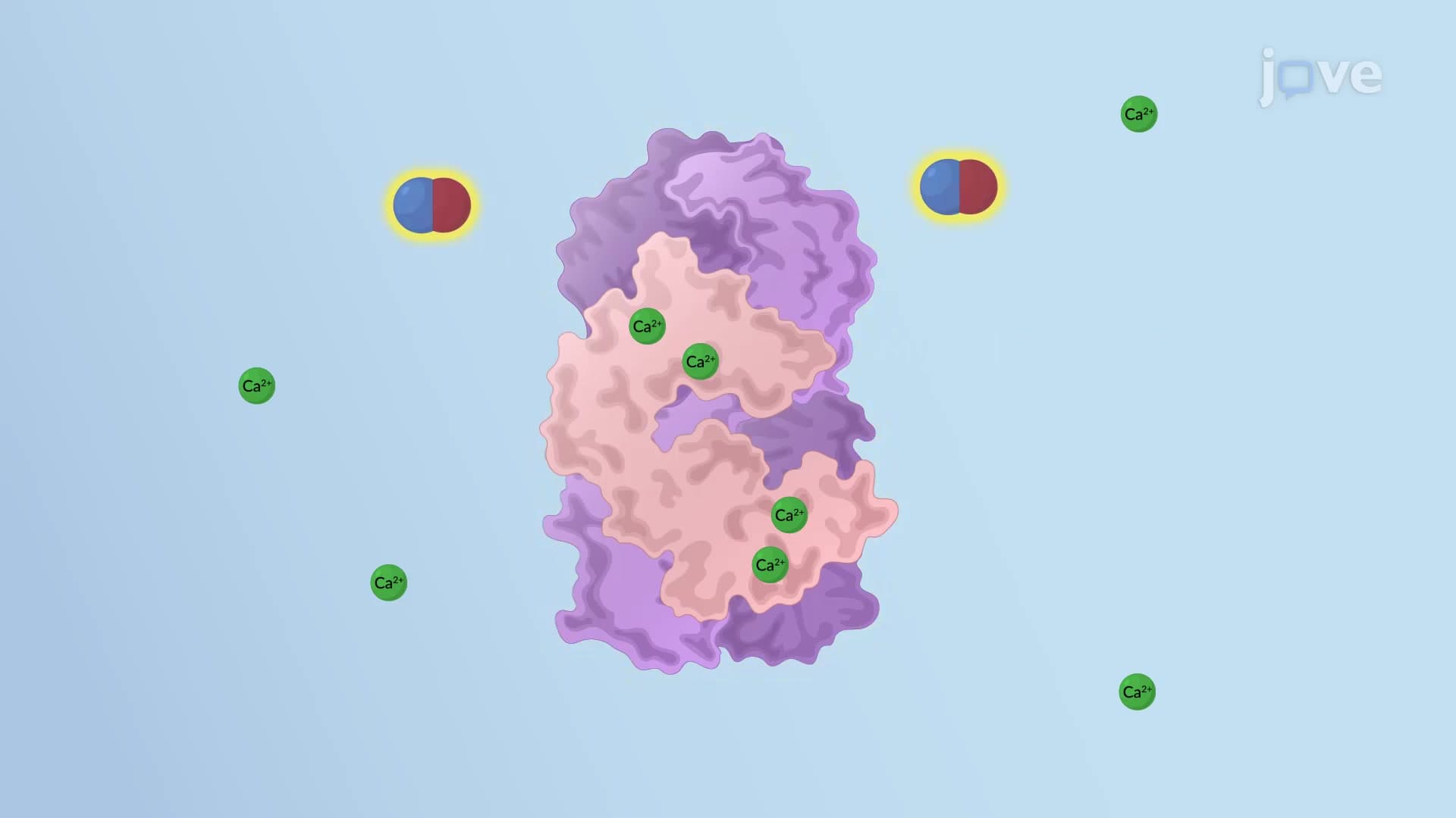
8. Calmodulin-Dependent Signaling Mechanisms Calcium-sensing protein calmodulin undergoes conformational changes upon binding four calcium ions, enabling high-affinity interactions with target proteins throughout the cell. The calcium-calmodulin complex activates diverse enzymes including calcium pumps, phosphodiesterases, and protein kinases to coordinate cellular responses. This versatile signaling system regulates muscle contraction, neurotransmitter release, and immune cell activation. Calmodulin also provides cross-talk between signaling pathways by activating phosphodiesterase to degrade cAMP, demonstrating how cells integrate multiple simultaneous signals. These mechanisms are essential for understanding neurological function and muscle physiology in health science curricula.
9. Nitric Oxide Signaling and Vascular Function Nitric oxide serves as a unique gaseous second messenger that diffuses between cells to regulate smooth muscle relaxation and blood vessel dilation. Endothelial cells produce NO through calcium-calmodulin activation of NO synthase, and this molecule crosses cell membranes to activate guanylyl cyclase in neighboring smooth muscle cells. The resulting cGMP activates protein kinase G, which promotes muscle relaxation through myosin dephosphorylation and calcium channel inhibition. This pathway explains how blood vessels dilate during exercise to increase tissue perfusion and how medications like nitroglycerin treat heart conditions by promoting coronary artery dilation.