Loading video…


 14 Concepts
 25.7K views

Video Summary: Assessment Cardiovascular System Iv Auscultation Explained
Did you know that a skilled nurse can detect heart failure just by listening to specific sounds through a stethoscope? Assessment Cardiovascular System IV Auscultation involves systematically listening to heart sounds at precise chest locations to identify normal and abnormal cardiac function. At Johns Hopkins Hospital, emergency room physicians use this technique daily to rapidly assess patients with chest pain, distinguishing between normal S1 and S2 sounds and pathological S3 and S4 sounds that indicate serious conditions. Watch the full video on JoVE Coach to master this concept with expert-led visuals and step-by-step explanations.
Assessment Cardiovascular System IV Auscultation represents a cornerstone skill in healthcare education, combining anatomical knowledge with practical clinical technique. This systematic approach to cardiac assessment requires understanding both the underlying physiology of heart valve function and the precise methodology for detecting abnormalities. Students studying for the MCAT or preparing for nursing school through the HESI A2 exam must master this concept as it bridges theoretical cardiovascular knowledge with hands-on clinical application.
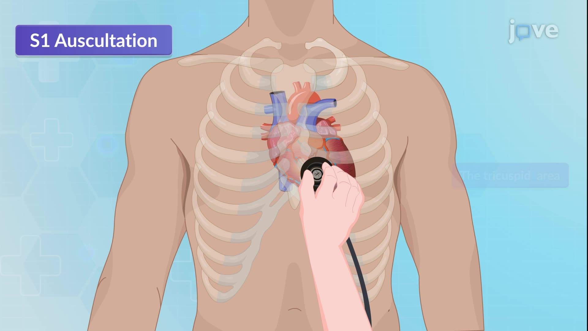
The systematic nature of cardiac auscultation relies on specific anatomical landmarks that correspond to optimal listening points for different heart valves. The tricuspid area, located at the fourth left intercostal space, provides the clearest assessment of the tricuspid valve, while the mitral area at the fifth intercostal space along the left midclavicular line offers optimal mitral valve evaluation. Advanced Placement Biology students often struggle with memorizing these locations, but understanding that these sites represent areas where sound transmission is most direct helps with retention.
The aortic and pulmonic areas, both positioned in the second intercostal space on the right and left respectively, allow healthcare providers to assess semilunar valve function. At Massachusetts General Hospital, medical students learn that Erb's point in the third left intercostal space serves as a unique location where S1 and S2 sounds achieve equal intensity, making it invaluable for distinguishing between these fundamental heart sounds.
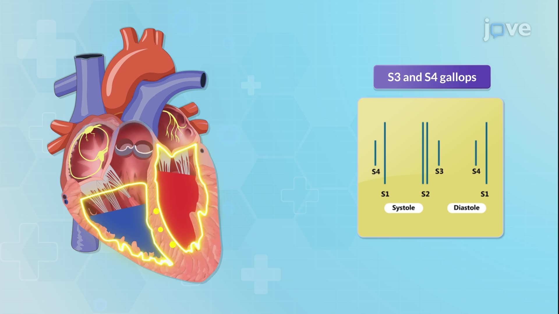
The distinction between normal and abnormal heart sounds carries profound clinical implications that extend far beyond academic study. S3 heart sounds, often called "gallop rhythms," indicate volume overload conditions like congestive heart failure, a condition affecting over 6 million Americans according to the CDC. This low-pitched sound requires the stethoscope's bell for detection and represents a critical diagnostic finding that can guide treatment decisions in emergency departments across the United States.
S4 heart sounds signal decreased ventricular compliance, commonly associated with left ventricular hypertrophy resulting from chronic hypertension. For students preparing for the NCLEX examination, understanding that S4 sounds indicate a "stiff" ventricle helps connect pathophysiology with clinical findings. These abnormal sounds typically manifest at the cardiac apex, making proper patient positioning crucial for accurate assessment.
Successful cardiac auscultation demands technical proficiency with stethoscope components and patient positioning. The diaphragm component detects high-frequency sounds like normal S1 and S2, while the bell captures low-frequency sounds including pathological S3 and S4. Students at UCLA Medical School learn that proper seal and pressure application significantly impact sound quality and diagnostic accuracy.
Patient positioning enhances sound transmission, with the left lateral decubitus position bringing the heart closer to the chest wall, particularly beneficial for detecting mitral valve abnormalities. This positioning technique proves especially valuable during USMLE Step 2 CS examinations, where students must demonstrate clinical competency under standardized conditions.
Related Micro-courses